. 2014 Dec 11;9(5):1718-1728.
doi: 10.1016/j.celrep.2014.11.011. Epub 2014 Dec 4.

A distal locus element mediates IFN-γ priming of lipopolysaccharide-stimulated TNF gene expression

Affiliations

A distal locus element mediates IFN-γ priming of lipopolysaccharide-stimulated TNF gene expression

Nancy A Chow et al. Cell Rep. .

Abstract

Interferon γ (IFN-γ) priming sensitizes monocytes and macrophages to lipopolysaccharide (LPS) stimulation, resulting in augmented expression of a set of genes including TNF. Here, we demonstrate that IFN-γ priming of LPS-stimulated TNF transcription requires a distal TNF/LT locus element 8 kb upstream of the TNF transcription start site (hHS-8). IFN-γ stimulation leads to increased DNase I accessibility of hHS-8 and its recruitment of interferon regulatory factor 1 (IRF1), and subsequent LPS stimulation enhances H3K27 acetylation and induces enhancer RNA synthesis at hHS-8. Ablation of IRF1 or targeting the hHS-8 IRF1 binding site in vivo with Cas9 linked to the KRAB repressive domain abolishes IFN-γ priming, but does not affect LPS induction of the gene. Thus, IFN-γ poises a distal enhancer in the TNF/LT locus by chromatin remodeling and IRF1 recruitment, which then drives enhanced TNF gene expression in response to a secondary toll-like receptor (TLR) stimulus.


Figures

Figure 1
Figure 1
IFN-γ priming promotes chromatin accessibility at hHS-8 in the TNF/LT locus. (A) IFN-γ priming enhances TNF mRNA levels in THP-1 cells stimulated with LPS. Cells were stimulated with IFN-γ alone for 3h, LPS alone for 1h, and both IFN-γ and LPS (IFN-γ for 2h followed by LPS for 1h). TNF mRNA levels were measured post LPS stimulation by q-PCR. (**) p ≤ 0.01, data is represented as mean ± SEM. (B, C) IFN-γ increases chromatin accessibility hHS-8. DHAs using the restriction enzyme ScaI (B) and BamHI (C) allowed for examination of hHS-8 and the TNF promoter, respectively, in resting and IFN-γ-treated THP-1 cells. (D) IFN-γ priming enhances TNF mRNA levels in primary human MDMs stimulated with LPS. MDMs were stimulated as in (A) and RNA was collected 1h after LPS stimulation. Data from 3 separate donors, (*) p ≤ 0.05, data is represented as mean ± SEM. (E, F) DHAs were performed in resting and IFN-γ-treated primary human MDMs as in (B, C). (G) Map of the human TNF/LT locus. DH sites and positions and directions of transcription of the TNF, LTA, and LTB genes are shown. Positions of the parental ScaI, parental BamHI, and DNase I digestion products for the DHAs are indicated. (H, I) IFN-γ and LPS decreases nucleosome occupancy at the TNF promoter and hHS-8. ChIP using THP-1 cells (H) and primary human MDMs (representative donor, I) measures nucleosome occupancy (total H3 levels) at both the TNF promoter and hHS-8. Although not significant, IFN-γ alone decreases total H3 levels at the TNF promoter (p=0.054) in THP-1 cells; this was not repeated in the MDM donor. Data from 3 separate experiments, (*) p ≤ 0.05 and (***) p ≤ 0.001, data are represented as mean ± SEM.
Figure 2
Figure 2
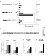
IRF1 binds to hHS-8 in an IFN-γ-inducible manner. (A) TNF/LT locus with partial sequences and binding site positions of transcription factors for both the TNF promoter and hHS-8. (B) rIRF1 binds to the TNF promoter. Quantitative DNase I footprinting analysis of the TNF promoter (−200 to +1) and increasing concentrations of rIRF1. Sense and anti-sense strand with G/A ladder and BSA control. Bars mark areas of rIRF1 binding at −172 to -136. (C) rIRF1 binds to hHS-8. Quantitative DNase I footprinting analysis of hHS-8 (−7031 to −6782) was performed as in (B). Bars mark areas of rIRF1 binding at −6833 to −6782. (D) IRF1 is recruited to hHS-8 in an IFN-γ-inducible manner. ChIP using primary human MDMs and analyzing IRF1 recruitment to the TNF promoter and hHS-8. Data from 3 separate donors, (*) p ≤ 0.05 and (**) p ≤ 0.01, data is represented as mean ± SEM. (E) IRF1 binding sites in hHS-8 are highly conserved in all primate species examined. Critical 5′-GAAA-3′ motifs for IRF1 binding are highlighted. (F, G) Enhanced TNF expression induced by IFN-γ priming is abrogated in IRF1-deficient murine BMDMs. Wild-type (F) and Irf1−/− (G) BMDMs were stimulated, and TNF protein levels in supernatants were measured by ELISA post LPS stimulation. Data from 3 separate experiments each with N=3, (*) p ≤ 0.05 and (***) p ≤ 0.001, data is represented as mean ± SEM. (H) IRF1 mRNA levels induced by IFN-γ are silenced by IRF1 shRNA. THP-1 cells that constitutively express lentivirally delivered shRNA targeting IRF1 or control shRNA encoding a scrambled sequence were stimulated with IFN-γ alone for 3h. Data from 3 separate experiments, (*) p ≤ 0.05, data is represented as mean ± SEM. (I) Enhanced TNF gene expression induced by IFN-γ priming is abrogated in human monocytes where IRF1 expression is silenced. THP-1 cells expressing IRF1 and control shRNA were stimulated, and TNF mRNA levels were measured (shown relative to LPS values). Data from 3 separate experiments, (*) p ≤ 0.05, data is represented as mean ± SEM.
Figure 3
Figure 3
hHS-8 functions as an IFN-γ-inducible, IRF1-dependent enhancer of TNF gene expression. (A) Disruption of IRF1 binding to hHS-8 abolishes inducible enhancer function and thus enhanced TNF gene expression induced by IFN-γ priming. Constructs using the pGL3-Basic luciferase vector were transfected into J774 cells and stimulated with IFN-γ alone for 8h, LPS alone for 6h, and both IFN-γ and LPS (IFN-γ 2h followed by LPS for 6h). “TNF” is the TNF promoter, “hHS-8” is the entire sequence of hHS-8 (1250bp), and “muthHS-8” is hHS-8 with mutations that disrupt IRF1 binding. Data from 3 separate experiments; (***) indicates p ≤ 0.001; data is represented as mean ± SEM. (B) Nucleotide changes in the critical 5′-GAAA-3′ motifs disrupt rIRF1 binding. EMSA was performed with rIRF1 and wild-type and mutant radiolabeled P32 oligonucleotides (sequences for positions −6838 to −6785). (C) Activation of hHS-8 enhancer function corresponds with increased H3K27ac prevalence at hHS-8. ChIP analyzing H3K27ac prevalence was performed using THP-1 cells and analyzing H3K27ac prevalence at the TNF promoter and hHS-8. Data from 3 separate experiments, (*) p ≤ 0.05, data is represented as mean ± SEM. (D, E) IFN-γ + LPS induces hHS-8 eRNA transcription. hHS-8 eRNA (RNA sequence containing IRF1 binding sites) were measured post LPS stimulation in THP-1 cells (D) and primary human MDMs (representative donor, E) Data from 3 separate experiments; (*) indicates p ≤ 0.05; data are represented as mean ± SEM.
Figure 4
Figure 4
hHS-8 IRF1 binding sites are required for IFN-γ priming of TNF gene expression in vivo. (A) Flow cytometry data demonstrating that >95% of THP-1 cells were successfully transduced with the CRISPR-Ctrl, CRISPR-TNFp, and CRISPR-hHS-8 lentiviruses at the time of experimental analysis. (B) Targeting of the TNF promoter with dCas9-KRAB. CRISPR-Ctrl and CRISPR-TNFp THP-1 cells were mock-stimulated or stimulated with LPS for 1h and TNF and IL-6 mRNA were quantitated after normalization to the housekeeper cyclophilin B. Data from at least 3 independent experiments are shown; (***) indicates p ≤ 0.001; data are represented as mean ± SD. (C) Targeting hHS-8 with dCas9-KRAB blocks priming of TNF. CRISPR-Ctrl and CRISPR-hHS-8 THP-1 cells were mock-stimulated, stimulated with LPS for 1h, or stimulated with IFN-γ for 2h and LPS for 1h. For analysis of TNF expression, data are presented as fold inductions over unstimulated TNF mRNA levels to control for baseline constitutive TNF transcription in THP-1 cells, while for analysis of IL6 expression data are presented as fold induction of primed versus non-primed conditions due to the absence of detectable IL-6 transcripts in the absence of stimulation. Data from 3 independent experiments are presented; (**) indicates p<0.01; data are represented as mean ± SD

References

    1. Baena A, Mootnick AR, Falvo JV, Tsytskova AV, Ligeiro F, Diop OM, Brieva C, Gagneux P, O’Brien SJ, Ryder OA, Goldfeld AE. Primate TNF promoters reveal markers of phylogeny and evolution of innate immunity. PloS one. 2007;2:e621. - PMC - PubMed
    1. Barthel R, Goldfeld AE. T cell-specific expression of the human TNF-alpha gene involves a functional and highly conserved chromatin signature in intron 3. J Immunol. 2003;171:3612–3619. - PubMed
    1. Biglione S, Tsytsykova AV, Goldfeld AE. Monocyte-specific accessibility of a matrix attachment region in the tumor necrosis factor locus. J Biol Chem - PMC - PubMed
    1. Calo E, Wysocka J. Modification of enhancer chromatin: what, how, and why? Molecular cell. 2013;49:825–837. - PMC - PubMed
    1. Cotney J, Leng J, Oh S, DeMare LE, Reilly SK, Gerstein MB, Noonan JP. Chromatin state signatures associated with tissue-specific gene expression and enhancer activity in the embryonic limb. Genome research. 2012;22:1069–1080. - PMC - PubMed

Publication types

LinkOut - more resources