Review
. 2015;9(3):175-82.
doi: 10.4161/19336918.2014.983808. Epub 2014 Nov 14.

Cadherins as regulators of neuronal polarity

Affiliations
Review

Cadherins as regulators of neuronal polarity

Annette Gärtner et al. Cell Adh Migr. 2015.

Abstract

A compelling amount of data is accumulating about the polyphonic role of neuronal cadherins during brain development throughout all developmental stages, starting from the involvement of cadherins in the organization of neurulation up to synapse development and plasticity. Recent work has confirmed that specifically N-cadherins play an important role in asymmetrical cellular processes in developing neurons that are at the basis of polarity. In this review we will summarize recent data, which demonstrate how N-cadherin orchestrates distinct processes of polarity establishment in neurons.

Keywords: N-cadherin; adhesion; axon; migration; neuronal polarity.


Figures
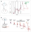
Figure 1.
Figure 1.
Schematic representation of polarity stages in pyramidal neurons in vivo and hippocampal neurons in vitro. (A) Excitatory cortical neurons derive from a radial glia cell, which divides asymmetrically giving rise to a supporting cell (gray) and a neuron (red). Following division, neurons have a polar phenotype with a leading pole (LP) oriented toward the pia and a trailing pole (TP) oriented toward the ventricular/subventricular zone (VZ/SVZ). In the intermediate zone (IZ) some cells show a multipolar stage characterized by several short processes that are not aligned with the radial glial fibers, which may initiate axon extension. Finally, neurons re-acquire a bipolar axis with the LP directed toward the pia, completing their migration to their final destination in the cortical plate (CP). Acquisition of mature polarized features, including the terminal differentiation of dendrites and somatic compartment is represented in green. (B) Hippocampal neurons are dissected from embryonic rodent hippocampus, trypsinized and plated on coverslips grown with the help of a glial feeder layer. Shortly after plating round neurons have a polarized organization of the cytoplasm, with the organelle pole not fixed in one direction (intracellular polarity; stage 0). An adhesion-dependent mechanism stabilizes the organelle pole and supports the sprout of the first neurite (pre-polarity stage 1, monopolar neuron). Note that the first neurite has the highest chances to become the axon at later stages. Subsequently, neurons acquire a bipolar axis with the growth of a second neurite from the pole in the opposite side of the first sprout (bipolar stage 2). Several other neurites grow, giving rise to a multipolar phenotype (multipolar stage 2). During the multipolar stage 2, rapid growth occurs from one of the 2 predisposed neurites of the bipolar axis (stage 3).
Figure 2.
Figure 2.
Schematic representation of domains in classical cadherins, main interactors and most relevant signaling pathways. Classical cadherins mediate homophilic interactions with other cadherins in the extracellular space, while intracellularly they organize the actin cytoskeleton and integrate several signaling pathways. The extracellular domains of cadherins are characterized by the repetition of several copies (5) of the cadherin motif, which mediates cell adhesion in a Ca2+ dependent manner. Several interactions can be established with receptors and other adhesion molecules (for a review see refs.28,29). Proteolytic processing of cadherin is regulated by the gamma-secretase/ADAM complex and mediates the release of intracellular and extracellular domains. The intracellular domain of cadherin interacts with p120-catenin and with the β-catenin core complex. The protein p120-catenin stabilizes adhesions and regulates interactions with other adhesion molecules and membrane remodelling complexes (for a review see ref.64). The core β-catenin complex has the dual function of regulating both adhesion and gene transcription. Adhesion is mediated by the interaction with α-catenin, which contacts actin and vinculin. These interactions are at the basis of actin/cytoskeletal remodelling events. Beta catenin is also implicated in the activation of wnt signaling pathways (for a review see ref.65).
Figure 3.
Figure 3.
Schematic representation of neuronal polarity steps regulated by N-cadherin during different developmental steps. (A) The clustering of N-cadherin molecules in round neurons is able to recruit the Golgi complex and the MTOC. In dissociated neurons N-cadherin promotes orientation and elongation of neurites. (B) In vivo during the establishment of polarity in pyramidal neurons, N-cadherin orients the Golgi complex in multipolar neurons in a Dab1-Rap1 dependent mechanism that is induced by a reelin gradient established by the Cajal-Retzius cells in the marginal zone. In bipolar neurons N-cadherin contributes to the radial orientation that is necessary for migration, and supports the radial glia locomotion of neurons mediated by Rab5-Rab11 membrane rearrangement. Once neurons reach the Cajal-Retzius cells in the marginal zone, N-cadherin promotes soma translocation.

References

    1. Gleeson JG. Neuronal migration disorders. Ment Retard Dev Disabil Res Rev 2001; 7:167-71; PMID:11553932; http://dx.doi.org/10.1002/mrdd.1024 - DOI - PubMed
    1. Gotz M, Huttner WB. The cell biology of neurogenesis. Nat Rev Mol Cell Biol 2005; 6:777-88; PMID:16314867; http://dx.doi.org/10.1038/nrm1739 - DOI - PubMed
    1. Franco SJ, Muller U. Shaping our minds: stem and progenitor cell diversity in the mammalian neocortex. Neuron 2013; 77:19-34; PMID:23312513; http://dx.doi.org/10.1016/j.neuron.2012.12.022 - DOI - PMC - PubMed
    1. Paridaen JT, Huttner WB. Neurogenesis during development of the vertebrate central nervous system. EMBO Rep 2014; 15:351-64; PMID:24639559; http://dx.doi.org/10.1002/embr.201438447 - DOI - PMC - PubMed
    1. Angevine JB, Jr., Sidman RL. Autoradiographic study of cell migration during histogenesis of cerebral cortex in the mouse. Nature 1961; 192:766-8; PMID:17533671; http://dx.doi.org/10.1038/192766b0 - DOI - PubMed