. 2014 Dec 22;31(6):690-706.
doi: 10.1016/j.devcel.2014.11.002. Epub 2014 Dec 4.

The left-right Pitx2 pathway drives organ-specific arterial and lymphatic development in the intestine

Affiliations

The left-right Pitx2 pathway drives organ-specific arterial and lymphatic development in the intestine

Aparna Mahadevan et al. Dev Cell. .

Abstract

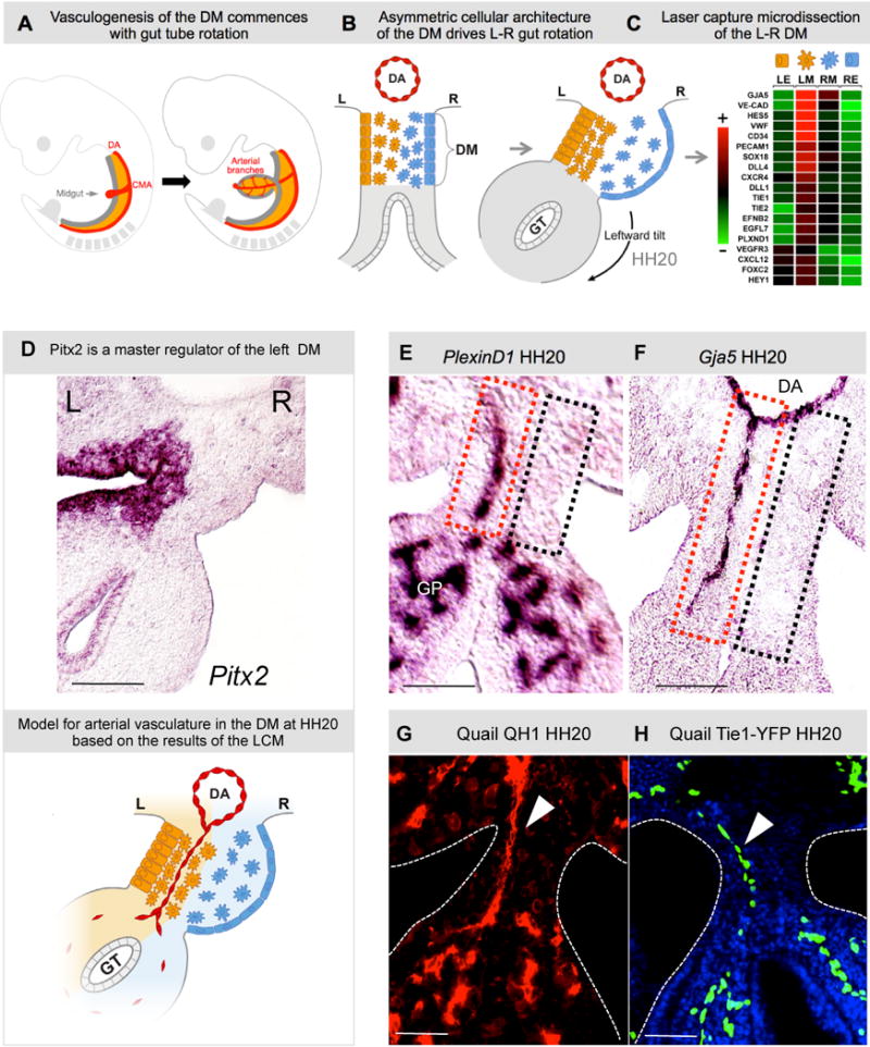
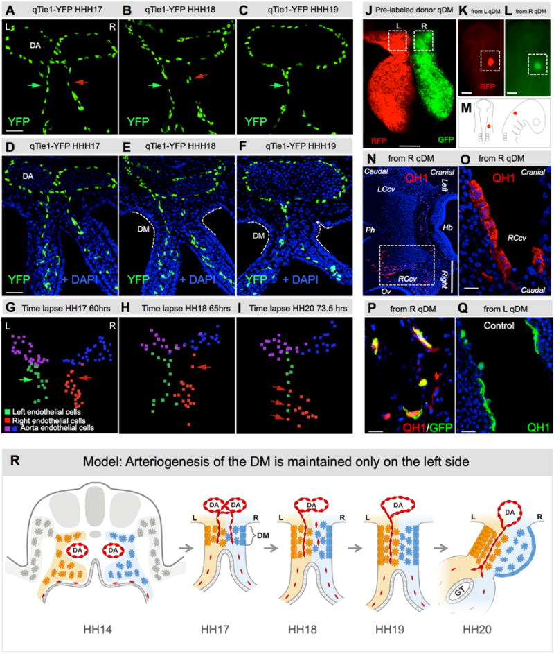
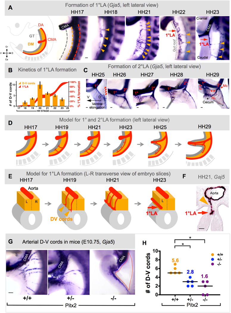
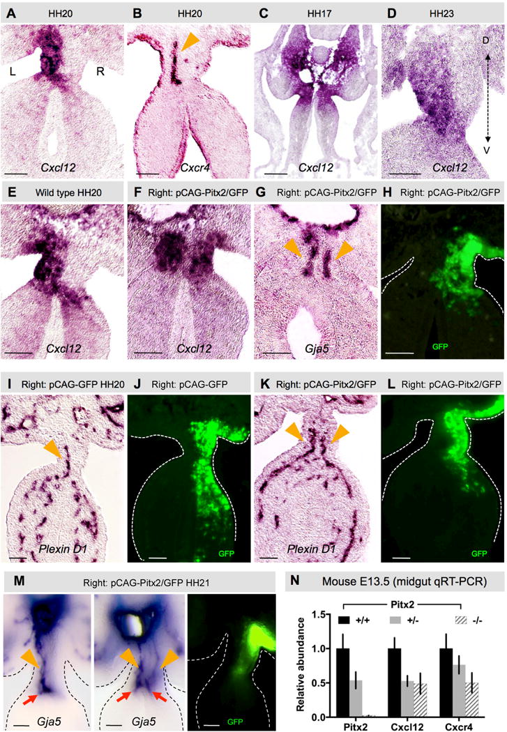
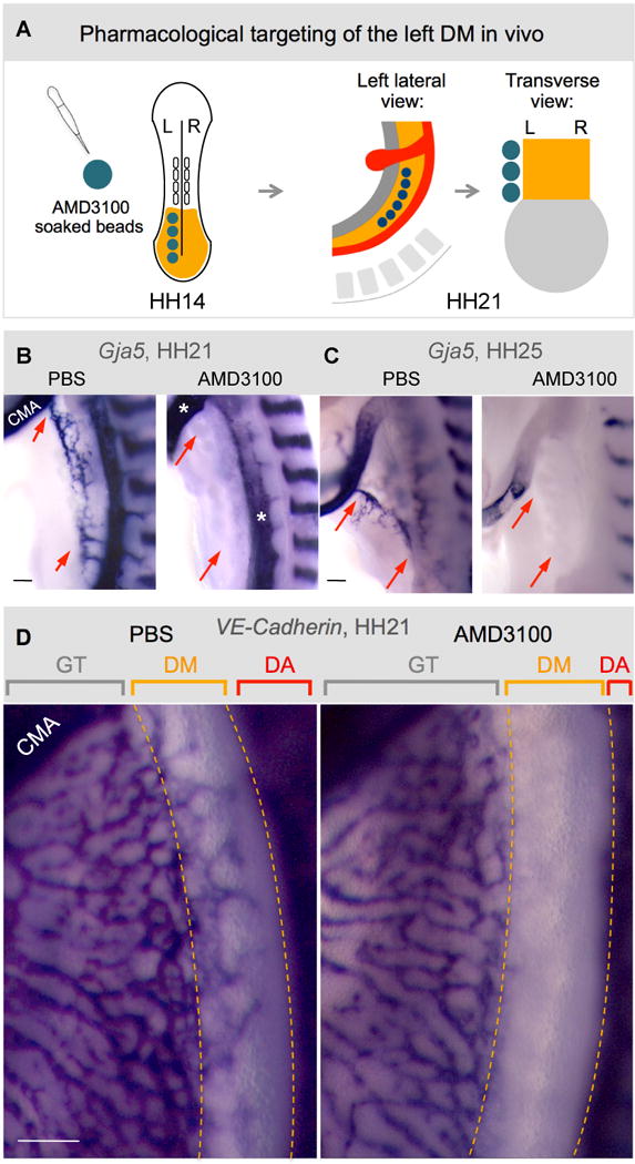
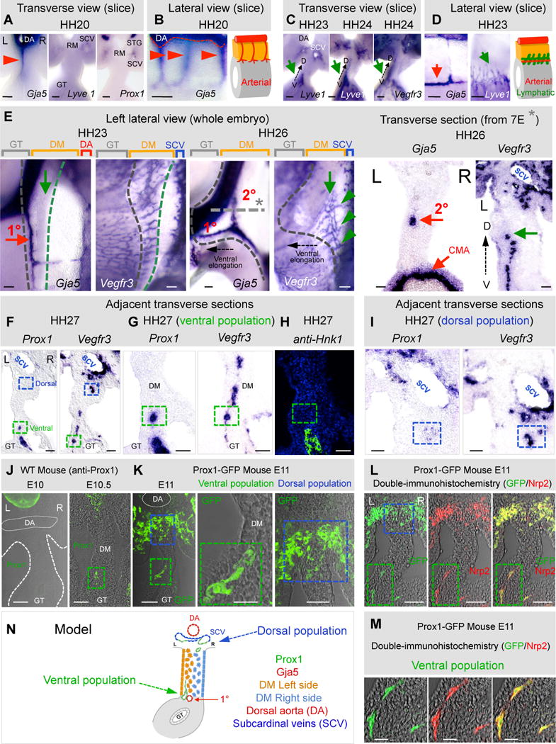
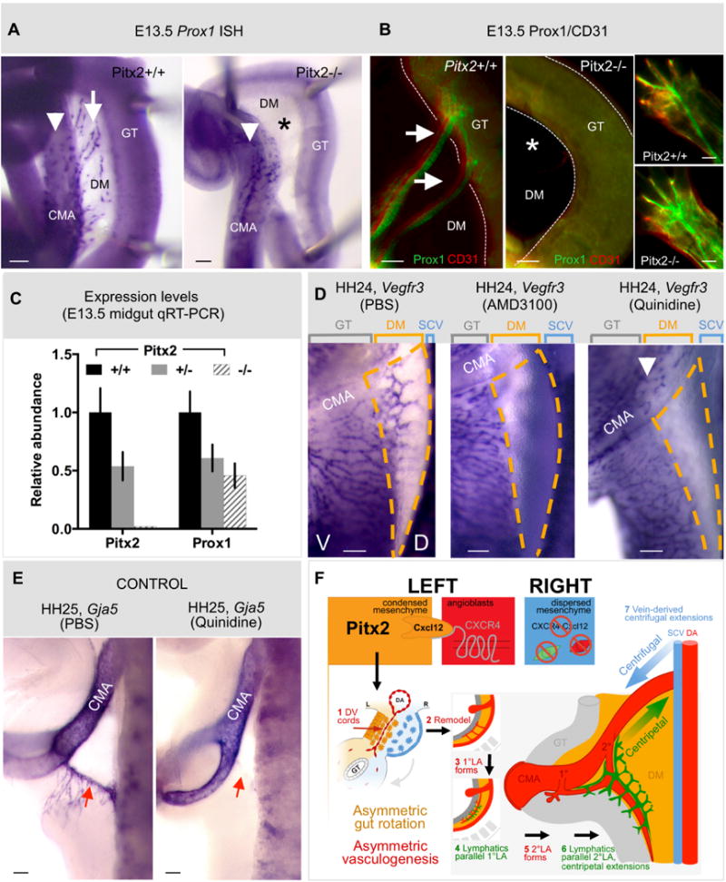
The dorsal mesentery (DM) is the major conduit for blood and lymphatic vessels in the gut. The mechanisms underlying their morphogenesis are challenging to study and remain unknown. Here we show that arteriogenesis in the DM begins during gut rotation and proceeds strictly on the left side, dependent on the Pitx2 target gene Cxcl12. Although competent Cxcr4-positive angioblasts are present on the right, they fail to form vessels and progressively emigrate. Surprisingly, gut lymphatics also initiate in the left DM and arise only after-and dependent on-arteriogenesis, implicating arteries as drivers of gut lymphangiogenesis. Our data begin to unravel the origin of two distinct vascular systems and demonstrate how early left-right molecular asymmetries are translated into organ-specific vascular patterns. We propose a dual origin of gut lymphangiogenesis in which prior arterial growth is required to initiate local lymphatics that only subsequently connect to the vascular system.


Figures

Fig. 1
Fig. 1. Arterial development in the DM is restricted to the left side
A Midgut arteriogenesis commences at chicken HH20 (mouse E10) in the dorsal mesentery (DM, orange) concomitant with rotation of the gut tube (GT, gray), as arterial branches of the cranial mesenteric artery (CMA, a branch of dorsal aorta, DA) first connect to the gut plexus. B Gut tube is suspended by DM; Pitx2-drives L-R cellular asymmetries to initiate leftward rotation. C Heat map of L-R differentially expressed genes (LE, left epithelia; LM, left mesenchyme; RM, right mesenchyme; RE, right epithelia) in the DM leads to a model for the role of Pitx2 (D, ISH, purple) in DM vascular patterning. E,F PlexinD1 and Gja5 ISH show presence (left, red box) or absence (right, black box) of arterial D-V cords. G,H QH1+ and Tie1+ cords in quail embryos (arrowhead). Scale bars: E–H (50 μm). See also Figure S1.
Fig. 2
Fig. 2. The right side of the DM becomes avascular during midgut rotation
A–F Transgenic quail midgut DM transverse sections with Tie1:H2B-eYFP (green) and DAPI (blue) staining. A L-R bilateral vascular endothelial plexus is present in the DM at HH17 (green and red arrows). B Vascular plexus on the DM right side begins to regress (red arrow). By HH19 (C) the right side is avascular and only the left-sided Tie1+ D-V cords remain (green arrow). G–I Still frames from live time-lapse tracking of Tie1:H2b-eYFP cells, illustrating right-sided (red) regression of vascular endothelium. Nuclei of the left, right and the dorsal aorta endothelium are represented as green, red and blue and purple spots, respectively (Imaris). J–Q Chick-quail chimera show endothelial progenitors with competence to form vessels are present on both sides of DM. J Dual-labeling of left (RFP) and right (GFP) donor quail DM (HH18). K,L,M Marked fragment is grafted onto chicken (HH9/10) where indicated, and scored at HH21 (N–O: Hb, hindbrain; Ov, otic vesicle; Ph, pharynx; LCcv, left cranial cardinal vein; RCcv, right cranial cardinal vein). Boxed region of N is magnified in O, where right/GFP-derived donor angioblasts (QH1, red; GFP co-stain in P) contribute to new vessels indistinguishable from control left/RFP-derived cells (QH1, green, Q). R Cartoon of right-sided vascular endothelial regression leading to the left-sided bias of vascular patterning in the midgut DM. Scale bars: A–F (50 μm); J (100 μm); K,L (200 μm); N (500 μm); O–Q (20 μm). See also Movies S1–3 and Fig. S2.
Fig. 3
Fig. 3. Left-sided D-V cords become the major arteries supplying the midgut and are Pitx2-driven
A Arteriogenesis time course (1°LA formation) via wholemount ISH (Gja5) in chicken DM (D-V cords, orange arrows; 1°LA, red arrow), quantified in B (orange bars show number of D-V cords; red curves show percent longitudinal extension of 1°LA). C Time course (2°LA formation): 1°LA connection to CMA is established (1°, ileocolic artery, Ic) and precedes initiation of 2°LA (2°, middle colic artery, Mc) as an outgrowth of the 1°LA (red arrowheads in left panel). Subsequent anastomosis of 1° and 2° form the right colic artery (Rc in right panel). D,E Model of 1° and 2°LA formation as seen from left lateral view (D) or in transverse (E). F Arteriogenesis in the DM initiates on the left (HH21, transverse view) where D-V cords remodel into the 1°LA G, Arterial cords ablated in E10.75 Pitx2−/− DM (dashed region); quantified in H (column scatter plot shows individual data and median (line); mean number is represented above each data point). Scale bars: A (100 μm); C (100 μm); F (50 μm); G (200 μm). See also Figure S3.
Fig. 4
Fig. 4. Cxcr4/Cxcl12 axis is regulated by Pitx2
Cxcl12 (A) and receptor Cxcr4 (B) expression in left mesenchyme and endothelial cells, respectively (ISH). C Cxcl12 is bilateral (HH17) prior to DM asymmetries then (D) develops a left ventro-dorsal gradient (HH23). E–L Right DM Pitx2 misexpression drives ectopic Cxcl12 expression (F vs. E) and D-V arterial cords (G, Gja5; K, PlexinD1), compared to GFP-only control (K vs. I; GFP (H,J,L) labels electroporated cells). Orange arrowheads mark arterial cords (B,G,I,K). M ‘Double left’ vascular phenotype upon right-sided Pitx2 misexpression (orange arrowheads point to D-V cords, red arrows point to 1°LA). N Relative expression levels of Cxcl12 and Cxcr4 in E13.5 Pitx2−/− DM. Scale bars are 50 μm. See also Figure S4.
Fig. 5
Fig. 5. The Cxcr4/Cxcl12 axis is necessary for arterial vascular development in the DM
A Targeting of the left DM with AMD3100. Beads are placed into the left coelomic cavity (HH14) prior to DM formation to target splanchnic mesoderm. Beads remain intact at HH21 when embryos are analyzed. Compared to PBS-control beads (B–D), AMD3100-beads ablate left arterial cords (B, HH21, ISH Gja5, red arrows) and 1°LA (C, HH25, red arrows), but leave adjacent gut vascular plexus, CMA, and DA, unaffected (D, VE-cadherin, HH21). CMA, cranial mesenteric artery; GT, gut tube; DA, dorsal aorta. Scale bars are 100 μm. See also Figure S5.
Fig. 6
Fig. 6. Cxcl12 expression is not sufficient to drive D-V cord formation in the absence of Pitx2
A–E Exogenous left (or right) Cxcl12 expression accelerates left-sided D-V remodeling and 1°LA formation. A Lateral views of DM arteriogenesis in WT, left-side (C) and right-side (D) Cxcl12 electroporated embryos (Wholemount ISH Gja5), quantified in B (bars show number of D-V cords, red curves show percent longitudinal extension of 1°LA). E Transverse sections of left-side (top) or right-side (bottom) Cxcl12 electroporated embryos (Gja5 ISH, GFP labels targeted cells) show left-sided (top) or right-sided (bottom) location bias in the formation 1°LA (red arrow). Dashed boxes mark the presumptive location of the normal left-sided (red) and ectopically formed right-sided (black) D-V arterial cords. Cartoon shows proposed model for the left-side dependent formation of the ectopic right-sided 1°LA, tested in a dual L-R targeting experiment in F. DM dual targeting (HH25, wholemount ISH Gja5, right lateral view of embryos, summarized in cartoons): (1) left-sided AMD3100-soaked beads with right-sided Cxcl12; (2) PBS beads and right-sided Cxcl12; or (3) left-sided AMD3100 beads with right-sided Pitx2 (bottom) demonstrate the requirement of the left side for 1°LA formation. Scale bars: A,C,D (100 μm); E (50 μm); F (100μm). See also Figure S6.
Fig. 7
Fig. 7. Lymphangiogenesis in the DM is left-sided and initiates locally
ISH during DM arteriogenesis with probes specific for arterial (Gja5) and lymphatic (Lyve1, Vegfr3, and Prox1) markers: transverse (A,C,E right column, F–I) and lateral views (B,D,E) of DM/gut tube vibratome slices (B,C,D) or wholemount embryos (E) and transverse sections (E). A,B At HH20 only arterial cords are present (red arrowhead and cartoon). C,D At HH23–24, Lyve1 and Vegr3 mark lymphatics in the ventral left DM (C,D, green arrows; D, cartoon) while 1°LA has formed (red arrow). E Left lateral views, HH23 showing lymphatic field (green arrow, Vegfr3) forming along the 1°LA (red arrow, Gja5). Lymphatic field (green arrow) at HH26 moves dorsally and parallels the 2°LA now formed (green arrowheads show dorsal connections of the lymphatic field with the SCV). Gray dashed line/asterisk marks location of transverse section. Black dashed arrow shows ventral elongation of the midgut loop. Transverse sections of ISH showing left-sided location of the 2°LA (Gja5) and lymphatic field (Vegfr3). F Distinct ventral (green box) and dorsal (blue box) populations of chicken DM lymphatics, revealed by Prox1 and Vegfr3 ISH on adjacent sections; higher magnifications: ventral (G) and dorsal (I) domains. H Prox1+ avian Nerve of Remak in the ventral domain stains Hnk1+, distinct from Prox1+, Hnk1− lymphatics (green box). JK Distinct lymphatic populations (E10–11) in WT mice (J, anti-Prox1) and in Prox1-GFP transgenic mice (K, anti-GFP). LM Double-IH of Prox1-GFP mice with Nrp2 (M shows higher magnification of ventral population from L) N Cartoon model summary of ventral left and dorsal lymphatic populations. Scale bars: A–D, E right column, I,J,K (left, right panels), L, M (50 μm); E left column, F,G,H (100 μm); K (middle panel) (20 μm). See also Figure S7.
Fig. 8
Fig. 8. Local lymphangiogenesis in the left DM requires the preceding arterial program, driven by Pitx2
A,B Prox1+ mesenteric lymphatic vessels (white arrows, Pitx2+/+) are absent in embryos lacking Pitx2 (asterisk in Pitx2−/− panels), as revealed by (A) wholemount ISH and (B) immunostaining for Prox1/CD31 at E13.5 in the mouse. Prox1+ cells alongside the CMA wall (white arrowhead in A) or of dermal lymphatics (B, insets) are unaffected by Pitx2 loss. C Quantitative RT-PCR reveals loss of Prox1 expression in whole gut isolates of Pitx2 +/− and Pitx2−/− mouse embryos. D Compared to PBS (left panel), the DM (orange dashed regions) is devoid of lymphatic vessels (ISH Vegfr3) upon pharmacological inhibition of arteriogenesis, either targeting Cxcr4 (AMD3100, middle panel) or Gja5/Cx43 (quinidine, right panel). E Confirmation that arteries (ISH Gja5) in the DM are lost upon quinidine treatment (right panel) compared to PBS controls (left panel). F Proposed model: Pitx2 directs Cxcl12 in the left DM. This induces endothelial chemotaxis via Cxcr4 and D-V cord formation, remodeling and formation of CMA arterial branches. A lymphatic plexus initiates in the ventral left DM and, via centripetal extension, connects with centrifugal branches of the systemic lymphatics of venous descent. Scale bars are 100 μm. See also Figure S8.

Similar articles

Cited by

References

    1. Alexander JS, Chaitanya GV, Grisham MB, Boktor M. Emerging roles of lymphatics in inflammatory bowel disease. Annals of the New York Academy of Sciences. 2010;1207(Suppl 1):E75–85. - PubMed
    1. Ara T, Tokoyoda K, Okamoto R, Koni PA, Nagasawa T. The role of CXCL12 in the organ-specific process of artery formation. Blood. 2005;105:3155–3161. - PubMed
    1. Aselli G. De lactibus, sive Lacteis venis, quarto vasorum mesaraicorum genere novo invento dissertatio. Medioloni, Milan; 1627.
    1. Betterman KL, Paquet-Fifield S, Asselin-Labat ML, Visvader JE, Butler LM, Stacker SA, Achen MG, Harvey NL. Remodeling of the lymphatic vasculature during mouse mammary gland morphogenesis is mediated via epithelial-derived lymphangiogenic stimuli. The American journal of pathology. 2012;181:2225–2238. - PubMed
    1. Bulow HE, Hobert O. The molecular diversity of glycosaminoglycans shapes animal development. Annu Rev Cell Dev Biol. 2006;22:375–407. - PubMed

Publication types

MeSH terms