. 2014 Dec 22;31(6):747-60.
doi: 10.1016/j.devcel.2014.10.024. Epub 2014 Dec 4.

Mesenchymal chemotaxis requires selective inactivation of myosin II at the leading edge via a noncanonical PLCγ/PKCα pathway

Affiliations

Mesenchymal chemotaxis requires selective inactivation of myosin II at the leading edge via a noncanonical PLCγ/PKCα pathway

Sreeja B Asokan et al. Dev Cell. .

Abstract

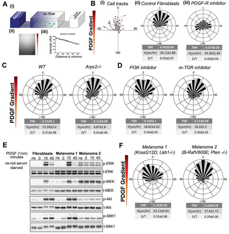
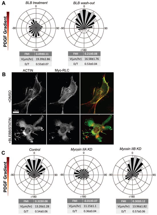
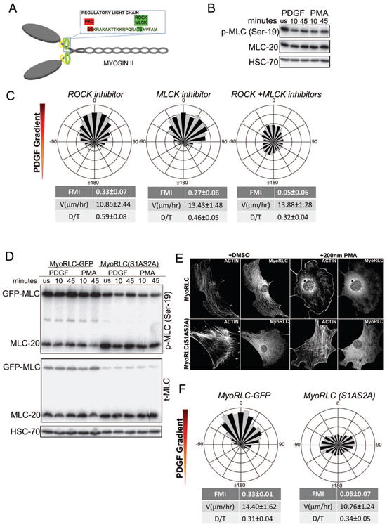
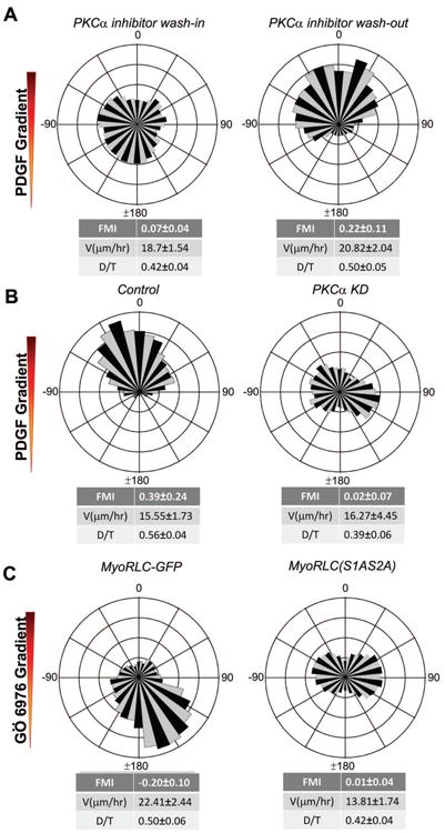
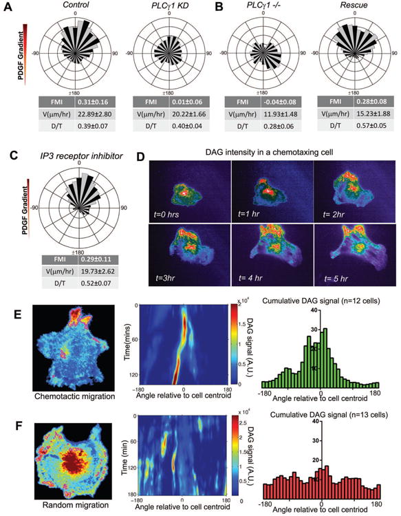
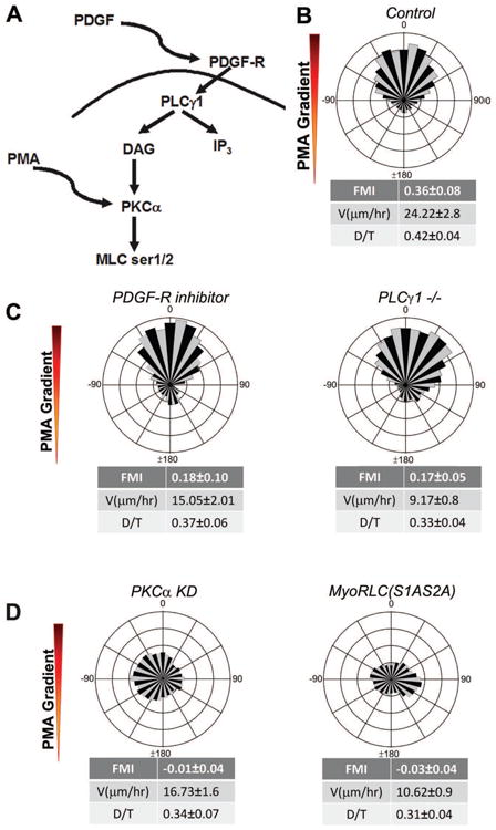
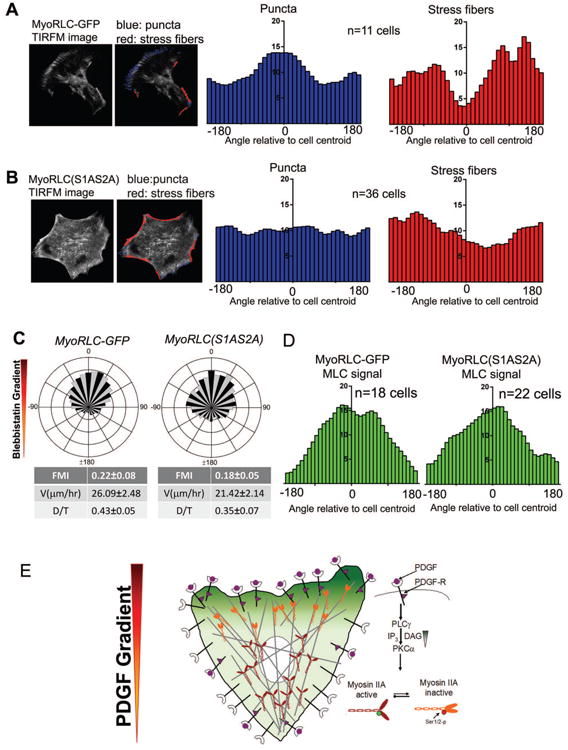
Chemotaxis, migration toward soluble chemical cues, is critical for processes such as wound healing and immune surveillance and is exhibited by various cell types, from rapidly migrating leukocytes to slow-moving mesenchymal cells. To study mesenchymal chemotaxis, we observed cell migration in microfluidic chambers that generate stable gradients of platelet-derived growth factor (PDGF). Surprisingly, we found that pathways implicated in amoeboid chemotaxis, such as PI3K and mammalian target of rapamycin signaling, are dispensable for PDGF chemotaxis. Instead, we find that local inactivation of Myosin IIA, through a noncanonical Ser1/2 phosphorylation of the regulatory light chain, is essential. This site is phosphorylated by PKCα, which is activated by an intracellular gradient of diacylglycerol generated by PLCγ. Using a combination of live imaging and gradients of activators/inhibitors in the microfluidic chambers, we demonstrate that this signaling pathway and subsequent inhibition of Myosin II activity at the leading edge are required for mesenchymal chemotaxis.


Figures

Figure 1
Figure 1. Pathways that are dispensable for mesenchymal chemotaxis
A. (i) A 3D schematic of the microfluidic chamber used for chemotaxis experiments. (ii) The fluorescent micrograph shows the formation of Cy5 dextran gradient in the channel. (iii) A line scan of fluorescent intensity of the gradient indicates that a linear gradient is formed in the chamber with an approximate slope of 43%. B. (i) Migratory tracks of IA32 fibroblast chemotaxing to a PDGF gradient (ii) Cell tracks of chemotaxing cells represented as a wind-rose plot (number of cell tracked (n) = 62). Each leaflet represents the count frequency of cells migrating in the corresponding angular bin. Forward Migration Index (FMI), persistence (D/T) and velocity values are indicated below the plot, +/- 95% C.I. (iii) Wind-rose plot showing inhibition of chemotaxis to PDGF in the presence of a uniform concentration of PDGF-R inhibitor, AG1296 (1 μM) (n = 68). C. Wind-rose plots of PDGF chemotaxis of WT (pre-Cre deletion) (n=82) and Arpc2 KO (n = 71) MEFs. D. Directional migration response of fibroblasts to PDGF gradient in the presence of uniform PI3Kα inhibitor IV (5 μM) (n = 74) or m-TOR inhibitor AZD8055 (1 μM) (n = 56). E. Western blot analysis of fibroblasts and melanoma cells stimulated with PDGF for the indicated incubation times, ns indicates not serum-starved. Phosphorylation of ERK, MEK, Akt and S6K1 were assessed. F. Chemotaxis of K-Ras/Lkb1 null (Melanoma 1) (n = 72) and B-Raf/Pten null (Melanoma 2) (n = 81) tumor cell lines to a PDGF gradient.
Figure 2
Figure 2. Myosin IIA is required for chemotaxis, whereas Myosin IIB is dispensable
A. Rose plot showing loss of chemotaxis in the presence of uniform concentration of blebbistatin (BLB, 15 μM) (n = 123) and recovery of chemotactic migration after wash-out of BLB (n = 97). B. Immunofluorescence of IA32 fibroblasts shows myosin light chain (GFP) and F-actin (red, Phalloidin) in cells before and after uniform BLB treatment. C. Circular histograms showing chemotaxing control cells, loss of chemotactic migration in Myosin IIA KD (n = 80) cells and intact chemotaxis in Myosin IIB KD cells (n = 61).
Figure 3
Figure 3. Chemotaxis requires regulation of Myosin II function via non-canonical RLC phosphorylation
A. Schematic of Myosin Regulatory Light Chain (Myo RLC) showing phospho-regulatory sites Ser1/Ser2 and Thr18/Ser19. Ser1/Ser2 are inhibitory phosphorylation sites regulated by conventional PKCα. Thr18/Ser19 are activating phosphorylation sites regulated by ROCK and MLCK. B. Western blot analysis to probe the extent of Ser19 phosphorylation of Myosin II RLC, which shows no change in response to PDGF or PMA stimulation. C. Wind-rose plots of cell migration show PDGF chemotaxis in the presence of either ROCK inhibitor Y27632 (15 μM) (n = 161) or MLCK inhibitor peptide 18 (10 μM) (n = 104). When added together, the ROCK and MLCK inhibitors impede chemotactic migration to PDGF (n = 94). D. Western blot analysis of Ser19 phosphorylation of Myosin II RLC after PDGF or PMA stimulation of both MyoRLC-GFP and MyoRLC(S1AS2A) cells shows no change in Ser19 phosphorylation of either the endogenous or GFP-tagged RLC. E. Immunofluorescence images of MyoRLC-GFP and MyoRLC(S1AS2A)-GFP cells show disassembly of actin stress fibers in wild-type cells and no disassembly in myosin mutant cells upon PMA stimulation. D. Wind-rose plots showing chemotaxing control MyoRLC-GFP cells (n=61) and non-chemotaxing mutant MyoRLC(S1AS2A)-GFP cells (n= 73) in a PDGF gradient.
Figure 4
Figure 4. PKCα is essential for chemotaxis
A. Wind-rose plots of fibroblasts in a PDGF gradient show inhibition of chemotaxis in the presence of a uniform concentration of PKCα inhibitor, Gö6967 (1 μM) (n=162) and recovery of chemotaxis after drug wash-out (n=88). B. Circular histograms show chemotaxis of wild-type fibroblasts in a PDGF gradient and the lack of chemotactic response of PKCα KD cells (n=68) in the same chamber. C. Chemo-repulsion of wild-type cells to gradients of Gö6967 (n=101). Mutant MyoRLC (S1AS2A) cells do not exhibit directional migration in a gradient of Gö6967 (n=89).
Figure 5
Figure 5. PLCγ is essential for PDGF chemotaxis and produces an asymmetric pattern of intracellular DAG
A. Wind-rose plot of control cells migrating in a PDGF (n=67) gradient and loss of directional migration of PLCγ KD cells (n=70) in the same chamber. B. Fibroblasts derived from PLCγ-null (PLCγ1-/-) (n=73) mice show a loss of chemotactic response to PDGF. Upon rescue of PLCγ expression, these fibroblasts regained their ability to chemotax (n=101). C. Circular histogram of cells chemotaxing to PDGF gradients in a uniform concentration of Xestospongin-(c) (1 μM) (n=69). D. Montage of an IA32 fibroblast transfected with GFP-tagged tandem C1 domain [(C1)2-GFP] chemotaxing to PDGF for more than five hours. E. Enriched pixels in a chemotaxing cell are shown outlined in magenta. The intensity values of the segmented pixels are summed within angular bins at each time point to create a signaling “map” with the angle plotted on the horizontal axis and time on the vertical axis. Histogram showing the cumulative intracellular DAG distribution in chemotaxing cells expressing (C1)2-GFP (n=12 cells). F. Enriched pixels in a randomly migrating cell are shown outlined in magenta. The intensity values of the segmented pixels are summed within angular bins at each time point to create a signaling “map” with the angle plotted on the horizontal axis and time on the vertical axis. Histogram showing the cumulative intracellular DAG distribution in randomly migrating cells expressing (C1)2-GFP (n=13 cells).
Figure 6
Figure 6. Phorbol ester (PMA) gradients bypass the need for PDGF receptors and PLCγ, but not PKCα or Myosin II function
A. Wind-rose plot of IA32 fibroblasts chemotaxing in PMA gradients (n=89). B. Wind-rose plots show that PMA gradients rescue chemotaxis of both IA32 fibroblasts in a uniform concentration of PDGF-R inhibitor AG1296 (1 μM) (n=45) and that of PLCγ1 -/- fibroblasts (n=61). C. Circular histograms showing no rescue of chemotaxis of PKCα KD (n=85) or mutant Myo-RLC(S1AS2A) cells (n=187) in PMA gradients.
Figure 7
Figure 7. Asymmetric Myosin II organization and activity are hallmarks of chemotactic migration in mesenchymal cells
A. TIRF microscopy movies of chemotaxing MyoRLC-GFP cells are analyzed to identify puncta (blue) and stress fiber regions (red). The localization of puncta and stress fibers relative to the cell centroid were averaged across multiple cells (n=11 cells) and presented as histograms. B. Analysis of puncta and stress fiber distributions, quantified as in A, in MyoRLC(S1AS2A)-GFP cells (n=36cells). C. Circular histograms of MyoRLC-GFP (n=162) and MyoRLC(S1AS2A) (n=67) cells chemotaxing in a gradient of BLB. D. Intensity of MyoRLC-GFP and MyoRLC(S1AS2A)-GFP during chemotaxis in a BLB gradient, presented as a histogram. E. Proposed model of PDGF chemotaxis: PDGF binds to PDGFR, recruits PLCγ to produce a localized intracellular gradient of DAG. The asymmetric DAG phosphorylates RLC at Ser1/2 via PKCα, inactivating MyoIIA at the leading edge of the chemotaxing cell. This localized Myosin II inactivation provides the asymmetry of force needed for directional migration.

Comment in

  • Unleashing mesenchymal chemotaxis.
    Aguilar-Cuenca R, Vicente-Manzanares M. Aguilar-Cuenca R, et al. Dev Cell. 2014 Dec 22;31(6):669-70. doi: 10.1016/j.devcel.2014.12.005. Dev Cell. 2014. PMID: 25535912

Similar articles

Cited by

References

    1. Andrae J, Gallini R, Betsholtz C. Role of platelet-derived growth factors in physiology and medicine. Genes Dev. 2008;22:1276–1312. - PMC - PubMed
    1. Beach JR, Licate LS, Crish JF, Egelhoff TT. Analysis of the role of Ser1/Ser2/Thr9 phosphorylation on myosin II assembly and function in live cells. BMC Cell Biol. 2011;12:52. - PMC - PubMed
    1. Bear J, Loureiro J, Libova I, Fassler R, Wehland J, Gertler F. Negative regulation of fibroblast motility by Ena/VASP proteins. Cell. 2000;101:717–728. - PubMed
    1. Bear JE, Haugh JM. Directed migration of mesenchymal cells: where signaling and the cytoskeleton meet. Curr Opin Cell Biol. 2014;30C:74–82. - PMC - PubMed
    1. Bear JE, Svitkina TM, Krause M, Schafer DA, Loureiro JJ, Strasser GA, Maly IV, Chaga OY, Cooper JA, Borisy GG, et al. Antagonism between Ena/VASP Proteins and Actin Filament Capping Regulates Fibroblast Motility. Cell. 2002;109:509–521. - PubMed

Publication types

LinkOut - more resources