. 2015 Jun;17(6):819-31.
doi: 10.1111/cmi.12403. Epub 2015 Jan 28.

Investigating the function of Fc-specific binding of IgM to Plasmodium falciparum erythrocyte membrane protein 1 mediating erythrocyte rosetting

Affiliations

Investigating the function of Fc-specific binding of IgM to Plasmodium falciparum erythrocyte membrane protein 1 mediating erythrocyte rosetting

Liz Stevenson et al. Cell Microbiol. 2015 Jun.

Abstract

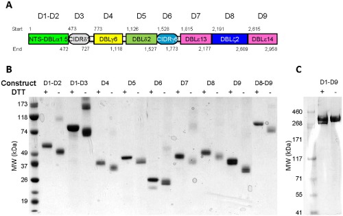
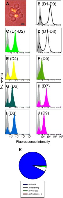
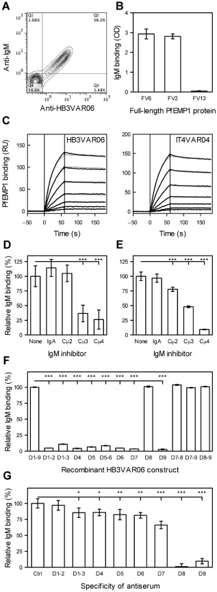
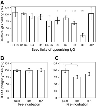
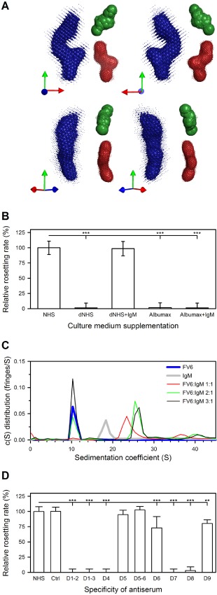
Acquired protection from Plasmodium falciparum malaria takes years to develop, probably reflecting the ability of the parasites to evade immunity. A recent example of this is the binding of the Fc region of IgM to VAR2CSA-type PfEMP1. This interferes with specific IgG recognition and phagocytosis of opsonized infected erythrocytes (IEs) without compromising the placental IE adhesion mediated by this PfEMP1 type. IgM also binds via Fc to several other PfEMP1 proteins, where it has been proposed to facilitate rosetting (binding of uninfected erythrocytes to a central IE). To further dissect the functional role of Fc -mediated IgM binding to PfEMP1, we studied the PfEMP1 protein HB3VAR06, which mediates rosetting and binds IgM. Binding of IgM to this PfEMP1 involved the Fc domains Cμ3-Cμ4 in IgM and the penultimate DBL domain (DBLζ2) at the C-terminus of HB3VAR06. However, IgM binding did not inhibit specific IgG labelling of HB3VAR06 or shield IgG-opsonized IEs from phagocytosis. Instead, IgM was required for rosetting, and each pentameric IgM molecule could bind two HB3VAR06 molecules. Together, our data indicate that the primary function of Fc -mediated IgM binding in rosetting is not to shield IE from specific IgG recognition and phagocytosis as in VAR2CSA-type PfEMP1. Rather, the function appears to be strengthening of IE-erythrocyte interactions. In conclusion, our study provides new evidence on the molecular details and functional significance of rosetting, a long-recognized marker of parasites that cause severe P. falciparum malaria.


Figures

Figure 1
Figure 1
Recombinant HB3VAR06 constructs. Schematic representation of HB3VAR06 showing individual DBL and CIDR domains (domain start and end boundaries given above and below individual domains), named and colour coded as proposed by Rask et al. (2010) (A). Coomassie stain of an SDS‐PAGE with recombinant HB3VAR06 constructs under non‐reducing (+) and reducing (−) conditions (B and C).
Figure 2
Figure 2
HB3VAR06‐positive P . falciparum parasites. Fluorescence micrograph of rosette around an erythrocyte infected by in vitro‐selected P. falciparum HB3. Error bar: 5 μm (A). Labelling of HB3VAR06+ IEs by antisera raised against different HB3VAR06 recombinant constructs measured by flow cytometry. Domains included in the constructs used for immunization are shown in brackets and background labelling (pre‐immunization sera) is shown by grey shading. Colour coding corresponds to that used in Fig. 1A, except for multidomain constructs including several domain subtypes (shown as black outlines) (B–J). Transcription profile of var genes in P. falciparum HB3 selected in vitro for expression of HB3VAR06 measured by quantitative real‐time PCR (K).
Figure 3
Figure 3
Binding of non‐specific IgM to HB3VAR06. Binding of non‐specific IgM and FV6‐specific antiserum to HB3‐infected IEs selected for expression of HB3VAR06 measured by flow cytometry of magnet‐purified late stage P. falciparum HB3‐infected erythrocytes (A). Binding of IgM to recombinant full‐length proteins representing HB3VAR06 (FV6), IT4VAR04 (FV2) and IT4VAR13 (FV13), respectively, measured by ELISA (B). Affinity of IgM for FV6 (left) and FV2 (right) measured by SPR. The SPR sensorgram data (black) and fits (grey) at five concentrations [1.125 (bottom trace); 2.25, 4.5, 9 and 18 nM (top trace)] are shown (C). Interference of non‐specific IgA and monoclonal antibodies specific for Cμ2 (HB57), Cμ3 (5D7) or Cμ4 (1G6) with IgM binding to HB3VAR06+ IEs measured by flow cytometry (D). Interference of IgA and monoclonal antibodies specific for Cμ2 (HB57), Cμ3 (5D7) or Cμ4 (1G6) with IgM binding to recombinant full‐length HB3VAR06 measured by ELISA (E). Binding of IgM to recombinant HB3VAR06 single‐, double‐ and triple‐domain constructs relative to IgM binding to FV6 measured by ELISA (F). Interference with IgM binding to HB3VAR06+ IEs by antisera raised against recombinant HB3VAR06 single‐ and double‐domain constructs measured by flow cytometry (G). Means and standard deviation and values statistically significant different (*P < 0.05; **P < 0.01; ***P < 0.001) from control values in the leftmost bar of each panel are shown (B, D–G). Control values were results obtained in the absence of any IgM inhibitor (D and E) using FV6 as the coating antigen (F) or in the presence of pre‐immunization serum (G). All experiments were repeated at least three times with similar results.
Figure 4
Figure 4
Non‐specific antibody interference with opsonization and phagocytosis. Interference with HB3VAR06 domain‐specific IgG recognition of HB3VAR06+ IEs by non‐specific IgM measured by flow cytometry (A). Phagocytosis of IEs opsonized by FV6‐specific antiserum (B) or human immune plasma (C) after or without pre‐incubation of IEs with non‐specific IgM or IgA measured by flow cytometry. Means and standard deviation, and statistically significant values (*P < 0.05; **P < 0.01; ***P < 0.001) relative to results obtained with pre‐immunization serum (A) or without IgM pre‐incubation (B and C) are shown.
Figure 5
Figure 5
The three‐dimensional conformation of HB3VAR06, IgM dependency of rosetting and stoichiometry of the FV6:IgM interaction. SAXS modelling of the envelopes of FV6 (blue), the N‐terminal domains D1–D3 (green) and the C‐terminal domains D7–D9 (red) as seen from four different angles. The relative point of view in each panel is indicated by arrows in the bottom left panel corner (A). Rosetting rates of HBVAR06+ IEs in culture medium supplemented with non‐immune human serum (NHS), IgM‐depleted NHS (dNHS), dNHS plus IgM (dNHS + IgM), Albumax or Albumax plus IgM (Albumax + IgM) are presented as in Fig. 3 (B). Analytical ultracentrifugation of recombinant full‐length HB3VAR06 (FV6; blue) or IgM (IgM; grey), and of FV6 and IgM together at molar ratios of 1:1 (red), 2:1 (green) and 3:1(black) (C). Rosetting rates of HBVAR06+ IEs in the presence of HB3VAR06 domain‐specific antisera or pre‐immunization serum (Ctrl), relative to rosetting in NHS. Data presented as in Fig. 3G (D).

Similar articles

Cited by

References

    1. Avril, M. , Tripathi, A.K. , Brazier, A.J. , Andisi, C. , Janes, J.H. , Soma, V.L. , et al (2012) A restricted subset of var genes mediates adherence of Plasmodium falciparum‐infected erythrocytes to brain endothelial cells. Proc Natl Acad Sci USA 109: E1782–E1790. - PMC - PubMed
    1. Barfod, L. , Dalgaard, M.B. , Pleman, S.T. , Ofori, M.F. , Pleass, R.J. , and Hviid, L. (2011) Evasion of immunity to Plasmodium falciparum malaria by IgM masking of protective IgG epitopes in infected erythrocyte surface‐exposed PfEMP1. Proc Natl Acad Sci USA 108: 12485–12490. - PMC - PubMed
    1. Barragan, A. , Spillmann, D. , Kremsner, P.G. , Wahlgren, M. , and Carlson, J. (1999) Plasmodium falciparum: molecular background to strain‐specific rosette disruption by glycosaminoglycans and sulfated glycoconjugates. Exp Parasitol 91: 133–143. - PubMed
    1. Barragan, A. , Fernandez, V. , Chen, Q. , von Euler, A. , Wahlgren, M. , and Spillmann, D. (2000a) The duffy‐binding‐like domain 1 of Plasmodium falciparum erythrocyte membrane protein 1 (PfEMP1) is a heparan sulfate ligand that requires 12 mers for binding. Blood 95: 3594–3599. - PubMed
    1. Barragan, A. , Kremsner, P.G. , Wahlgren, M. , and Carlson, J. (2000b) Blood group A antigen is a co‐receptor in Plasmodium falciparum rosetting. Infect Immun 68: 2971–2975. - PMC - PubMed

Publication types

MeSH terms

Substances