. 2014;13(24):3921-6.
doi: 10.4161/15384101.2014.973324.

USP7 controls Chk1 protein stability by direct deubiquitination

Affiliations

USP7 controls Chk1 protein stability by direct deubiquitination

Ignacio Alonso-de Vega et al. Cell Cycle. 2014.

Abstract

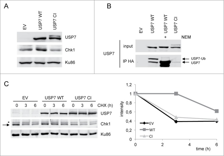
Chk1, an essential checkpoint kinase in the DNA damage response pathway (DDR), is tightly regulated by both ATR-dependent phosphorylation and proteasome-mediated degradation. Here we identify ubiquitin hydrolase USP7 as a novel regulator of Chk1 protein stability. USP7 was shown before to regulate other DDR proteins such as p53, Hdm2 and Claspin, an adaptor protein in the ATR-Chk1 pathway required for Chk1 activation. Depletion or inhibition of USP7 leads to lower Chk1 levels. The decreased Chk1 protein after USP7 knock down cannot be rescued by simultaneously elevating Claspin levels, demonstrating that the effect of USP7 on Chk1 is independent of its known effect on Claspin. Conversely, overexpression of USP7 wild type, but not a catalytic mutant version, elevates Chk1 levels and increases the half-life of Chk1 protein. Importantly, wild type, but not catalytic mutant USP7 can deubiquitinate Chk1 in vivo and in vitro, confirming that USP7 directly regulates Chk1 protein levels. Finally we show that USP7 catalytic mutant is (mono-)ubiquitinated, which suggests auto-deubiquitination by this ubiquitin hydrolase, possibly important for its regulation.

Keywords: CI, catalytic inactive; Chk1; DDR, DNA damage response; DUB, deubiquitylating enzyme; USP, ubiquitin specific peptidase; USP7; WT, wild type; claspin; ubiquitin hydrolase.


Figures

Figure 1.
Figure 1.
USP7 depletion results in decreased Chk1 protein levels. (A) U2OS cells were transfected twice with Luciferase (Luc) or different USP7 siRNA oligos. 72 h later, cells were lysed for analysis by western blot with the indicated antibodies. (B) 293T and U2OS cells were treated with PD22077. At the indicated time points, cells were lysed for analysis by western blot with the indicated antibodies. (C) U2OS cells were transfected twice with the indicated siRNA oligos (20 μM). Of Claspin siRNA oligos, lower amounts were used (0.1 or 0.02 μM). Cells were lysed and analyzed as in (A). (D) U2OS cells were transfected twice with the indicated siRNA oligos. 72 h later, cells were lysed for analysis by western blot with the indicated antibodies. Right panel demonstrates USP29 downregulation in representative samples. (E) U2OS cells were transfected with Luc or USP7 siRNA oligos. After 24 h, cells were simultaneously transfected with empty vector (EV) or HA-Claspin and Luc or USP7 siRNA oligos. The next day, cells were lysed and analyzed as in (A). In all panels: the arrow points to Chk1. The asterisk indicates an aspecific band.
Figure 2.
Figure 2.
Overexpression of USP7 stabilizes Chk1 levels. (A) 293T cells were transfected with empty vector (EV), USP7 wild type (WT) or USP7 catalytic mutant (CI) expression vector. After 40 h, whole cell extracts were prepared and analyzed by western blotting using the indicated antibodies. (B) 293T cells were transfected with the different USP7 expression vectors. After 40 h, cells were lysed, incubated with HA-Ub-VS for 1 h at 37°C, in presence or absence of NEM, followed by an anti-HA immunoprecipitation and western blotting with the indicated antibodies. USP7-Ub is the active form of the enzyme. (C) 293T cells were transfected as in (A). After 40 h, cells were left untreated or incubated with CHX for the indicated times. Whole cell extracts were prepared and analyzed by western blotting using the indicated antibodies. The arrow points to Chk1, the asterisk indicates an aspecific band. Right panel represents the quantification of the Chk1 western. Chk1 was compared to loading control Ku86 and the amount of Chk1 at t = 0 h was put to 1, in each of the conditions (EV, USP7 WT and USP7 CI).
Figure 3.
Figure 3.
USP7 deubiquitinates Chk1 and itself in vivo. (A) In vivo Chk1 ubiquitination. 293T cells were transfected with EV or His-Ub, alone or together with Flag-USP7 WT. Cells were incubated for 16 h with MG132 before lysis. Then a pull down was performed with Ni-NTA beads followed by western blot analysis with the indicated antibodies. (B) In vitro Chk1 deubiquitination assay. 293T cells were transfected with EV or His-Ub. Cells were incubated for 16 h with MG132, lysed and a Ni-NTA pull down was performed. Subsequently, beads were washed and incubated with purified USP7 WT or CI, in the absence or presence of NEM. (C) 293T cells were transfected with USP7 WT or CI, together with His-Ub. Cells were lysed and an anti-Flag immunoprecipitation was performed followed by western blot analysis with the indicated antibodies.

References

    1. Bartek J, Bartkova J, Lukas J. DNA damage signalling guards against activated oncogenes and tumour progression. Oncogene 2007; 26:7773-9; PMID:18066090; http://dx.doi.org/10.1038/sj.onc.1210881 - DOI - PubMed
    1. Smits VAJ, Warmerdam DO, Martín Y, Freire R. Mechanisms of ATR-mediated checkpoint signalling. Front Biosci 2010; 15:840-53; http://dx.doi.org/10.2741/3649 - DOI - PubMed
    1. Smith J, Tho LM, Xu N, Gillespie DA. The ATM-Chk2 and ATR-Chk1 pathways in DNA damage signaling and cancer. Adv Cancer Res 2010; 108:73-112; PMID:21034966; http://dx.doi.org/10.1016/B978-0-12-380888-2.00003-0 - DOI - PubMed
    1. Smits VAJ, Reaper PM, Jackson SP. Rapid PIKK-dependent release of Chk1 from chromatin promotes the DNA-damage checkpoint response. Curr Biol 2006; 16:150-9; PMID:16360315; http://dx.doi.org/10.1016/j.cub.2005.11.066 - DOI - PubMed
    1. Smits VAJ. Spreading the signal: dissociation of Chk1 from chromatin. Cell Cycle 2006; 5:1039-43; PMID:16721053; http://dx.doi.org/10.4161/cc.5.10.2761 - DOI - PubMed

Publication types

MeSH terms