. 2014;13(22):3602-10.
doi: 10.4161/15384101.2014.962957.

C/EBPα regulates CRL4(Cdt2)-mediated degradation of p21 in response to UVB-induced DNA damage to control the G1/S checkpoint

Affiliations

C/EBPα regulates CRL4(Cdt2)-mediated degradation of p21 in response to UVB-induced DNA damage to control the G1/S checkpoint

Jonathan R Hall et al. Cell Cycle. 2014.

Abstract

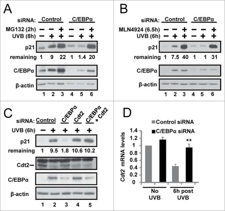
The bZIP transcription factor, C/EBPα is highly inducible by UVB and other DNA damaging agents in keratinocytes. C/EBPα-deficient keratinocytes fail to undergo cell cycle arrest in G1 in response to UVB-induced DNA damage and mice lacking epidermal C/EBPα are highly susceptible to UVB-induced skin cancer. The mechanism through which C/EBPα regulates the cell cycle checkpoint in response to DNA damage is unknown. Here we report untreated C/EBPα-deficient keratinocytes have normal levels of the cyclin-dependent kinase inhibitor, p21, however, UVB-treated C/EBPα-deficient keratinocytes fail to up-regulate nuclear p21 protein levels despite normal up-regulation of Cdkn1a mRNA levels. UVB-treated C/EBPα-deficient keratinocytes displayed a 4-fold decrease in nuclear p21 protein half-life due to the increased proteasomal degradation of p21 via the E3 ubiquitin ligase CRL4(Cdt2). Cdt2 is the substrate recognition subunit of CRL4(Cdt2) and Cdt2 mRNA and protein levels were up-regulated in UVB-treated C/EBPα-deficient keratinocytes. Knockdown of Cdt2 restored p21 protein levels in UVB-treated C/EBPα-deficient keratinocytes. Lastly, the failure to accumulate p21 in response to UVB in C/EBPα-deficient keratinocytes resulted in decreased p21 interactions with critical cell cycle regulatory proteins, increased CDK2 activity, and inappropriate entry into S-phase. These findings reveal C/EBPα regulates G1/S cell cycle arrest in response to DNA damage via the control of CRL4(Cdt2) mediated degradation of p21.

Keywords: C/EBPα; C/EBPα, CCAAT/enhancer binding protein α; CRL, Cullin-RING ubiquitin ligases; CRL4Cdt2; DCAF, DDB1/CUL4-associated factor; Cdt2, Cdc10 dependent transcript 2; G1/S DNA damage checkpoint; SCF, Skp, Cullin, F-box containing complex; UVB, ultraviolet B; keratinocytes, p21.


Figures

Figure 1.
Figure 1.
C/EBPα is required for UVB-induced upregulation of p21 protein levels. (A) Immunoblot analysis of untreated control and C/EBPα siRNA treated Balb/MK2 keratinocytes and K5Cretg/+ and K5Cretg/+;C/EBPαflox/flox epidermal lysates. (B) Immunoblot analysis of lysates from Balb/MK2 keratinocytes treated with C/EBPα siRNA or control siRNA and collected at the indicated times after exposure to 5 mJ/cm2 UVB. (C) IHC staining for p21 in formalin-fixed paraffin-embedded sections of mouse skin from K5Cretg/+ and K5Cretg/+;C/EBPαflox/flox mice 4 h after treatment with 50 mJ/cm2 UVB. (D) Immunoblot analysis of p21, and C/EBPα from lysates isolated from K5Cretg/+ and K5Cretg/+;C/EBPαflox/flox mouse epidermis collected at the indicated times following exposure to 100 mJ/cm2 UVB. (E and F) Relative Cdkn1a (p21) mRNA levels in (E) Balb/MK2 cells treated with C/EBPα siRNA or control siRNA and (F) K5Cre and K5Cre;C/EBPαflox/flox in mouse epidermis collected at the indicated times following UVB. Data are expressed as the mean normalized to Gapdh ±S .D. (N ≥ 3). There were no statistically significant differences between control and C/EBPα-deficient keratinocytes in culture or in vivo as measured and calculated by Student's T test. (G) Immunoblot analysis of lysates of NHEK cells treated with C/EBPα siRNA or control siRNA and collected 10 h post 10 mJ/cm2 UVB. (H) Immunoblot analysis of Balb/MK2 treated with C/EBPα siRNA or control siRNA and collected 6 or 12 h after 25 μM MNNG treatment. (I) Immunoblot analysis of Balb/MK2 treated with C/EBPα siRNA or control siRNA and collected 8 h after exposure to 5 mJ/cm2 UVB.
Figure 2.
Figure 2.
C/EBPα regulates nuclear p21 protein stability following UVB-induced DNA damage. (A) Immunoblot analysis of cytosolic and nuclear fractions prepared from siRNA treated Balb/MK2 keratinocytes collected 8 h post 5 mJ/cm2 UVB. (B) Immunoblot analysis of nuclear fractions prepared from siRNA treated Balb/MK2 collected at the indicated times post UVB. (C) Immunoblot analysis of p21 and C/EBPα in UVB and CHX treated nuclear extracts. Five h post 5 mJ/cm2 UVB cells were treated with 40 μg/mL CHX (t = 0) and collected at the indicated times. Values below the p21 image represent the fraction of p21 remaining at that time point as measure by densitometry normalized to β-actin. Values are also plotted to the right. (D) Immunoblot analysis of p21 and C/EBPα in CHX treated nuclear extracts. C/EBPα or control siRNA cells were treated with CHX. Values below the p21 image represent the fraction of p21 remaining at that time point as measure by densitometry normalized to β-actin. (E) Immunoblot analysis of p21 co-IP from nuclear extracts 6 h post 5 mJ/cm2 UVB.
Figure 3.
Figure 3.
C/EBPα regulates CRL4Cdt2-mediated degradation of p21 in response to UVB. (A) Immunoblot analysis of control and C/EBPα siRNA nuclear extracts 8 h after 5 mJ/cm2 UVB. Ten μM MG132 was added 2 h before collection. (B) Immunoblot analysis of control and C/EBPα siRNA nuclear extracts 6 h after exposure to 5 mJ/cm2 UVB. Cells were treated with 800 nM MLN4924 30 min before UVB exposure. (C) Immunoblot analysis of control, C/EBPα, and Cdt2 siRNA nuclear extracts 6 h after 5 mJ/cm2 UVB. (D) Relative Cdt2 mRNA levels in control and C/EBPα siRNA 6 h following 5 mJ/cm2 UVB. Data are expressed as the mean normalized to Gapdh ±S .D. (N = 3). **= P value < 0.01 as calculated using Student's T test.
Figure 4.
Figure 4.
Failure to up-regulate nuclear p21 protein levels in UVB-exposed C/EBPα-deficient keratinocytes leads to functional consequences on cell cycle protein complexes, CDK2 activity and the G1/S checkpoint. (A) Immunoblot analysis of CDK2 co-IP from control and C/EBPα siRNA nuclear extracts 6 h following 5 mJ/cm2 UVB. (B) Phosphorylation of H1 by CDK2 immunoprecipitated from control and C/EBPα siRNA nuclear extracts 5 mJ/cm2 after UVB exposure. (C) Bar plot of peptide abundance for unique peptides for the proteins of interest across the 2 technical p21 IP replicates. Peptide abundances are expressed as a percent of the most abundant for a specific protein. **For PCNA, we could not confidently integrate a peptide signal in the siRNA replicates. A second peptide for each protein shows similar results (Fig. S3). (D)3H-thymidine incorporation in synchronized control and C/EBPα siRNA treated keratinocytes after 5 mJ/cm2 UVB exposure. Data represents the mean ± S.D., * = P value < 0.01 as calculated using Student's T test (N = 3) and are representative of 3 independent experiments.

Similar articles

Cited by

References

    1. American Cancer Society 2013 Cancer Facts and Figures . Atlanta: American Cancer Society. http://cancer.org.
    1. Brash DE. Sunlight and the onset of skin cancer. Trends Genet 1997; 13:410-4; PMID:9351343; http://dx.doi.org/10.1016/S0168-9525(97)01246-8 - DOI - PubMed
    1. de Gruijl FR, van Kranen HJ, Mullenders LH. UV-induced DNA damage, repair, mutations and oncogenic pathways in skin cancer. J Photochem Photobiol 2001; 63:19-27; PMID:11684448; http://dx.doi.org/10.1016/S1011-1344(01)00199-3 - DOI - PubMed
    1. Brash DE, Rudolph JA, Simon JA, Lin A, McKenna GJ, Baden HP, Halperin AJ, Ponten J. A role for sunlight in skin cancer: UV-induced p53 mutations in squamous cell carcinoma. Proc Natl Acad Sci U S A 1991; 88:10124-8; PMID:1946433; http://dx.doi.org/10.1073/pnas.88.22.10124 - DOI - PMC - PubMed
    1. Kastan MB, Bartek J. Cell-cycle checkpoints and cancer. Nature 2004; 432:316-23; PMID:15549093; http://dx.doi.org/10.1038/nature03097 - DOI - PubMed

Publication types

MeSH terms