. 2015 Jan 20;6(2):1231-48.
doi: 10.18632/oncotarget.2840.

Aspartate β-Hydroxylase expression promotes a malignant pancreatic cellular phenotype

Affiliations

Aspartate β-Hydroxylase expression promotes a malignant pancreatic cellular phenotype

Xiaoqun Dong et al. Oncotarget. .

Erratum in

Abstract

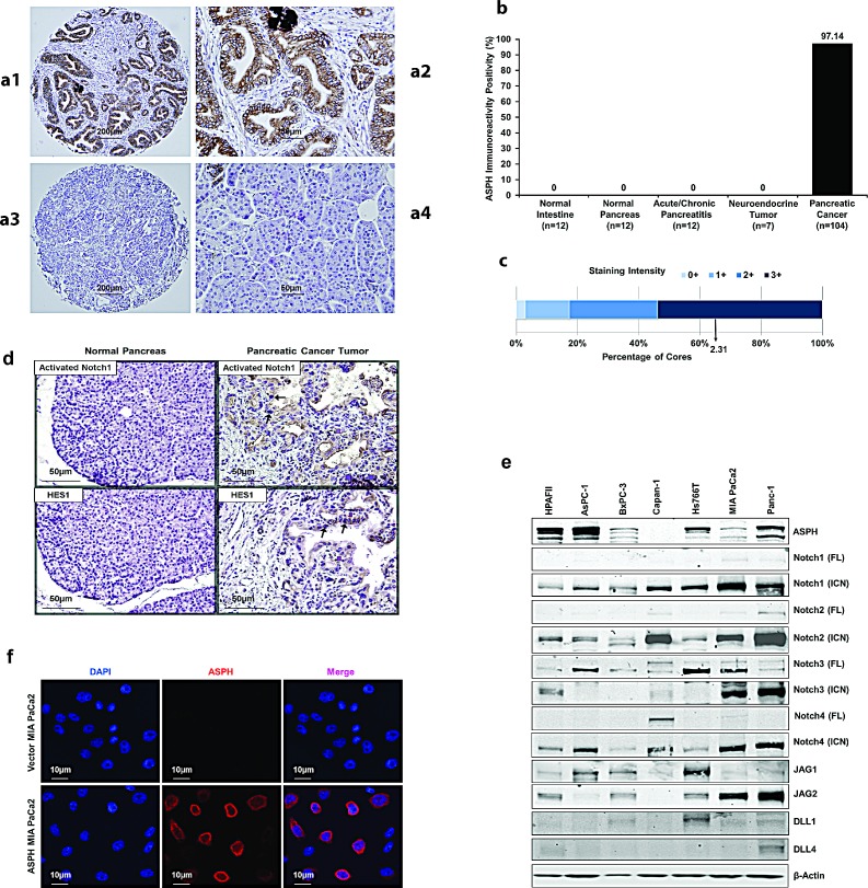
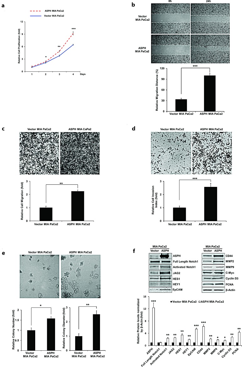
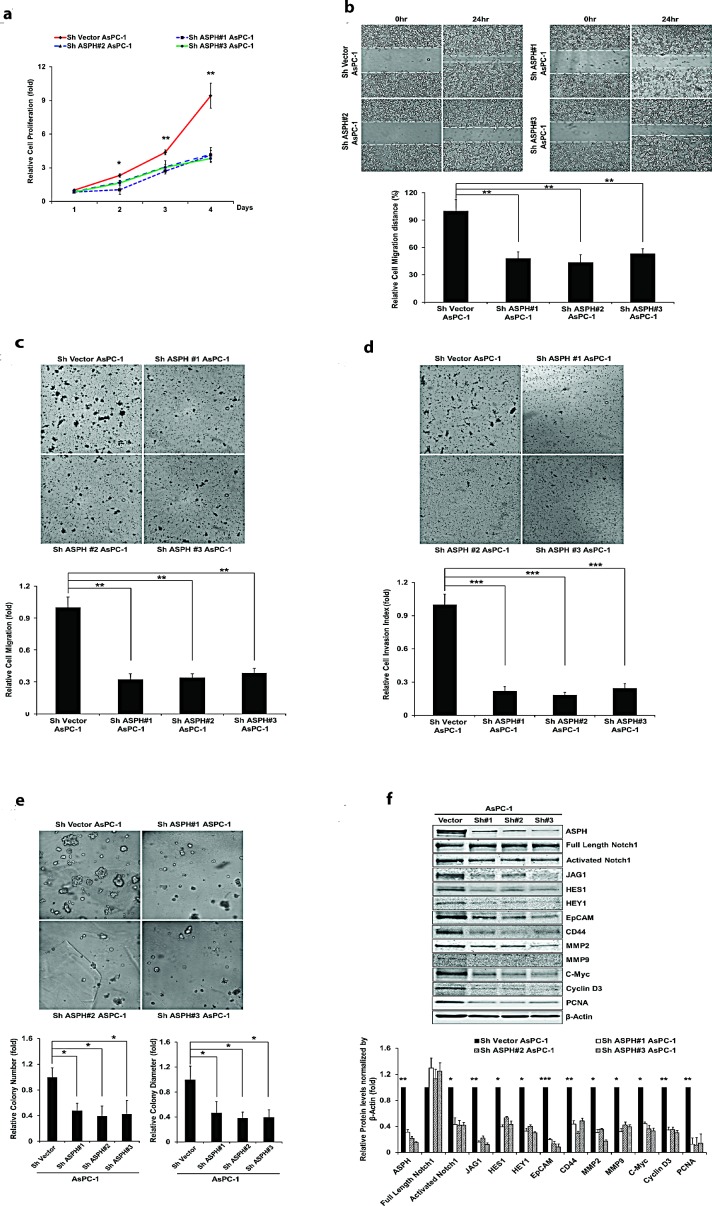
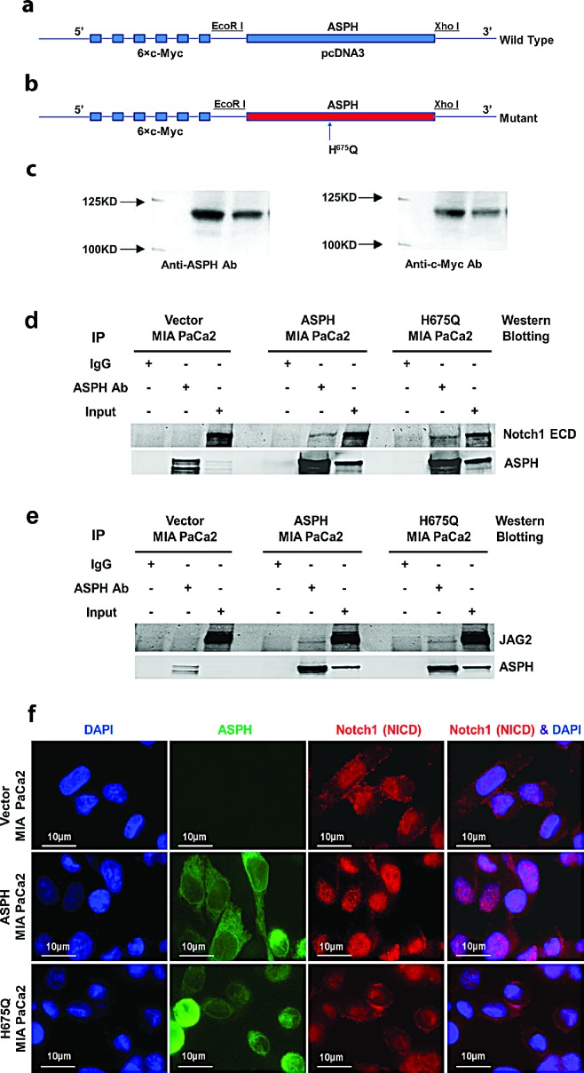
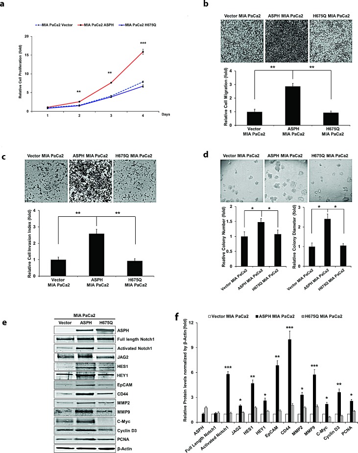
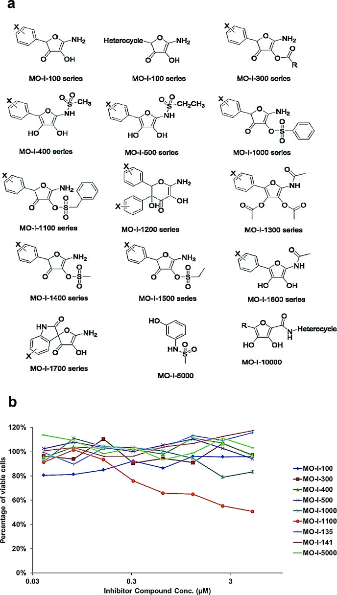
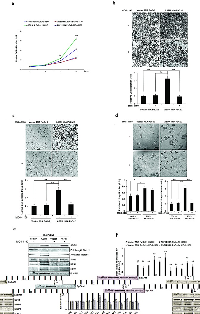
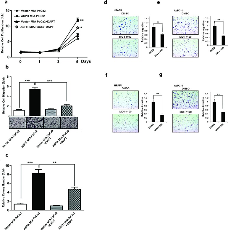
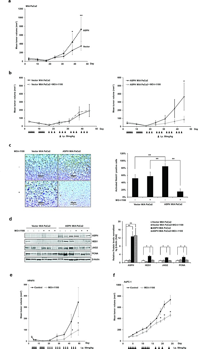
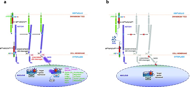
Pancreatic cancer (PC) is one of the leading causes of cancer related deaths due to aggressive progression and metastatic spread. Aspartate β-hydroxylase (ASPH), a cell surface protein that catalyzes the hydroxylation of epidermal growth factor (EGF)-like repeats in Notch receptors and ligands, is highly overexpressed in PC. ASPH upregulation confers a malignant phenotype characterized by enhanced cell proliferation, migration, invasion and colony formation in vitro as well as PC tumor growth in vivo. The transforming properties of ASPH depend on enzymatic activity. ASPH links PC growth factor signaling cascades to Notch activation. A small molecule inhibitor of β-hydroxylase activity was developed and found to reduce PC growth by downregulating the Notch signaling pathway. These findings demonstrate the critical involvement of ASPH in PC growth and progression, provide new insight into the molecular mechanisms leading to tumor development and growth and have important therapeutic implications.


Conflict of interest statement

Declaration

There is no conflict of interest among the authors.

Figures

Fig. 1
Fig. 1. ASPH expression in human PDAC, pancreatic neuroendocrine tumor, acute and chronic pancreatitis, normal pancreas and normal intestine
(a1) Typical ASPH expression in PDAC at 40× in tissue microarrays (TMAs) from 104 tumors. (a2) A representative example of IHS with mAb FB-50 (400×). (a3) A representative PDAC at 40× stained with a negative control mAb against hepatitis B surface antigen of the same isotype as mAb FB-50. (a4) Normal adjacent pancreas stained with the anti-ASPH FB-50 mAb (400×). (b) A summary of positive and negative expression of ASPH by IHS in normal tissues, pancreatitis and pancreatic tumors. (c) A bar graph of staining intensity with a mean score of 2.31 for PDACs where 0 = no, 1 = weak, 2 = moderate, 3 = strong expression of ASPH by IHS. Fig. 1. (d) Activated Notch1 and HES1 were also overexpressed in the cytoplasm and nuclei of tumor tissue compared to adjacent normal pancreas of PC patients (400×). The arrows highlight the positive staining of activated Notch1 and HES1 in the nuclei of tumor cells in representative examples. (e) Expression profiling of ASPH and Notch signaling proteins in 7 human PC cell lines. Most PC cell lines showed upregulation and activation of C-terminal intracellular Notch (ICN) 1, 2, 4 and to a lesser extent, Notch 3 as determined by Western blot analysis. The Notch ligands Jagged (JAG) 1 and 2 were expressed in 4/7 and 5/7 of the cell lines, respectively. In contrast, Delta (DLL) 1 and 4 were infrequently upregulated. (f) Lentiviral transfection and stable expression of ASPH in MIA PaCa2 cells. Note the cell surface localization of ASPH after stable overexpression by lentivirus as depicted in the middle panel as compared to the empty vector stable transfected control.
Fig. 2
Fig. 2. Stable overexpression of ASPH in MIA PaCa2 cells promotes cell proliferation
(a), wound healing (b), migration (c), invasion (d), and colony formation (e) as indices of changes in the malignant phenotype. ASPH overexpression also enhances the expression of activated Notch1, JAG2, and Notch signaling downstream target genes including HES1, HEY1, EpCAM, CD44, c-Myc, MMP2/9, cyclin D3 and PCNA as shown in Western blot analysis (f). *p<0.05; **p<0.01; ***p<0.001.
Fig. 3
Fig. 3. Knockdown of ASPH by specific shRNAs in AsPC-1 cells inhibited
(a) proliferation, (b) wound healing, (c) migration, (d) invasion, and (e) colony formation as indices of changes in the malignant phenotype. (f) Endogenous overexpression of ASPH was associated with upregulation of activated Notch1 and JAG2, as well as Notch signaling downstream target genes HES1, HEY1, EpCAM, CD44, c-Myc, MMP2/9, cyclin D3 and PCNA in a lentiviral transfected stable cell line expressing a nonspecific scrambled shRNA. The AsPC-1 cell lines stable transfected with ASPH specific shRNAs demonstrated downregulation of activated Notch1, JAG2, and Notch activated genes as determined by Western blot analysis. *p<0.05; **p<0.01; ***p<0.001.
Fig. 4
Fig. 4. ASPH gene was subcloned into pcDNA3 with 5′ c-Myc tagged vector. Inclusion of a c-Myc-tag allowed discrimination of exogenous expressed protein from endogenous cellular levels
(a) “Wild type” (WT) ASPH construct; (b) ASPH mutant (H675Q) construct where enzymatic activity was reduced to 20% of “wild type” protein level; (c) Western blot analysis of WT and mutant proteins with antibodies to ASPH and c-Myc. A direct physical interaction of ASPH with (d) Notch1 or (e) JAG2 by co-immunoprecipitation (IP) was observed in MIA PaCa2 cells. ASPH, Notch1, and JAG2 immunoprecipitates were fractionated by SDS-PAGE and analyzed by Western blot using the FB-50 monoclonal antibody. The negative control (Neg) corresponds to an IP result using non-relevant antibody to hepatitis B virus. (f) Microscopy showing increased nuclear translocation of activated Notch1 ICN in the nuclei of MIA PaCa2 cells stably expressing WT-ASPH. Note that the mutant H675Q ASPH substantially reduces nuclear localization of Notch1 ICN comparable to that in vector transfected MIA PaCa2 cells, which suggests that the enzymatic activity of the protein is critical for Notch activation.
Fig. 5
Fig. 5. The β-hydroxylase activity of ASPH is required for its transforming activity
MIA PaCa2 cells were stably transfected with empty vector (EV), “wild type” (WT)-ASPH, or H675Q-ASPH mutant and the levels of expression were confirmed by Western blot. There is low-level endogenous ASPH in MIA PaCa2 cells as shown here and in Fig. 1. Effects of EV, WT-ASPH and H675Q-ASPH on (a) cell proliferation, (b) migration, (c) invasion, and (d) colony formation are significantly different. (e and f) represents a Western blot demonstrating WT-ASPH induced activation of Notch signaling as determined by increased levels of Notch1 ICN, JAG2, as well as increased expression of downstream responsive genes HES1, HEY1, EpCAM, CD44, c-Myc, MMP2/9, cyclin D3 and PCNA. In contrast, the mutant H675Q ASPH construct shows significantly reduced activated Notch1 ICN, JAG2, as well as HES1, HEY1, EpCAM, CD44, c-Myc, MMP2/9, cyclin D3 and PCNA. The results suggest that the β-hydroxylase activity of ASPH is essential for Notch signaling activation. *p<0.05; **p<0.01; ***p<0.001.
Fig. 6
Fig. 6
(a) Candidate parent compounds selected and evaluated as potential inhibitors of ASPH β-hydroxylase activity were synthesized on the basis of the crystal structure of the catalytic site in the C-terminal region of ASPH using computer assisted drug design. (b) Effects of SMIs on cell viability of FOCUS HCC cells that highly expressed ASPH on the cell surface as a drug screen for biological activity. Note that MO-I-1100 demonstrated a dose dependent effect on cell viability over a range of 0.03 – 3 μM whereas other compounds such as MO-I-100, 300 and 400 for example, showed little, if any, activity.
Fig. 7
Fig. 7. Effect of SMI MO-I-1100 on the PC phenotype induced by exogenous or endogenous high-level of WT-ASPH expression
MIA PaCa2 cells stably transfected with empty vector or the “wild type” ASPH construct via lentiviral transfection depicted in Fig. 2. The inhibitory effects of MO-I-1100 on (a) proliferation, (b) migration, (c) invasion, and (d) colony formation of MIA PaCa2 cells were observed. There is a significant reduction in the expression of Notch1 ICN, JAG2, as well as downstream responsive genes HES1, HEY1, EpCAM, CD44, c-Myc, MMP2/9, cyclin D3 and PCNA induced by MO-I-1100 compared to the DMSO treatment (e and f). *p<0.05; **p<0.01; ***p<0.001.
Fig. 8
Fig. 8. The γ-secretase inhibitor DAPT (at a concentration of 10 μM) reversed the aggressive malignant phenotype exhibited by ASPH overexpression in MIA PaCa2 cells
DAPT attenuated ASPH induced (a) cell proliferation, (b) migration and (c) colony formation through inhibition of Notch1 ICN signaling. A SMI (MO-I-1100) exerted inhibitory effects on the PC phenotype induced by endogenous high-level of ASPH expression. A significant reduction in (d, e) migration and (f, g) invasion was observed in HPAFII and AsPC-1 PC cells with high endogenous levels of ASPH (Fig. 1) treated with MO-I-1100 at 5 μM. *p<0.05; **p<0.01; ***p<0.001 when compared with control.
Fig. 9
Fig. 9. Effect of a SMI (MO-I-1100) on PC tumor growth in subcutaneous (s.c.) tumor model of nude mice
(a) ASPH transfected MIA PaCa2 cells had high tumorigenicity compared to MIA PaCa2 cells transfected with empty vector. Growth curve on 8 animals in each group indicated enhanced tumor development induced by ASPH overexpression. (b) represents tumor growth curves derived from MIA PaCa2 cells stably transfected with empty vector or a WT-ASPH expression construct as delivered by lentivirus. Note that anti-tumor effects exhibited by MO-I-1100 were observed only in tumors with exogenous ASPH overexpression. Each group had 10 animals. Effect of MO-I-1100 on Notch signaling in vivo has been observed. Tumors taken from the growth curve that were responsive to MO-I-1100 treatment depicted in (b) were analyzed for Notch signaling compared to untreated control. There were 3 tumors taken from the MO-I-1100 treatment group and 3 from the untreated control. (c) demonstrates that the expression of the Notch1 ICN is substantially downregulated by the SMI of APSH β-hydroxylase activity by IHS. (d) represents reduction in the expression of JAG2, as well as Notch activated genes HES1 and PCNA following MO-I-1100 treatment. Similar inhibitory effects of MO-I-1100 on PC tumor growth in vivo were observed in (e) HPAFII and (f) AsPC-1 cells induced s.c. tumors. These human PC cell lines had high endogenous expression of ASPH as shown in Fig. 1a. *p < 0.05; **p < 0.01.
Fig. 10
Fig. 10. Hypothesis of how ASPH may be involved in the pathogenesis of PC progression through Notch activation [6]
The structure of ASPH molecule consists of cytoplasmic (green), transmembrane (purple), luminal region (blue) and catalytic domain (red), which contains the catalytic site (M670HPGTH675). (a) In the presence of β-hydroxylase activity of ASPH, binding of the JAG ligand (green) on one cell to the Notch receptor (purple) on another cell results in two proteolytic cleavages of the receptor. The ADAM10 or TACE (TNF-α-converting enzyme; ADAM17) metalloprotease (black) catalyzes the S2 cleavage and generates a substrate for S3 cleavage by the γ-secretase complex (red). This proteolytic processing mediates release of the Notch intracellular domain (NICD), which enters the nucleus and interacts with the DNA-binding CSL (CBF1, Su(H) and LAG-1) protein. The co-activator Mastermind (Mam) and other transcription factors are recruited to the CSL complex, whereas co-repressors (Co-R) are released. Thus, the Notch signaling is on and the downstream target genes are expressed: CD44 and EpCAM are markers for cancer stem cells; PCNA, cyclin D3, c-Myc, MMP2/9, HES1 and HEY1 are involved in cell proliferation, migration, invasion, tumor growth and metastasis in PC. (b) SMIs block the catalytic site of ASPH, which leads to loss of β-hydroxylase activity and failure of JAG2 binding to Notch1. Thus, Notch signaling is off and its oncogenic effector is inhibited in PC.

References

    1. Siegel R, Naishadham D, Jemal A. Cancer statistics, 2013. CA: a cancer journal for clinicians. 2013;63(1):11–30. - PubMed
    1. Vincent A, Herman J, Schulick R, Hruban RH, Goggins M. Pancreatic cancer. Lancet. 2011;378(9791):607–620. - PMC - PubMed
    1. Jia S, VanDusen WJ, Diehl RE, Kohl NE, Dixon RA, Elliston KO, Stern AM, Friedman PA. cDNA cloning and expression of bovine aspartyl (asparaginyl) beta-hydroxylase. J Biol Chem. 1992;267(20):14322–14327. - PubMed
    1. Lanzoni G, Oikawa T, Wang Y, Cui CB, Carpino G, Cardinale V, Gerber D, Gabriel M, Dominguez-Bendala J, Furth ME, Gaudio E, Alvaro D, Inverardi L, Reid LM. Concise review: clinical programs of stem cell therapies for liver and pancreas. Stem cells. 2013;31(10):2047–2060. - PMC - PubMed
    1. Engel J. EGF-like domains in extracellular matrix proteins: localized signals for growth and differentiation? FEBS letters. 1989;251(1-2):1–7. - PubMed

Publication types

MeSH terms

Substances