Review
. 2014;5(2):9.
doi: 10.4161/21541248.2014.973757.

Vav family exchange factors: an integrated regulatory and functional view

Affiliations
Review

Vav family exchange factors: an integrated regulatory and functional view

Xosé R Bustelo. Small GTPases. 2014.

Abstract

The Vav family is a group of tyrosine phosphorylation-regulated signal transduction molecules hierarchically located downstream of protein tyrosine kinases. The main function of these proteins is to work as guanosine nucleotide exchange factors (GEFs) for members of the Rho GTPase family. In addition, they can exhibit a variety of catalysis-independent roles in specific signaling contexts. Vav proteins play essential signaling roles for both the development and/or effector functions of a large variety of cell lineages, including those belonging to the immune, nervous, and cardiovascular systems. They also contribute to pathological states such as cancer, immune-related dysfunctions, and atherosclerosis. Here, I will provide an integrated view about the evolution, regulation, and effector properties of these signaling molecules. In addition, I will discuss the pros and cons for their potential consideration as therapeutic targets.

Keywords: Ac, acidic; Ahr, aryl hydrocarbon receptor; CH, calponin homology; CSH3, most C-terminal SH3 domain of Vav proteins; DAG, diacylglycerol; DH, Dbl-homology domain; Dbl-homology; GDP/GTP exchange factors; GEF, guanosine nucleotide exchange factor; HIV, human immunodeficiency virus; IP3, inositoltriphosphate; NFAT, nuclear factor of activated T-cells; NSH3, most N-terminal SH3 domain of Vav proteins; PH, plekstrin-homology domain; PI3K, phosphatidylinositol-3 kinase; PIP3, phosphatidylinositol (3,4,5)-triphosphate; PKC, protein kinase C; PKD, protein kinase D; PLC-g, phospholipase C-g; PRR, proline-rich region; PTK, protein tyrosine kinase; Phox, phagocyte oxidase; Rho GTPases; SH2, Src homology 2; SH3, Src homology 3; SNP, single nucleotide polymorphism; TCR, T-cell receptor; Vav; ZF, zinc finger region; cGMP, cyclic guanosine monophosphate; cancer; cardiovascular biology; disease; immunology; nervous system; signaling; therapies.


Figures

Figure 1.
Figure 1.
Examples of the multidomain structure of some Vav family proteins. Abbreviations for domains have been indicated in the main text. The shape of the DH tries to mimic the 3 dimensional structure of this domain. Phosphorylation sites are shown as yellow circles. Amino acid numbers correspond to the primary sequence of mouse Vav1. An Ac α helix involved in the stabilization of the autoinhibited structure of Vav proteins is shown as a blue box. Cel, C. elegans; Dm, D. melanogaster.
Figure 2.
Figure 2.
The Vav family interactome. Examples of some of the Vav family interacting proteins and domains involved. Effector molecules are shown in blue. Positive and negative regulators are shown in green and red, respectively. Proteins whose interaction was only described for Vav2 or Vav3 are indicated by asterisks. Please, note that some of these proteins may exert several functions in the context of Vav signaling.
Figure 3.
Figure 3.
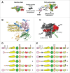
Mechanism of activation of Vav family proteins. (A) A model for the phosphorylation-mediated activation of Vav proteins based on recently described structural and biochemical data. The autoinhibited state of nonphosphorylated Vav proteins is stabilized by extensive contacts of the CH domain and 2 tyrosine residues of the Ac region (Tyr, Tyr160) with the DH and PH domains; an α helix present in the Vav Ac domain (which contains the Tyr174 phosphosite) with amino acids located in the GTPase binding interface of the DH domain; and additional residues of the Ac with the PH domain. In addition, 2 independent amino acid stretches of the CSH3, which do not include the canonical PRR binding site, establish interactions with both the DH and PH domains (see also panel C below). Upon phosphorylation of indicated residues, the autoinhibited structure is released. Note that the 3 dimensional structure of inactive and active full-length Vav proteins is unknown, so the conformations shown for the 2 functional states of Vav proteins are hypothetical. Nonphosphorylated and phosphorylated tyrosine residues are shown as blue and yellow circles, respectively. The 2 sites of the CSH3 domain of Vav proteins involved in formation of the autoinhibited structure are depicted as dark blue and red boxes. Color codes for Vav domains are those used in Figure 1. (B) Crystal structure of the autoinhibited Vav1 CH-Ac-DH-PH-ZF region. Stretches of the Ac region that could not be crystallized are shown as broken lanes. (C) The Vav1 CSH3 structure showing the areas that establish the intramolecular interaction with indicated domains (red color). C, domain C-terminus; N, domain N-terminus; PRRBS, PRR binding site. The area corresponding to the canonical CSH3 PRR binding site is shown in green. (D and E) Examples of some hyperactive Vav mutants (proteins 1 to 5) generated by either domain truncation (D) or amino acid mutations (E). The full-length protein (WT) is included in both panels to facilitate an easy understanding of the type of mutations made. In E, the specific amino acid residues that had been mutated have been highlighted using a cross sign. Mutants are ordered from top to bottom following the order of discovery. The first mutation ever found in the Vav family is indicated in D (mutant 1). The truncated mutant proteins labeled as 2, 3, 4, and 5 shown in panel D were first described by Schuebel et al. (1996), Schuebel et al. (1998), Movilla et al. (1999), and Barreira et al. (2014), respectively. In E, mutant proteins labeled as 1–2 and 3–5 were first described by Lopez-Lago et al. (2000) and Barreira et al. (2014), respectively.
Figure 4.
Figure 4.
Signaling steps for the stimulation of Vav proteins in lymphocytes. For simplicity, we only show the phosphorylation step mediated by a Syk family member. Gray and green arrows represent translocation and phosphorylation steps, respectively. This step should include SH2-mediated interactions with the kinase (not shown in steps b and c). ITAM, immunoreceptor tyrosine-based activation motif.

References

    1. Katzav S, Martin-Zanca D, Barbacid M. Vav, a novel human oncogene derived from a locus ubiquitously expressed in hematopoietic cells. EMBO J 1989; 8:2283-90; PMID:2477241 - PMC - PubMed
    1. Henske EP, Short MP, Jozwiak S, Bovey CM, Ramlakhan S, Haines JL, Kwiatkowski DJ. Identification of VAV2 on 9q34 and its exclusion as the tuberous sclerosis gene TSC1. Ann Hum Genet 1995; 59:25-37; PMID:7762982; http://dx.doi.org/10.1111/j.1469-1809.1995.tb01603.x - DOI - PubMed
    1. Schuebel KE, Bustelo XR, Nielsen DA, Song BJ, Barbacid M, Goldman D, Lee IJ. Isolation and characterization of murine vav2, a member of the vav family of proto-oncogenes. Oncogene 1996; 13:363-71; PMID:8710375 - PubMed
    1. Movilla N, Bustelo XR. Biological and regulatory properties of Vav-3, a new member of the Vav family of oncoproteins. Mol Cell Biol 1999; 19:7870-85; PMID:10523675 - PMC - PubMed
    1. Bustelo XR. Regulatory and signaling properties of the Vav family. Mol Cell Biol 2000; 20:1461-77; PMID:10669724; http://dx.doi.org/10.1128/MCB.20.5.1461-1477.2000 - DOI - PMC - PubMed

Publication types

MeSH terms

Substances