

Editorial
doi: 10.1002/uog.14746.
Cell-free fetal DNA: the new tool in fetal medicine
Affiliations
- PMID: 25483938
- PMCID: PMC5029578
- DOI: 10.1002/uog.14746
Item in Clipboard
Editorial
Cell-free fetal DNA: the new tool in fetal medicine
Ultrasound Obstet Gynecol.
2015 May.
No abstract available
Figures
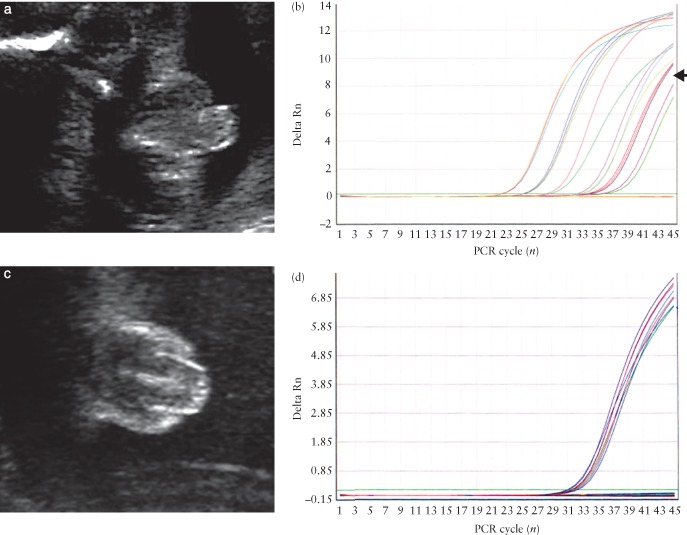
Genital ambiguity in a male fetus (a), as evidenced by amplification (arrow) of SRY sequences in cell‐free fetal DNA (cffDNA) (b), and in a female fetus (c), in which there is amplification only of the control DNA sequences (d). PCR, polymerase chain reaction; Rn, normalized reporter.
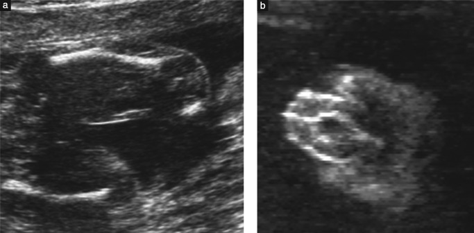
Features of campomelic dysplasia detectable on ultrasound include shortened ‘bowed’ limbs (a) and ambiguous genitalia (b).
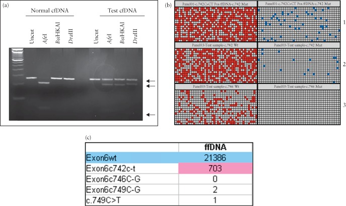
Detection of a mutation in the fibroblast growth factor receptor 3 (FGFR3 ) gene causing thanatophoric dysplasia, showing the increasing ease of interpretation between polymerase chain reaction (PCR)‐based method (a), digital PCR (b) and digital readout obtained from sequencing (c). PCR‐based method (a) relies on subjective interpretation; very faint bands for mutant alleles in affected cell‐free (cf) DNA can be seen (bottom arrows). The wild‐type (normal) allele is strongly present in all samples (upper arrow). This compares with digital PCR (b) for detection of the mutant allele c.742 C > T (blue dot) and wild‐type alleles (red dot). Each row represents one sample. Wild‐type signals are present in all samples but the mutant allele is only present in the positive control (panel 1) and test sample (panel 2). Panel 3 is the result obtained from a normal pregnancy and shows only wild‐type alleles present. The digital readout obtained from sequencing (c) reveals a very high wild‐type allele count (blue), as this represents both maternal and fetal alleles, and a lower mutant allele (pink) count, but is still very high compared with the counts for other disease‐causing mutations, indicating that the fetus has thanatophoric dysplasia as a result of the c.742 C > T mutation.
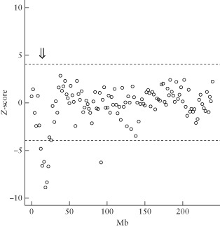
Detection of chromosomal rearrangements in cell‐free fetal DNA (cffDNA) using standard aneuploidy sequencing. A small deletion ( ) of chromosome 2, confirmed as 46,XY,del(2) (p23p25.1), is indicated when the expected number of reads falls outside a
 Z ‐score of ± 4 ( ).
).
) of chromosome 2, confirmed as 46,XY,del(2) (p23p25.1), is indicated when the expected number of reads falls outside a ).References
-
- Lo YM, Corbetta N, Chamberlain PF, Rai V, Sargent IL, Redman CW, Wainscoat JS. Presence of fetal DNA in maternal plasma and serum. Lancet 1997. ; 350 : 485–487. - PubMed
-
- Alberry M, Maddocks D, Jones M, Abdel Hadi M, Abdel‐Fattah S, Avent N, Soothill PW. Free fetal DNA in maternal plasma in anembryonic pregnancies: confirmation that the origin is the trophoblast. Prenat Diagn 2007. ; 27 : 415–418. - PubMed
-
- Lunn FM, Chiu RW, Allen Chan KC, Yeung Leung T, Kin Lau T, Dennis Lo YM. Microfluidics digital PCR reveals a higher than expected fraction of fetal DNA in maternal plasma. Clin Chem 2008. ; 54 : 1664–1672. - PubMed
Publication types
MeSH terms
Substances
Grants and funding
LinkOut - more resources
Full Text Sources
Other Literature Sources
Medical
