Review
. 2015;21(4):431-48.
doi: 10.2174/138161282104141204124129.

Targeting cardiomyocyte Ca2+ homeostasis in heart failure

Affiliations
Review

Targeting cardiomyocyte Ca2+ homeostasis in heart failure

Asmund T Roe et al. Curr Pharm Des. 2015.

Abstract

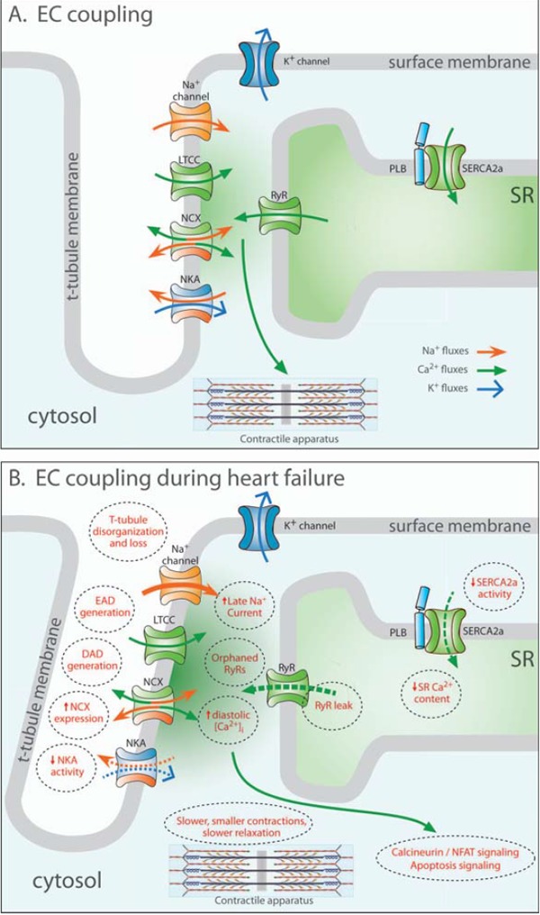
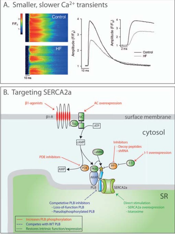
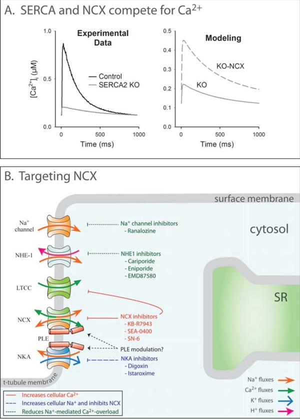
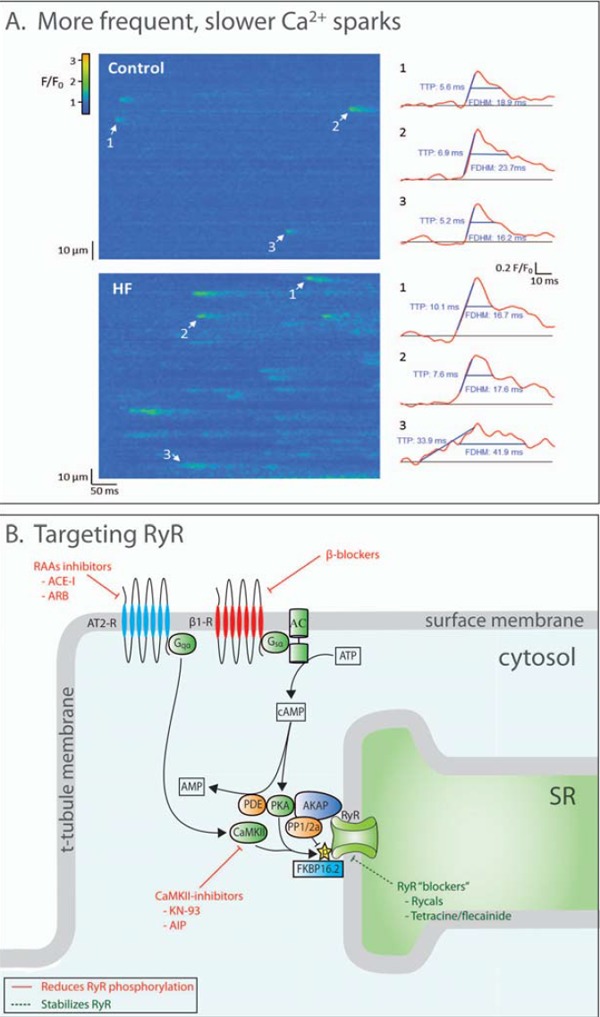
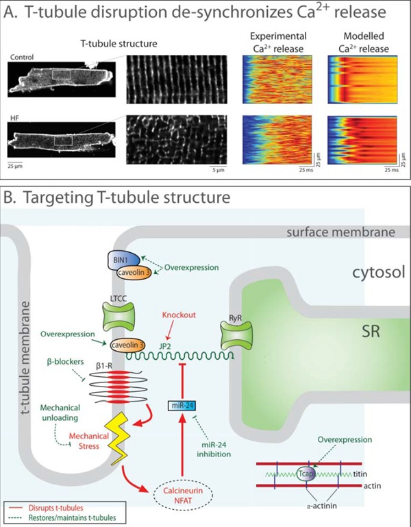
Improved treatments for heart failure patients will require the development of novel therapeutic strategies that target basal disease mechanisms. Disrupted cardiomyocyte Ca(2+) homeostasis is recognized as a major contributor to the heart failure phenotype, as it plays a key role in systolic and diastolic dysfunction, arrhythmogenesis, and hypertrophy and apoptosis signaling. In this review, we outline existing knowledge of the involvement of Ca(2+) homeostasis in these deficits, and identify four promising targets for therapeutic intervention: the sarcoplasmic reticulum Ca(2+) ATPase, the Na(+)-Ca(2+) exchanger, the ryanodine receptor, and t-tubule structure. We discuss experimental data indicating the applicability of these targets that has led to recent and ongoing clinical trials, and suggest future therapeutic approaches.


Figures

Fig. (1)
Fig. (1)
Excitation-contraction (EC) contraction coupling in the normal and failing heart. In healthy cardiomyocytes (A), the action potential propagates into the t-tubules, opening voltage-gated L-type Ca2+ channels (LTCCs). The resulting Ca2+ influx triggers the opening of Ca2+ release channels (ryanodine receptors, RyRs) in the membrane of the sarcoplasmic reticulum (SR). Released Ca2+ initiates contraction as it binds to the contractile apparatus, and relaxation occurs as Ca2+ is recycled into the SR by the SR Ca2+ ATPase 2a (SERCA2a), and removed from the cell via the Na+-Ca2+ exchanger (NCX). During heart failure (B), Ca2+ release is impaired, leading to slower and smaller contractions. Reduced SR Ca2+ content results from decreased SERCA2a activity, increased RyR leak and, in some cases, increased NCX activity. Systolic dysfunction also results from disruption of T-tubule structure, which functionally “orphans” some RyRs from LTCCs. Slowed and incomplete Ca2+ removal from the cytosol impairs cardiomyocyte relaxation, and promotes hypertrophy and apoptosis signaling. Triggered cardiac arryhythmia has been linked to RyR leak, and removal of released Ca2+ by NCX, causing DADs. EADs may result from inappropriate re-opening of Ca2+ channels. Deficient Ca2+ cycling is also linked to altered Na+ homeostasis, following downregulation of the Na+-K+ ATPase (NKA) and increased late Na+ current.
Fig. (2)
Fig. (2)
Disrupted Ca2+ homeostasis in failing cardiomyocytes and therapeutic targeting of SERCA2a. A: Representative confocal line-scan images (left) show that Ca2+ release is de-synchronized across failing cells, resulting in smaller and slower Ca2+ transients (centre panel, magnified in inset at right). Unpublished data are from a rat model of heart failure following myocardial infarction and sham-operated controls (fluo-4 AM loading). Smaller transients additionally result from declining SR content due to reduced SERCA2a activity. B: SERCA2a activity may be therapeutically increased in heart failure by gene therapy-mediated overexpression or pharmacological stimulation. Phospholamban (PLB)-dependent inhibition of SERCA could be relieved by increasing PLB phosphorylation by: 1) elevating cAMP levels (stimulating β-adrenergic signaling or preventing cAMP breakdown by phosphodiesterases (PDEs)), or 2) preventing PLB dephosphorylation by inhibiting protein phosphatase 1 (PP1), or increasing activity of inhibitor 1 (I-1). Alternatively, competitive inhibition of PLB could be employed.
Fig. (3)
Fig. (3)
Altered NCX activity as a therapeutic target in heart failure. A: Experimental Ca2+ transients from SERCA2 knockout mice are dramatically reduced (left panel). Modeling data predict that simultaneous NCX ablation increases Ca2+ transient magnitude (right). This indicates that NCX competes with SERCA2a for the same pool of Ca2+ and reduces SR Ca2+ content and release. Data are adapted from [227], with permission. B: NCX activity could be therapeutically modulated by direct targeting or altering electrochemical gradients. NCX inhibitors attenuate cellular Ca2+ extrusion and thereby increase cellular Ca2+ load and ultimately contractility. Inhibition of NKA similarly inhibits NCX-mediated Ca2+ extrusion by increasing cellular Na+ levels. However, prevention of Ca2+ overload is desirable in patients at risk for arrhythmia, and this may be attained by inhibition of Na+ influx pathways, which augments Ca2+ extrusion.
Fig. (4)
Fig. (4)
Altered Ca2+ sparks in failing cardiomyocytes and therapeutic targets of RyR activity. A: Line scan confocal imaging of failing cardiomyocytes (post-infarction mouse) reveals more frequent and slower Ca2+ sparks (temporal profiles of indicated sparks shown at right; reproduced from [6], with permission). Thus, disrupted RyR function in heart failure promotes SR Ca2+ leak and dyssynchronous Ca2+ release. B: RyR function is regulated by a large protein complex. Phosphorylation (by PKA and CaMKII) and dephosphorylation (by protein phosphatase 1 or 2a) are important regulatory pathways. Strategies to inhibit RyR phosphorylation, such as CaMKII inhibitors, are demonstrated to reduce SR Ca2+ leak. RyRs “blockers” such as rycals are an alternative approach to reducing leak.
Fig. (5)
Fig. (5)
T-tubule disruption in heart failure and pathways to restore t-tubule structure. A: Confocal images post-infarction murine cardiomyocytes stained with di-8-ANEPPS show disrupted t-tubule structure in heart failure (left panel, magnified in insets). Accordingly, SR Ca2+ release was desynchronized in comparison with sham-operated controls. Mathematical modeling quantitatively reproduced the dyssynchronous pattern of Ca2+ release when changes in t-tubule organization, RyR threshold, and SR Ca2+ content were accounted for (right panels). Data are adapted from [193], with permission. B: Ttubule integrity and function is dependent on several proteins. JP2, along with caveolin 3, anchors t-tubules to the SR membrane. Increased calcineurin-NFAT signaling resulting from mechanical stress downregulates JP2 and disrupts t-tubules. JP2 expression may be restored by mechanical unloading, blockade of calcineurin-NFAT signaling, or by overexpressing the stretch-sensitive protein Tcap. BIN1 is involved in t-tubule growth and is downregulated in heart failure. Overexpression of BIN1 may therefore attenuate t-tubule loss.

References

    1. Bers DM. Altered cardiac myocyte Ca regulation in heart failure. Physiology (Bethesda) 2006;21:380–7. - PubMed
    1. Louch WE, Sejersted OM, Swift F. There goes the neighborhood pathological alterations in T-tubule morphology and consequences for cardiomyocyte Ca2+ handling. J Biomed Biotechnol. 2010;2010:503906–0. - PMC - PubMed
    1. Bers DM. Cardiac excitation-contraction coupling. Nature. 2002;415:198–205. - PubMed
    1. Gomez AM, Valdivia HH, Cheng H, Lederer MR, Santana LF, Cannell MB, McCune SA, Altschuld RA, Lederer WJ. Defective excitation-contraction coupling in experimental cardiac hypertrophy and heart failure. Science. 1997;276:800–6. - PubMed
    1. Heinzel FR, Bito V, Biesmans L, Wu M, Detre E, von Wegner F, Claus P, Dymarkowski S, Maes F, Bogaert J, Rademakers F, D'Hooge J, Sipido K. Remodeling of T-tubules and reduced synchrony of Ca2+ release in myocytes from chronically ischemic myocardium. Circ Res. 2008;102:338–46. - PubMed

Publication types