

Isolation and characterization of islet stellate cells in rat
- PMID: 25483957
- PMCID: PMC4594200
- DOI: 10.4161/isl.28701
Isolation and characterization of islet stellate cells in rat
Abstract
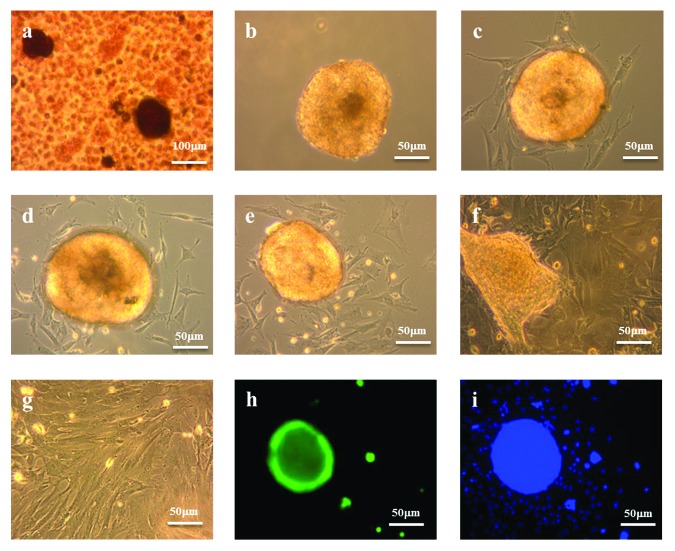
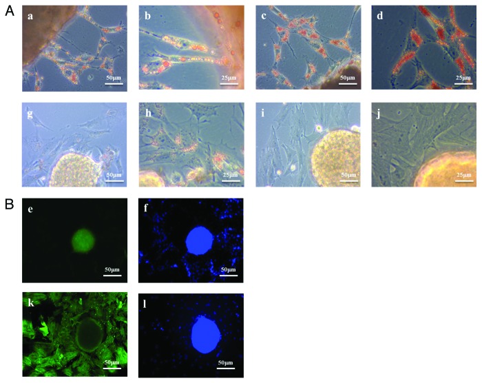
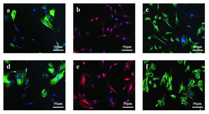
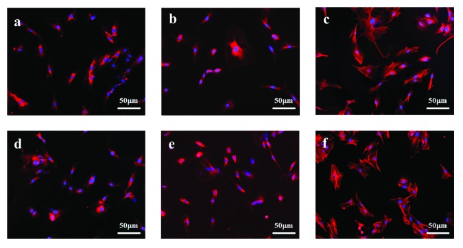
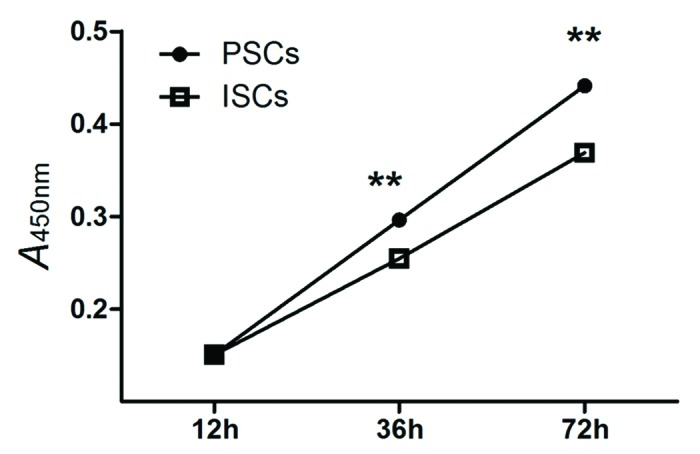
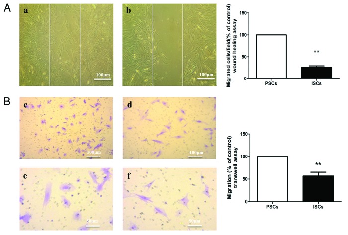
The central role of PSCs in pancreatic fibrogenesis is well established. However, the mechanism responsible for the islet fibrosis presenting in the late stage of T2DM has not been fully elucidated. This study was designed to determine whether the endocrine pancreatic islets contain cells resembling PSCs. PSCs were isolated from pancreas using standard explants techniques. A similar method was used to acquire ISCs. Adherent ISCs with a stellate, angular morphology migrated from the edge of cultured islets within 48 h of primary culture. ISCs contained fewer lipid droplets than equivalent PSCs, and their rapid disappearance accompanied by the increased expression of α-SMA suggested that ISCs were more rapidly activated than PSCs in vitro. They expressed α-SMA, vimentin, GFAP and were positive for ECM components col-I, col-III and FN, all of which are characteristics of classical PSCs. However, ISCs differed from PSCs by having reduced rates of proliferation and migration in vitro. Our in vitro study shows that isolated islets contain a population of stellate cells which are phenotypically similar but not identical to PSCs. In view of the established role of PSCs in pancreatic fibrosis, we suggest that these may contribute to islet fibrosis in T2DM.
Keywords: ISC; PSC; islet; islet stellate cell; pancreatic stellate cell.
Figures
Similar articles
-
Isolation and characterization of human islet stellate cells.Exp Cell Res. 2016 Feb 1;341(1):61-66. doi: 10.1016/j.yexcr.2015.11.002. Epub 2015 Nov 4. Exp Cell Res. 2016. PMID: 26546984
-
Oxidative stress plays a role in high glucose-induced activation of pancreatic stellate cells.Biochem Biophys Res Commun. 2013 Sep 20;439(2):258-63. doi: 10.1016/j.bbrc.2013.08.046. Epub 2013 Aug 22. Biochem Biophys Res Commun. 2013. PMID: 23973482
-
A role of pancreatic stellate cells in islet fibrosis and β-cell dysfunction in type 2 diabetes mellitus.Biochem Biophys Res Commun. 2017 Apr 1;485(2):328-334. doi: 10.1016/j.bbrc.2017.02.082. Epub 2017 Feb 21. Biochem Biophys Res Commun. 2017. PMID: 28232184
-
Pancreatic stellate cell: physiologic role, role in fibrosis and cancer.Curr Opin Gastroenterol. 2015 Sep;31(5):416-23. doi: 10.1097/MOG.0000000000000196. Curr Opin Gastroenterol. 2015. PMID: 26125317 Review.
-
Pancreatic stellate cells--multi-functional cells in the pancreas.Pancreatology. 2013 Mar-Apr;13(2):102-5. doi: 10.1016/j.pan.2012.12.058. Pancreatology. 2013. PMID: 23561965 Review.
Cited by
-
Genetic lineage tracing reveals stellate cells as contributors to myofibroblasts in pancreas and islet fibrosis.iScience. 2023 May 26;26(6):106988. doi: 10.1016/j.isci.2023.106988. eCollection 2023 Jun 16. iScience. 2023. PMID: 37378313 Free PMC article.
-
Regenerating islet-derived protein 1 inhibits the activation of islet stellate cells isolated from diabetic mice.Oncotarget. 2015 Nov 10;6(35):37054-65. doi: 10.18632/oncotarget.6163. Oncotarget. 2015. PMID: 26496027 Free PMC article.
-
Pancreatic stellate cell: Pandora's box for pancreatic disease biology.World J Gastroenterol. 2017 Jan 21;23(3):382-405. doi: 10.3748/wjg.v23.i3.382. World J Gastroenterol. 2017. PMID: 28210075 Free PMC article. Review.
-
A New Islet Transplantation Method Combining Mesenchymal Stem Cells with Recombinant Peptide Pieces, Microencapsulated Islets, and Mesh Bags.Biomedicines. 2020 Aug 21;8(9):299. doi: 10.3390/biomedicines8090299. Biomedicines. 2020. PMID: 32825661 Free PMC article.
-
Hypoxia Increases β-Cell Death by Activating Pancreatic Stellate Cells within the Islet.Diabetes Metab J. 2020 Dec;44(6):919-927. doi: 10.4093/dmj.2019.0181. Epub 2020 May 11. Diabetes Metab J. 2020. PMID: 32431113 Free PMC article.
References
Publication types
MeSH terms
Substances
LinkOut - more resources
Full Text Sources
Other Literature Sources
Medical
Miscellaneous