. 2014;6(6):1402-14.
doi: 10.4161/mabs.36389.

A general strategy for generating intact, full-length IgG antibodies that penetrate into the cytosol of living cells

Affiliations

A general strategy for generating intact, full-length IgG antibodies that penetrate into the cytosol of living cells

Dong-Ki Choi et al. MAbs. 2014.

Abstract

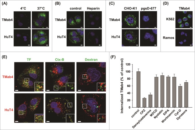
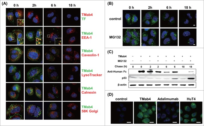
Full-length IgG antibodies cannot cross cell membranes of living cells; this limits their use for direct targeting of cytosolic proteins. Here, we describe a general strategy for the generation of intact, full-length IgG antibodies, herein called cytotransmabs, which internalize into living cells and localize in the cytosol. We first generated a humanized light chain variable domain (VL) that could penetrate into the cytosol of living cells and was engineered for association with various subtypes of human heavy chain variable domains (VHs). When light chains with humanized VL were co-expressed with 3 heavy chains (HCs), including 2 HCs of the clinically approved adalimumab (Humira®) and bevacizumab (Avastin®), all 3 purified IgG antibodies were internalized into the cytoplasm of living cells. Cytotransmabs primarily internalized into living cells by the clathrin-mediated endocytic pathway through interactions with heparin sulfate proteoglycan that was expressed on the cell surface. The cytotransmabs escaped into the cytosol from early endosomes without being further transported into other cellular compartments, like the lysosomes, endoplasmic reticulum, Golgi apparatus, and nucleus. Furthermore, we generated a cytotransmab that co-localized with the targeted cytosolic protein when it was incubated with living cells, demonstrating that the cytotransmab can directly target cytosolic proteins. Internalized cytotransmabs did not show any noticeable cytotoxicity and remained in the cytosol for more than 6 h before being degraded by proteosomes. These results suggest that cytotransmabs, which efficiently enter living cells and reach the cytosolic space, will find widespread uses as research, diagnostic, and therapeutic agents.

Keywords: CDR, complementarity-determining region; HC, heavy chain; IgG antibody; IgG, immunoglobulin G; LC, light chain; VH, heavy chain variable domain; VL, light chain variable domain; cell-penetrating antibody; cellular internalization; cytosolic protein targeting; endosomal release; intracellular trafficking; next-generation antibody.


Figures

Figure 1.
Figure 1.
Generation of humanized, cytosol-penetrating VL single domain antibodies by closely maintaining the CDR1 conformation of m3D8 VL. (A) Cellular internalization and localization of mouse m3D8 VL and humanized VLs (hT0, hT2, and hT3) in HeLa cells that were treated with VLs (10 μM) for 6 h at 37°C. The cells were then analyzed for internalized VLs by confocal fluorescence microscopy. Images show the merging of VLs (red) and Hochest33342-stained nuclei (blue) at the centered single confocal section. (B) Superposition of the homology modeled hT0, hT2, and hT3 structures with the crystal structure of m3D8 VL (PDB 3BD3) to compare their CDR conformations with those of m3D8 VL. Double-headed arrow indicates the distance between 2 Cα atoms of Arg-27f in the tip of CDR1 of m3D8 VL and hT0 VL. The image was generated using PyMol software (DeLano Scientific LLC). The primary sequences of all VLs are shown in Fig. S1A.
Figure 2.
Figure 2.
Generation of cytosol-penetrating intact IgG-format cytotransmabs, which internalize into living cells and then localize in the cytosol without cytotoxicity. (A) Schematic diagram showing the general strategy for generation of cytotransmabs by replacing endogenous VL of conventional IgG with the humanized cytosol-penetrating hT4 VL. (B and C) Biochemical characterizations of the purified parent mAbs and cytotransmabs by reducing and non-reducing SDS-PAGE (B) and SEC (C) analyses. In (B), the purified antibodies (5 μg) were separated on 12% SDS polyacrylamide gels and visualized by Coomassie brilliant blue staining (R-250). In (C), the purified antibodies were injected at 200 μg/mL in 70 μL of sample volume and monitored at 280 nm. Arrows indicate the elution positions of molecular weight standards. (D) Cellular internalization and localization of cytotransmabs (TMab4, HuT4, and AvaT4) and control mAbs (adalimumab and bevacizumab) in HeLa cells that were treated with the antibodies (1 μM) for 6 h at 37°C and then analyzed by confocal immunofluorescence microscopy. (E) Concentration-dependent internalization of TMab4 in HeLa cells that were treated with the indicated concentrations of TMab4 for 6 h at 37°C prior to confocal immunofluorescence microscopy analysis. (F) Time-dependent internalization of TMab4 in HeLa cells that were treated with TMab4 (1 μM) at 37°C for the indicated periods. In (D–F), after removing cell-surface bound antibodies with low pH glycine buffer (pH 2.5), internalized antibodies were visualized with FITC-conjugated anti-human IgG Fc antibody (green). Images show the merging of antibodies (green) and Hochest33342-stained nuclei (blue) at the centered single confocal section. Image magnification, 630×; scale bar, 10 μm. (G and H) Cytotoxicity of internalized cytotransmabs in HeLa cells that were treated with the antibodies (1 μM) at 37°C for 24 h or 48 h. In (G), the morphological features of cells were observed by phase-contrast microscopy after 48 h. Image magnification, 200×; scale bar, 10 μm. In (H), the cell viability was determined by MTT assay after 24 h or 48 h. The data represent mean ± SD compared with the PBS-treated controls.
Figure 3.
Figure 3.
Cytotransmabs internalize into living cells primarily by energy-dependent, clathrin-mediated endocytosis through interactions with cell-surface HSPG. (A) Effect of temperature on the internalization of cytotransmabs into HeLa cells that were incubated with TMab4 or HuT4 (1 μM) for 6 h at 4°C or 37°C. (B) Effect of an external heparin competitor on cytotransmab internalization into HeLa cells that were treated with heparin (300 IU/mL) for 30 min at 37°C before incubating TMab4 or HuT4 (1 μM) for an additional 6 h. (C) Internalization of cytotransmabs into CHO-K1 wild type and pgsD-677 mutant (HSPG-deficient) cells that were incubated with TMab4 or HuT4 (1 μM) for 6 h at 37°C. (D) Internalization of cytotransmabs into non-adherent K562 (HSPG-expressed) and Ramos (HSPG-deficient) cells that were treated with TMab4 (1 μM) for 6 h at 37°C. In (A–D), internalized cytotransmabs were observed by confocal microscopy after staining with FITC-conjugated anti-human IgG Fc antibody (green) and nuclei with Hoechst33342 (blue). (E) Co-localization of internalized cytotransmabs with endocytosis markers. HeLa cells were co-incubated with cytotransmabs (1 μM) and endocytosis markers including 10 μg/mL of Alexa Fluor 488-transferrin (TF, green), FITC-cholera toxin B (Ctx-B, green), and Oregon green-dextran (Dextran, green) for 30 min at 37°C. Subsequently, internalized cytotransmabs were stained with TRITC-conjugated anti-Human IgG Fc antibody (red). The regions in the white box were magnified for better imaging of co-localization. In (A–E), centered single confocal sections are shown. Image magnification, 630×; scale bar, 5 μm. (F) Effect of endocytosis inhibitors on TMab4 internalization. HeLa cells were pre-treated for 30 min with the indicated endocytosis inhibitors, and then incubated with TMab4 for additional 2 h, followed by immunofluorescence staining, as shown Fig. S6. The internalization levels of TMab4 in the presence of inhibitors are represented as mean percentage (%) ± SEM by comparing the fluorescence intensity from inside of cells (n > 100 cells per group) with that of the untreated ′control′. ***P < 0.001 compared with the control.
Figure 4.
Figure 4.
Intracellular trafficking and cytosolic release of internalized TMab4. (A) Pulse-chase intracellular trafficking of internalized TMab4 monitored by co-localization with TF, early endosome marker EEA-1, caveolin-1, late endosome/lysosome marker LysoTracker, ER marker calnexin, or Golgi marker 58K Golgi protein, as visualized by confocal immunofluorescence microscopy. Insets, enlarged images of the regions in the boxes. (B and C) Intracellular stability of internalized TMab4 with or without the proteasome inhibitor MG132 (30 μM) was analyzed by confocal microscopy (B) and western blotting (C). HeLa cells were pulsed with 3 μM of TMab4 for 30 min and incubated for the indicated times in the presence or absence of MG132 (30 μM) prior to analysis. In (B), TMab4 was visualized with FITC-conjugated anti-human IgG Fc antibody (green). In (C), equal amounts of cell lysates were loaded, and retained TMab4 was detected by protein gel blotting, using p53 as a positive control (revealing proteasome-involved degradation) and β-actin as a loading control. In (A–B): magnification, 630×; scale bar, 5 μm. (D) Cytotransmab-mediated cytosolic release of calcein. HeLa cells were untreated (′control′) or treated with 5 μM of TMab4, adalimumab, or HuT4 for 4 h and then incubated with 50 μM of calcein for additional 2 h at 37°C, followed by confocal analysis. Image magnification, 630×; scale bar, 20 μm. In (A, B, and D), images show the merging of markers/antibodies (indicated colors) and Hochest33342-stained nuclei (blue) at the centered single confocal section.
Figure 5.
Figure 5.
Anti-KRS KT4 internalizes into living cells and binds to the cytosolic KRS. (A) Diagram of IgG-format KT4 carrying KRS-binding C20 VH domain and cytosol-penetrating hT4 VL domain. (B) Direct ELISA to determine the binding specificity of C20 and KT4 IgG antibodies (100 nM) to plate-coated antigens (1 μg/mL), KRS, histidyl-tRNA synthetase (HRS), tryptophanyl-tRNA synthetase (WRS), or ARS-interacting multifunctional protein 1 (AIMP1). TMab4 was employed as a negative control. (C) Co-localization of internalized KT4 with KRS in GFP-fused KRS expressing HeLa cells, treated KT4 or TMab4 (20 μM) for 2 h at 37°C, and then determined by confocal microscopy. KT4 and TMab4 were visualized with TRITC-conjugated anti-Human IgG Fc antibody. Image magnification, 630×; scale bar, 5 μm. Images show the KRS (green), antibodies (red), and Hochest33342-stained nuclei (blue) at the centered single confocal section. Middle insets, enlarged images of the boxed regions. (D) Western blot analysis of immunoprecipitation (IP) of KRS by internalized KT4. The cell lysates prepared from HeLa cells that were treated with PBS (control), KT4 (6 μM), or TMab4 (6 μM) for 12 h at 37°C were precipitated using protein A agarose and analyzed by western blotting with β-actin as a loading control.

References

    1. Marschall AL, Frenzel A, Schirrmann T, Schungel M, Dubel S. Targeting antibodies to the cytoplasm. MAbs 2011; 3:3-16; PMID:21099369; http://dx.doi.org/10.4161/mabs.3.1.14110 - DOI - PMC - PubMed
    1. Marschall A, Zhang C, Frenzel A, Schirrmann T, Hust M, Perez F, Dübel S. Delivery of antibodies to the cytosol: Debunking the myths. MAbs 2014; 6:943-56; PMID:24848507 - PMC - PubMed
    1. Ivanov AA, Khuri FR, Fu H. Targeting protein-protein interactions as an anticancer strategy. Trends Pharmacol Sci 2013; 34:393-400; PMID:23725674; http://dx.doi.org/10.1016/j.tips.2013.04.007 - DOI - PMC - PubMed
    1. Freund G, Sibler AP, Desplancq D, Oulad-Abdelghani M, Vigneron M, Gannon J, Van Regenmortel MH, Weiss E. Targeting endogenous nuclear antigens by electrotransfer of monoclonal antibodies in living cells. MAbs 2013; 5:518-22; PMID:23765067; http://dx.doi.org/10.4161/mabs.25084 - DOI - PMC - PubMed
    1. Weill CO, Biri S, Adib A, Erbacher P. A practical approach for intracellular protein delivery. Cytotechnology 2008; 56:41-8; PMID:19002840; http://dx.doi.org/10.1007/s10616-007-9102-3 - DOI - PMC - PubMed

Publication types

MeSH terms

Substances