. 2015;11(1):88-99.
doi: 10.4161/15548627.2014.984277.

V-ATPase and osmotic imbalances activate endolysosomal LC3 lipidation

Affiliations

V-ATPase and osmotic imbalances activate endolysosomal LC3 lipidation

Oliver Florey et al. Autophagy. 2015.

Abstract

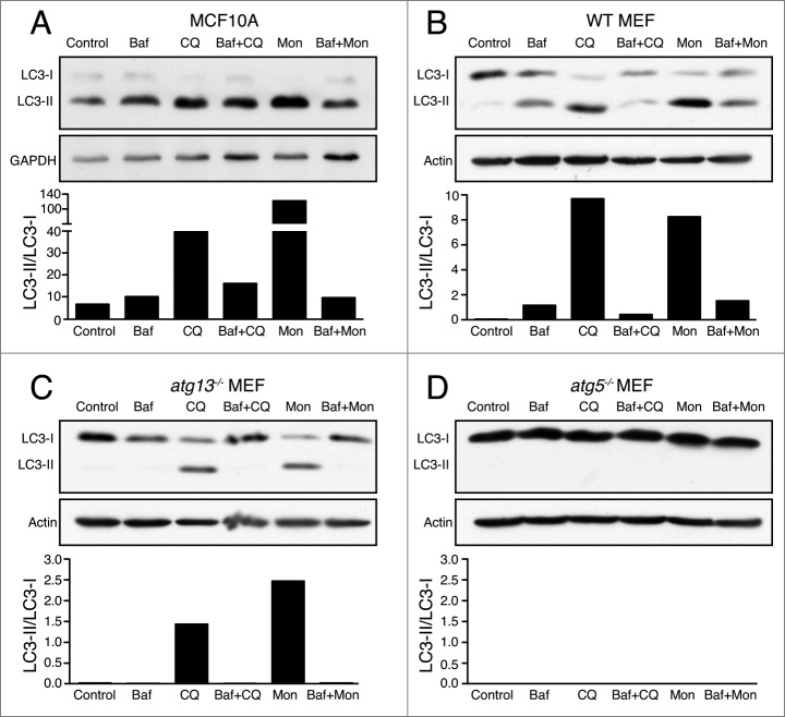
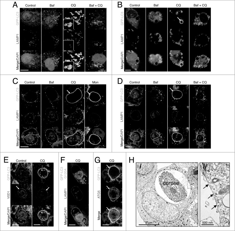
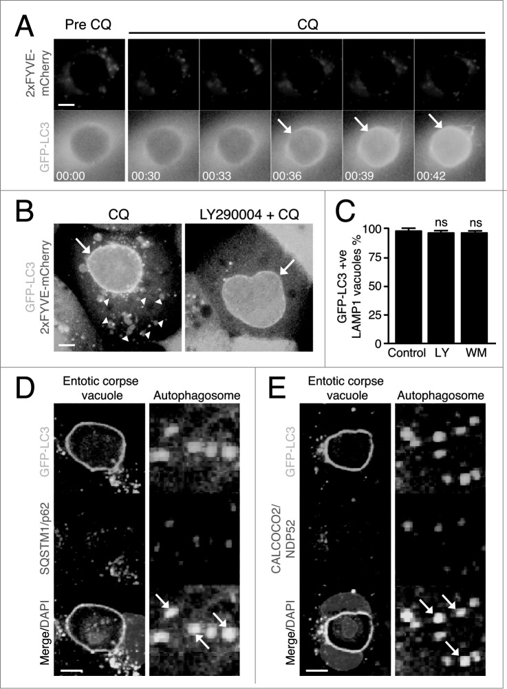
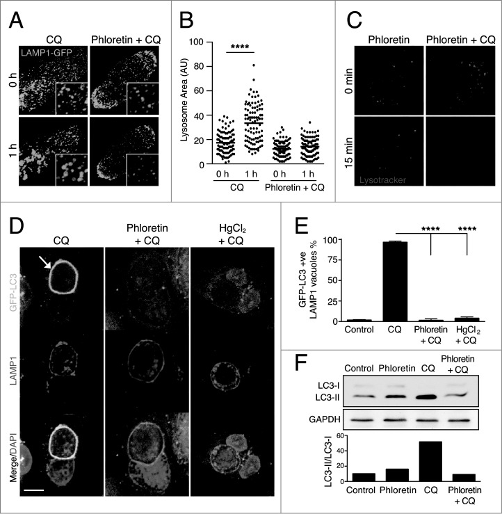
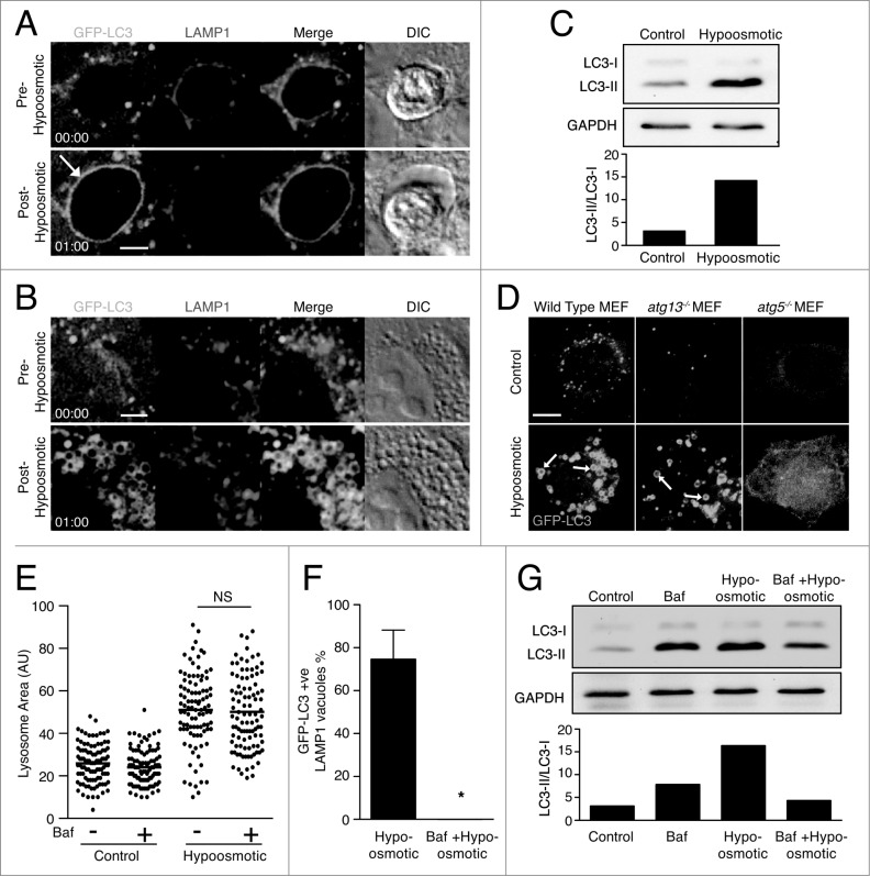
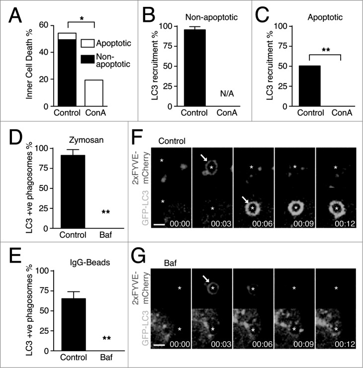
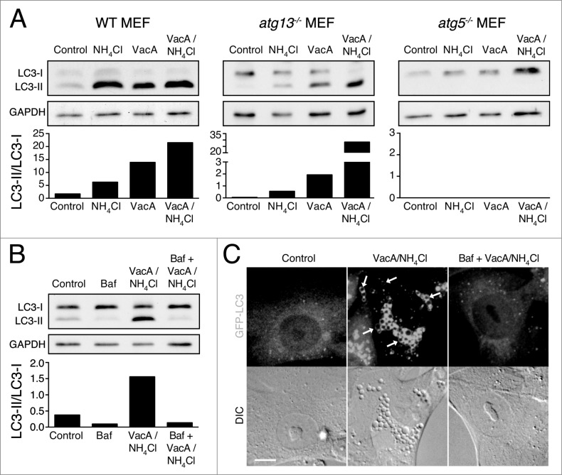
Recently a noncanonical activity of autophagy proteins has been discovered that targets lipidation of microtubule-associated protein 1 light chain 3 (LC3) onto macroendocytic vacuoles, including macropinosomes, phagosomes, and entotic vacuoles. While this pathway is distinct from canonical autophagy, the mechanism of how these nonautophagic membranes are targeted for LC3 lipidation remains unclear. Here we present evidence that this pathway requires activity of the vacuolar-type H(+)-ATPase (V-ATPase) and is induced by osmotic imbalances within endolysosomal compartments. LC3 lipidation by this mechanism is induced by treatment of cells with the lysosomotropic agent chloroquine, and through exposure to the Heliobacter pylori pore-forming toxin VacA. These data add novel mechanistic insights into the regulation of noncanonical LC3 lipidation and its associated processes, including LC3-associated phagocytosis (LAP), and demonstrate that the widely and therapeutically used drug chloroquine, which is conventionally used to inhibit autophagy flux, is an inducer of LC3 lipidation.

Keywords: ATG, autophagy-related; Baf, bafilomycin A1; CALCOCO2/NDP52, calcium binding and coiled-coil domain 2; CQ, chloroquine; ConA, concanamycin A; FYCO1, FYVE and coiled-coil domain containing 1; GFP, green fluorescent protein; Helicobacter pylori; LAMP1, lysosomal-associated membrane protein 1; LAP; LAP, LC3-associated phagocytosis; LC3; MAP1LC3/LC3, microtubule-associated protein 1 light chain 3; MTOR, mechanistic target of rapamycin; PIK3C3/VPS34, phosphatidylinositol 3-kinase; PtdIns3K, phosphatidylinositol 3-kinase; PtdIns3P, phosphatidylinositol 3-phosphate; RB1CC1/FIP200, RB1-inducible coiled-coil 1; SQSTM1/p62, sequestosome 1; TEM, transmission electron microscopy; TLR, toll-like receptor; ULK1/2, unc-51 like autophagy activating kinase 1/2; V-ATPase; V-ATPase, vacuolar-type H+-ATPase; VacA, vacuolating toxin A; autophagy; catalytic subunit type 3; chloroquine; entosis; lysosome; phagocytosis.


Figures

Figure 1.
Figure 1.
Chloroquine and monensin induce V-ATPase-dependent LC3 lipidation. (A to D) Representative western blots for LC3 and GAPDH on (A) MCF10A cells, (B) Wild-type MEFs (C), atg13−/− MEFs or (D) and atg5−/− MEFs treated with lysosome inhibitors bafilomycin A1 (Baf, 100 nM), chloroquine (CQ, 100 μM), monensin (Mon, 100 μM) for 1 h or with 15 min Baf pretreatment followed by CQ or Mon for 1 h. Ratios of lipidated LC3-II/unlipidated LC3-I were quantified and graphed. See Figure S2 for repeat protein gel blots and quantification. See also Figures S1 and S2.
Figure 2.
Figure 2.
Chloroquine and monensin induce LC3 lipidation onto multiple lysosomal compartments dependent on V-ATPase activity. (A and B) Confocal images of GFP-LC3 and LAMP1 immunostaining of (A) lysosomes in MCF10A and (B) uncoated latex bead phagosomes in J774 macrophage following treatment with Baf (100 nM), CQ (100 mM) or Baf + CQ for 1 h. Arrow indicates GFP-LC3 lipidation onto a phagosome. Bar = 6 μm. (C and D) Images of GFP-LC3 and LAMP1 on entotic corpse vacuoles in MCF10A cells treated with Baf, CQ, Mon (100 μM) or Baf + CQ for 1 h. Arrows indicate GFP-LC3 lipidation onto vacuoles. Bar = 10 mm. (E) GFP-LC3 and ATP6V0D1 (V0D1) staining on entotic corpse vacuoles with our without CQ treatment. Bar = 10 μm. (F) GFP-LC3G120A and LAMP1 staining on entotic corpse vacuoles following CQ treatment. Bar = 10 μm. (G) GFP-LC3 and ATG5 immunostaining on entotic corpse vacuole (arrows) following CQ treatment. Bar = 10 μM. (H) (i) Electron microscopy of corpse containing cell-in-cell structure treated with CQ (100 μM), (ii) entotic corpse vacuole has a single membrane (arrows). See also Figure S3; Movie S1.
Figure 3.
Figure 3.
Chloroquine-mediated LC3 recruitment is independent of PtdIns3P and autophagy receptor proteins. (A) Time-lapse microscopy of GFP-LC3 and 2xFYVE-mCherry on entotic corpse vacuoles following treatment with CQ (100 μM). Arrows indicate GFP-LC3 lipidation onto vacuole. Bar = 2 μm. (B) Confocal images of entotic corpse vacuoles treated with CQ (100 μM) with or without LY290004 (25 μM). Arrows indicate GFP-LC3 lipidation onto entotic vacuole, arrowheads indicate 2xFYVE-mCherry-positive vesicles. Bar = 2 μm. (C) Quantification of GFP-LC3 recruitment to LAMP1-positive entotic vacuoles with or without LY290004 (LY 25 μM) or wortmannin (WM, 200 nM); data are mean ± SEM from 3 independent experiments; NS, not significant. (D and E) Entotic corpse vacuoles treated with CQ (100 μM) for 1 h and immunostained for (D) SQSTM1 or (E) CALCOCO2. Arrows indicate autophagosomes with colocalized LC3 and SQSTM1 or CALCOCO2. Bar = 4 μm. See also Figure S4.
Figure 4.
Figure 4.
Chloroquine-mediated noncanonical LC3 lipidation is dependent on water flux. (A) Confocal images of LAMP1-GFP fluorescence in MCF10A cells before and after treatment with CQ (100 μM) with or without phloretin (180 μM). Insets show vesicles with LAMP1-GFP. (B) Quantification of LAMP1-GFP-labeled vesicle size; P < 0.01 **** by one-way ANOVA. (C) Confocal images of LysoTracker Red in MCF10A cells before and after treatment with phloretin with or without CQ for 15 min. (D) Confocal images of GFP-LC3 and LAMP1 immunostaining on entotic corpse vacuoles in MCF10A cells following treatment with CQ with or without phloretin (180 mM) or HgCl2 (15 mM). Arrow indicates GFP-LC3 lipidation onto a vacuole. Bar = 5 μm. (E) Quantification of GFP-LC3 lipidation onto LAMP1-positive entotic corpse vacuoles as in (D), data mean ± SEM from 3 independent experiments; P < 0.001 ****. (F) Western blot analysis of LC3 in MCF10A cells treated with phloretin, CQ or both for 1 h. Quantification of LC3-II/LC3-I graphed below. See also Movie S2.
Figure 5.
Figure 5.
Osmotic imbalances are sufficient to induce LC3 lipidation onto lysosomal compartments in a V-ATPase-dependent manner. (A and B) Confocal images of GFP-LC3 and LAMP1-RFP from time-lapse microscopy of (A) entotic corpse vacuoles or (B) lysosomes in MCF10A cells treated with hypotonic media. Arrow indicates GFP-LC3 lipidation onto entotic corpse vacuole. Bar = 2 μm. (C) Western blot analysis of LC3 in MCF10A cells cultured in control and hypotonic media for 1 h. Quantification of LC3-II/LC3-I graphed below. (D) Confocal images GFP-LC3 in wild-type, atg13−/− and atg5−/− MEFs cultured in control or hypotonic media for 30 min. Arrows indicate GFP-LC3 on vacuoles. Bar = 4 μm. (E) Quantification of LAMP1-GFP vesicle size in MCF10A cells under control or hypotonic conditions with or without Baf (100 nM); NS, not significant. (F) Quantification of hypo-osmotic induced LC3 lipidation onto LAMP1-positive entotic corpse vacuoles with or without Baf (100 nM). Data are mean ± SEM from 3 independent experiments; P < 0.01 *. (G) Western blot of LC3 in MCF10A cells in control or hypotonic media with or without Baf (100 nM). Quantification of LC3-II/LC3-I graphed below. See also Figure S5; Movie S3 and Movie S4.
Figure 6.
Figure 6.
LC3 recruitment during LAP and entosis is dependent on V-ATPase activity. (A) Quantification of entotic inner cell death over 20 h with or without Concanamycin A (ConA, 100 nM). Data are mean ± SEM of 3 separate experiments; P < 0.05 *. (B and C) Quantification of LC3 recruitment to entotic vacuole with or without ConA (100 μM) during (B) nonapoptotic or (C) apoptotic death of inner cells. Data are mean ± SEM of 3 separate experiments; P < 0.002 **; N/A, not applicable. (D and E) Quantification of GFP-LC3 recruitment to (D) zymosan phagosomes in RAW264.7 cells and (E) IgG-coated bead phagosomes in IFNG-treated RAW264.7 cells. Data are mean ± SEM of 3 separate experiments; P < 0.002 **. (F and G) Confocal time-lapse images of GFP-LC3 and 2xFYVE-mCherry in RAW264.7 cells during zymosan phagocytosis with or without Baf (100 μM). Arrows point to 2xFYVE recruitment and GFP-LC3 lipidation as indicated. Bar = 3 μm.
Figure 7.
Figure 7.
VacA activates noncanonical LC3 lipidation. (A) Western blot analysis of LC3 in wild-type, atg13−/− and atg5−/− MEFs treated with NH4Cl (5 mM), VacA (10 μM) or both for 2 h. Quantification of LC3-II/LC3-I graphed below. (B) Western blot analysis of LC3 in atg13−/− MEFs treated with Baf (100 nM), NH4Cl (5 mM) + VacA (10 μM) or Baf + NH4Cl + VacA for 2 h. Quantification of LC3-II/LC3-I graphed below. (C) Confocal images of differential interference contrast and GFP-LC3 in atg13−/− MEFs treated with NH4Cl (5 mM) + VacA (10 μM) or Baf + NH4Cl + VacA for 2 h. Arrows indicate GFP-LC3 on vacuoles. Bar = 5 μm.

References

    1. Choi AM, Ryter SW, Levine B. Autophagy in human health and disease. New Engl J Med 2013; 368:651-62; PMID:23406030; http://dx.doi.org/10.1056/NEJMra1205406 - DOI - PubMed
    1. Feng Y, He D, Yao Z, Klionsky DJ. The machinery of macroautophagy. Cell Res 2014; 24:24-41; PMID:24366339; http://dx.doi.org/ 10.1038/cr.2013.16 - DOI - PMC - PubMed
    1. Florey O, Overholtzer M. Autophagy proteins in macroendocytic engulfment. Trends Cell Biol 2012; 22:374-80; PMID:22608991; http://dx.doi.org/10.1016/j.tcb.2012.04.005 - DOI - PMC - PubMed
    1. Sanjuan MA, Dillon CP, Tait SW, Moshiach S, Dorsey F, Connell S, Komatsu M, Tanaka K, Cleveland JL, Withoff S, et al. . Toll-like receptor signalling in macrophages links the autophagy pathway to phagocytosis. Nature 2007; 450:1253-7; PMID:18097414; http://dx.doi.org/10.1038/nature06421 - DOI - PubMed
    1. Kim JY, Zhao H, Martinez J, Doggett TA, Kolesnikov AV, Tang PH, Ablonczy Z, Chan CC, Zhou Z, Green DR, et al. . Noncanonical autophagy promotes the visual cycle. Cell 2013; 154:365-76; PMID:23870125; http://dx.doi.org/10.1016/j.cell.2013.06.012 - DOI - PMC - PubMed

Publication types

MeSH terms