. 2015 Jan 5;25(1):61-8.
doi: 10.1016/j.cub.2014.10.052. Epub 2014 Dec 4.

Aurora kinases phosphorylate Lgl to induce mitotic spindle orientation in Drosophila epithelia

Affiliations

Aurora kinases phosphorylate Lgl to induce mitotic spindle orientation in Drosophila epithelia

Graham P Bell et al. Curr Biol. .

Abstract

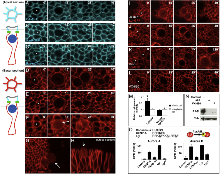
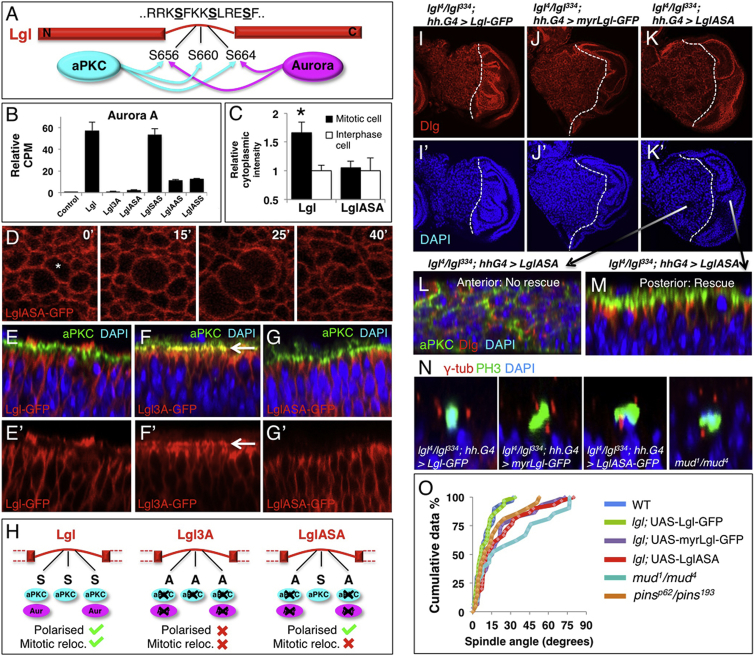
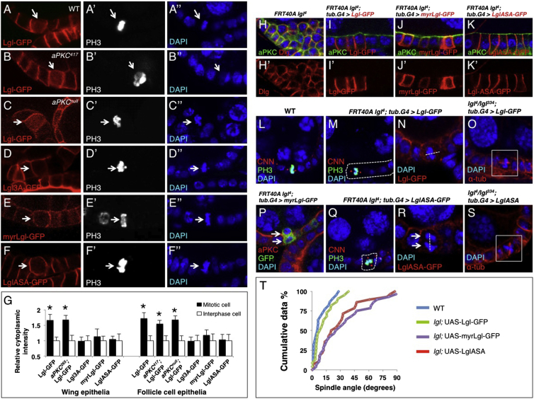
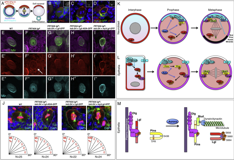
The Lethal giant larvae (Lgl) protein was discovered in Drosophila as a tumor suppressor in both neural stem cells (neuroblasts) and epithelia. In neuroblasts, Lgl relocalizes to the cytoplasm at mitosis, an event attributed to phosphorylation by mitotically activated aPKC kinase and thought to promote asymmetric cell division. Here we show that Lgl also relocalizes to the cytoplasm at mitosis in epithelial cells, which divide symmetrically. The Aurora A and B kinases directly phosphorylate Lgl to promote its mitotic relocalization, whereas aPKC kinase activity is required only for polarization of Lgl. A form of Lgl that is a substrate for aPKC, but not Aurora kinases, can restore cell polarity in lgl mutants but reveals defects in mitotic spindle orientation in epithelia. We propose that removal of Lgl from the plasma membrane at mitosis allows Pins/LGN to bind Dlg and thus orient the spindle in the plane of the epithelium. Our findings suggest a revised model for Lgl regulation and function in both symmetric and asymmetric cell divisions.


Figures

Figure 1
Figure 1
Aurora Kinases Phosphorylate Lgl to Relocalize It to the Cytoplasm during Mitosis in the Wing Epithelium (A–C) Live imaging of third instar wing imaginal discs. Fluorescently tagged Crumbs (Crb-GFP; A) and Par-6 (Par-6-GFP; B) remain apically localized during mitosis (apical section; diagrammed on the left). Baz-mCherry is partially downregulated but remains apical (C). (D and E) Fluorescently tagged Discs-large (Dlg-GFP; D) and Scribble (Scrib-GFP; E) remain basolaterally localized during mitosis (basal section; diagrammed on the left). (F–H) Fluorescently tagged Lethal giant larvae (Lgl-GFP) becomes cytoplasmic at mitosis (basal section; diagrammed on the left): note the complete relocalization at mitosis with no membrane staining (G). (I) Lgl-GFP relocalizes to the cytoplasm at mitosis normally in an aPKC417 kinase-dead MARCM clone in wing disc epithelia, indicating that aPKC phosphorylation is not responsible for regulating Lgl at mitosis. Other kinase-dead alleles of aPKC gave similar results (not shown). (J) Phosphomutant Lgl3A-GFP fails to relocalize to the cytoplasm during mitosis in wing disc epithelia. (K) Relocalization of Lgl-GFP to the cytoplasm in mitosis is strongly delayed in aurA87Ac-3/Df mutants. Similar results were obtained with other aurora A mutant alleles or with AurA RNAi (not shown). (L) Relocalization of Lgl-GFP to the cytoplasm in mitosis is blocked upon treatment of the epithelium with the Aurora A/B inhibitor VX-680. (M) Quantification of control and VX-680 treated wing discs expressing Lgl-GFP. (N) Western blotting analysis of actin5c.Gal4 UAS-Lgl-GFP wing discs showing absence of a strong phosphorylated Lgl band upon treatment of samples with λ-phosphatase or treatment of the epithelium with VX-680 prior to sample preparation. (O) Aurora A and B directly phosphorylate the Lgl phosphosite motif in an in vitro kinase assay. CENP-A is a positive control substrate that is known to be phosphorylated by both Aurora A and B. Error bars indicate 1 SD from the mean. See also Figures S1 and S3 and Movies S1, S2, and S3.
Figure 2
Figure 2
Aurora-Insensitive Lgl Rescues Cell Polarity in the Wing Epithelium but Fails to Relocalize during Mitosis and Disrupts Mitotic Spindle Orientation (A) Schematic of aPKC and Aurora A/B kinase phosphorylation of Lgl. (B) Aurora A directly phosphorylates Lgl on S656 and S664 of the tripartite motif, but not S660. (C) Quantification of cytoplasmic intensity of LglASA-GFP construct compared to Lgl-GFP. (D) LglASA-GFP does not relocalize to the cytoplasm in mitosis. (E–G) Lgl-GFP is localized basolaterally and does not overlap with aPKC in wing disc (E). Nonphosphorylatable Lgl3A-GFP spreads apically and colocalizes with aPKC (arrow; F). LglASA-GFP is localized basolaterally and does not overlap with aPKC (G). (H) Schematic of Lgl, Lgl3A, and LglASA constructs and their respective phosphorylation potential by aPKC or Aurora kinases. (I) lgl4/lgl334 mutant discs expressing Lgl-GFP in the posterior compartment show a rescue of polarity this compartment. (J and K) lgl4/lgl334 mutant discs expressing myrLgl-GFP (J) or LglASA-GFP (K) in the posterior compartment show a rescue of cell polarity in this compartment. (L) Cross-section of the anterior portion of the disc in (K) showing tissue disorganization and lack of polarity. (M) Cross-section of the posterior portion of the disc in (K) showing normal tissue organization and cell polarization. (N) Mitotic spindles in wild-type discs are oriented in the plane of the epithelium, whereas clones of lgl4 mutant cells expressing myrLgl-GFP or LglASA-GFP show misoriented spindles, similar to pinsp62/193 mutants (not shown) or mud1/4 mutants. (O) Quantification of mitotic spindle orientation relative to the plane of the epithelium in (N). Low angles reflect planar spindle orientation, whereas high angles reflect more apical-basal spindle orientation. n > 30 for each experiment. Error bars indicate 1 SD from the mean. See also Figure S2.
Figure 3
Figure 3
Aurora-Mediated Phosphorylation of Lgl Is Required for Mitotic Spindle Orientation in the Follicle Cell Epithelium (A) Lgl-GFP relocalizes to the cytoplasm at mitosis in the ovarian follicle cell epithelium. (B) Relocalization of Lgl to the cytoplasm during mitosis still occurs in aPKC417 kinase-dead MARCM clones. (C) Relocalization of Lgl to the cytoplasm during mitosis still occurs in aPKCK06403-null mutant MARCM clones. (D) Phosphomutant Lgl3A-GFP fails to relocalize to the cytoplasm during mitosis. (E) Membrane-tethered myrLgl-GFP fails to relocalize to the cytoplasm during mitosis. (F) Aurora-insensitive LglASA-GFP fails to relocalize to the cytoplasm during mitosis. (G) Quantification of cytoplasmic intensity for (A)–(F). Error bars indicate 1 SD from the mean. (H–K) Clones of lgl4 mutant follicle cells show loss of polarity and multilayering (H); this can be rescued by the expression of Lgl-GFP (I), myrLgl-GFP (J), or LglASA-GFP (K). (L–O) Mitotic spindles in wild-type follicle cell epithelia are oriented in the plane of the epithelium (L). Rescue of spindle orientation is achieved by expression of Lgl-GFP in clones of lgl4 mutant cells or lgl4/334 mutant animals (M–O). (P–S) Mitotic spindles fail to orient in the plane of the epithelium in clones of lgl4 mutant cells, or lgl4/334 mutant animals, expressing myrLgl-GFP (P), LglASA-GFP (Q and R), or untagged Lgl-ASA (S). (T) Quantification of spindle orientation in (L)–(S). n > 20 spindles for each experiment.
Figure 4
Figure 4
Aurora-Mediated Phosphorylation of Lgl Is Dispensable for Cell Polarity and Mitotic Spindle Orientation in Larval Brain Neuroblasts (A) Schematic of asymmetric cell division in larval brain neuroblasts. (B–D) Lgl-GFP is cytoplasmic in metaphase neuroblasts, whereas aPKC is found in an apical crescent (B). LglASA-GFP is polarized to the basal side of the neuroblast and does not affect aPKC localization (C). myrLgl-GFP is localized around the cell cortex and its expression drives aPKC around the cortex (D). (E–I) aPKC and Miranda form apical and basal crescents, respectively, in metaphase neuroblasts (E). lgl4 mutant neuroblasts show spreading of aPKC and cytoplasmic Miranda (F). lgl4 neuroblasts expressing Lgl-GFP (G) or LglASA-GFP (H) show normal aPKC and Miranda localization. lgl4 neuroblasts expressing myrLgl-GFP show aPKC spreading around the cell cortex and colocalizing with Miranda, indicating that aPKC kinase activity is inhibited by myrLgl-GFP (I). (J) Quantification of mitotic spindle orientation relative to the apical-basal axis of third-instar larval brain neuroblasts, as marked by either aPKC or Baz localization. Wild-type and lgl4 neuroblasts expressing Lgl-GFP, LglASA-GFP, or myrLgl-GFP all showed show normal spindle orientation. Baz was used to mark the apical-basal axis in the case of myrLgl-GFP because aPKC is no longer restricted apically when this construct is expressed. (K) Model of asymmetric cell division in neuroblasts. Neuroblasts are not obviously polarized during interphase. Aurora phosphorylates Par-6, Lgl, and Pins at mitosis, and the Baz-aPKC-Par-6 complex becomes apically localized with Insc, which binds to Baz. Phosphorylation of Par-6 promotes aPKC activity and correct segregation of cell fate determinants. Phosphorylation of Lgl on S656 and S664 relocalizes it to the cytoplasm. Pins phosphorylation on S436 and relocalization of Lgl to the cytoplasm promotes Dlg:Pins complex formation, but this complex is not strictly essential for spindle orientation due to Baz-Insc recruitment of Pins and subsequent formation of an apical Gαi:Pins:Mud complex to orient the spindle in the apical-basal axis. (L and M) Model of symmetric cell division in epithelial cells. Epithelial cells are polarized in interphase, with apical Crb and Baz complexes and basolateral Dlg and Lgl. aPKC-phosphorylated Lgl is removed from the apical membrane and can bind to Dlg at the basolateral membrane in interphase. At mitosis, Aurora phosphorylates Lgl on S656 and S664 to relocalize Lgl to the cytoplasm. Aurora also phosphorylates Pins on S436. These events promote formation of a Dlg:Pins complex, which is essential for spindle orientation within the plane of the epithelium in follicle cells.

References

    1. Bryant P.J., Schubiger G. Giant and duplicated imaginal discs in a new lethal mutant of Drosophila melanogaster. Dev. Biol. 1971;24:233–263. - PubMed
    1. Mechler B.M., McGinnis W., Gehring W.J. Molecular cloning of lethal(2)giant larvae, a recessive oncogene of Drosophila melanogaster. EMBO J. 1985;4:1551–1557. - PMC - PubMed
    1. Jacob L., Opper M., Metzroth B., Phannavong B., Mechler B.M. Structure of the l(2)gl gene of Drosophila and delimitation of its tumor suppressor domain. Cell. 1987;50:215–225. - PubMed
    1. Lützelschwab R., Klämbt C., Rossa R., Schmidt O. A protein product of the Drosophila recessive tumor gene, l (2) giant gl, potentially has cell adhesion properties. EMBO J. 1987;6:1791–1797. - PMC - PubMed
    1. Bilder D., Li M., Perrimon N. Cooperative regulation of cell polarity and growth by Drosophila tumor suppressors. Science. 2000;289:113–116. - PubMed

Publication types