

Mesenchymal stromal cell labeling by new uncoated superparamagnetic maghemite nanoparticles in comparison with commercial Resovist--an initial in vitro study
- PMID: 25484583
- PMCID: PMC4245086
- DOI: 10.2147/IJN.S66986
Mesenchymal stromal cell labeling by new uncoated superparamagnetic maghemite nanoparticles in comparison with commercial Resovist--an initial in vitro study
Abstract
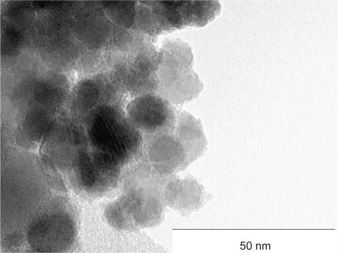
Objective: Cell therapies have emerged as a promising approach in medicine. The basis of each therapy is the injection of 1-100×10(6) cells with regenerative potential into some part of the body. Mesenchymal stromal cells (MSCs) are the most used cell type in the cell therapy nowadays, but no gold standard for the labeling of the MSCs for magnetic resonance imaging (MRI) is available yet. This work evaluates our newly synthesized uncoated superparamagnetic maghemite nanoparticles (surface-active maghemite nanoparticles - SAMNs) as an MRI contrast intracellular probe usable in a clinical 1.5 T MRI system.
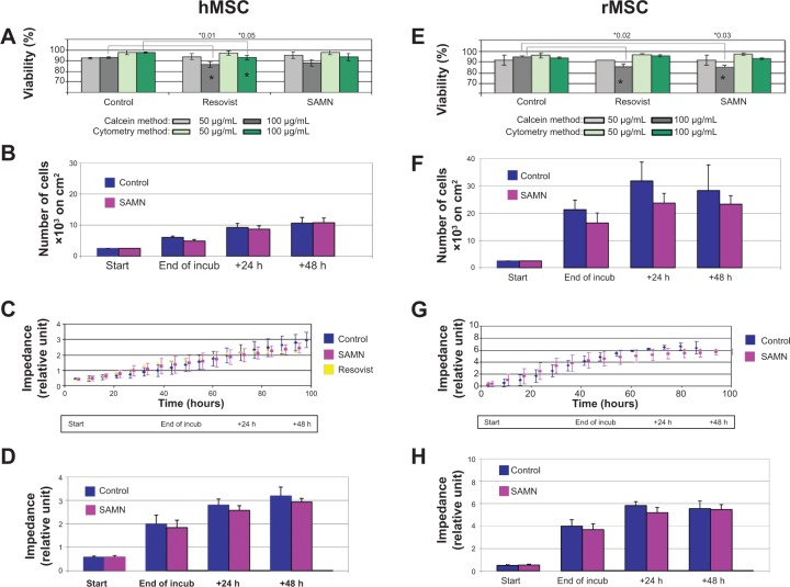
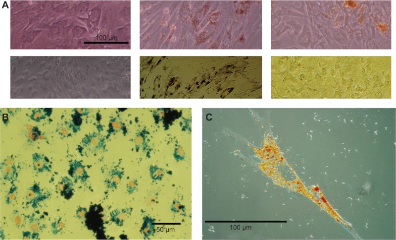
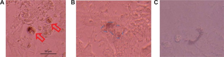
Methods: MSCs from rat and human donors were isolated, and then incubated at different concentrations (10-200 μg/mL) of SAMN maghemite nanoparticles for 48 hours. Viability, proliferation, and nanoparticle uptake efficiency were tested (using fluorescence microscopy, xCELLigence analysis, atomic absorption spectroscopy, and advanced microscopy techniques). Migration capacity, cluster of differentiation markers, effect of nanoparticles on long-term viability, contrast properties in MRI, and cocultivation of labeled cells with myocytes were also studied.
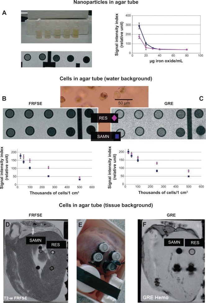
Results: SAMNs do not affect MSC viability if the concentration does not exceed 100 μg ferumoxide/mL, and this concentration does not alter their cell phenotype and long-term proliferation profile. After 48 hours of incubation, MSCs labeled with SAMNs show more than double the amount of iron per cell compared to Resovist-labeled cells, which correlates well with the better contrast properties of the SAMN cell sample in T2-weighted MRI. SAMN-labeled MSCs display strong adherence and excellent elasticity in a beating myocyte culture for a minimum of 7 days.
Conclusion: Detailed in vitro tests and phantom tests on ex vivo tissue show that the new SAMNs are efficient MRI contrast agent probes with exclusive intracellular uptake and high biological safety.
Keywords: magnetic resonance imaging; mesenchymal stromal cells; stem cell labeling; stem cell tracking; superparamagnetic iron oxide nanoparticles.
Figures
Similar articles
-
Rhodamine bound maghemite as a long-term dual imaging nanoprobe of adipose tissue-derived mesenchymal stromal cells.Eur Biophys J. 2017 Jul;46(5):433-444. doi: 10.1007/s00249-016-1187-1. Epub 2016 Nov 26. Eur Biophys J. 2017. PMID: 27889810
-
Amino-polyvinyl alcohol coated superparamagnetic iron oxide nanoparticles are suitable for monitoring of human mesenchymal stromal cells in vivo.Small. 2014 Nov 12;10(21):4340-51. doi: 10.1002/smll.201400707. Epub 2014 Jul 2. Small. 2014. PMID: 24990430
-
Optimized labeling of bone marrow mesenchymal cells with superparamagnetic iron oxide nanoparticles and in vivo visualization by magnetic resonance imaging.J Nanobiotechnology. 2011 Feb 9;9:4. doi: 10.1186/1477-3155-9-4. J Nanobiotechnology. 2011. PMID: 21542946 Free PMC article.
-
Poly(N,N-dimethylacrylamide)-coated maghemite nanoparticles for labeling and tracking mesenchymal stem cells.2009 Dec 23 [updated 2010 Feb 16]. In: Molecular Imaging and Contrast Agent Database (MICAD) [Internet]. Bethesda (MD): National Center for Biotechnology Information (US); 2004–2013. 2009 Dec 23 [updated 2010 Feb 16]. In: Molecular Imaging and Contrast Agent Database (MICAD) [Internet]. Bethesda (MD): National Center for Biotechnology Information (US); 2004–2013. PMID: 20641410 Free Books & Documents. Review.
-
Amine-modified silica-coated polyhedral superparamagnetic iron oxide nanoparticle–labeled rabbit bone marrow–derived mesenchymal stem cells.2009 Dec 11 [updated 2010 Jan 28]. In: Molecular Imaging and Contrast Agent Database (MICAD) [Internet]. Bethesda (MD): National Center for Biotechnology Information (US); 2004–2013. 2009 Dec 11 [updated 2010 Jan 28]. In: Molecular Imaging and Contrast Agent Database (MICAD) [Internet]. Bethesda (MD): National Center for Biotechnology Information (US); 2004–2013. PMID: 20641854 Free Books & Documents. Review.
Cited by
-
Real-time cell analysis system in cytotoxicity applications: Usefulness and comparison with tetrazolium salt assays.Toxicol Rep. 2020 Feb 7;7:335-344. doi: 10.1016/j.toxrep.2020.02.002. eCollection 2020. Toxicol Rep. 2020. PMID: 32090021 Free PMC article. Review.
-
The surface reactivity of iron oxide nanoparticles as a potential hazard for aquatic environments: A study on Daphnia magna adults and embryos.Sci Rep. 2018 Aug 29;8(1):13017. doi: 10.1038/s41598-018-31483-6. Sci Rep. 2018. PMID: 30158568 Free PMC article.
-
The Effect of Rhodamine-Derived Superparamagnetic Maghemite Nanoparticles on the Motility of Human Mesenchymal Stem Cells and Mouse Embryonic Fibroblast Cells.Molecules. 2019 Mar 27;24(7):1192. doi: 10.3390/molecules24071192. Molecules. 2019. PMID: 30934664 Free PMC article.
-
Quantitative Magnetic Particle Imaging Monitors the Transplantation, Biodistribution, and Clearance of Stem Cells In Vivo.Theranostics. 2016 Jan 1;6(3):291-301. doi: 10.7150/thno.13728. eCollection 2016. Theranostics. 2016. PMID: 26909106 Free PMC article.
-
Rhodamine bound maghemite as a long-term dual imaging nanoprobe of adipose tissue-derived mesenchymal stromal cells.Eur Biophys J. 2017 Jul;46(5):433-444. doi: 10.1007/s00249-016-1187-1. Epub 2016 Nov 26. Eur Biophys J. 2017. PMID: 27889810
References
-
- Martin-Rendon E, Brunskill SJ, Hyde CJ, Stanworth SJ, Mathur A, Watt SM. Autologous bone marrow stem cells to treat acute myocardial infarction: a systematic review. Eur Heart J. 2008;29:1807–1818. - PubMed
-
- Wakitani S, Mitsuoka T, Nakamura N, Toritsuka Y, Nakamura Y, Horibe S. Autologous bone marrow stromal cell transplantation for repair of full-thickness articular cartilage defects in human patellae: two case reports. Cell Transplant. 2004;13:595–600. - PubMed
-
- Kebriaei P, Robinson S. Treatment of graft-versus-host-disease with mesenchymal stromal cells. Cytotherapy. 2011;13:262–268. - PubMed
-
- Corot C, Robert P, Idée JM, Port M. Recent advances in iron oxide nanocrystal technology for medical imaging. Adv Drug Deliv Rev. 2006;58:1471–1504. - PubMed
Publication types
MeSH terms
Substances
LinkOut - more resources
Full Text Sources
Other Literature Sources
Molecular Biology Databases
