Metastasis to the proximal ureter from prostatic adenocarcinoma: A rare metastatic pattern
- PMID: 25485016
- PMCID: PMC4250253
- DOI: 10.5489/cuaj.2169
Metastasis to the proximal ureter from prostatic adenocarcinoma: A rare metastatic pattern
Abstract
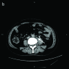
Prostate cancer is one of the most common male malignancies, but it rarely metastasizes to the proximal ureter. We report a case of a 76-year-old man who presented with flank pain and lower urinary tract symptoms. Abdominal computed tomography scan revealed multiple filling defects at the middle of the left ureter, enlarged retroperitoneal lymph nodes, and probable psoas invasion. The patient underwent nephroureterectomy with excision of a cuff of bladder, and was found to have an adhesion between the middle part of left ureter and psoas intraoperatively. The pathological examination displayed positive immunohistochemical staining with prostate-specific antigen and prostate acid phostate, supporting the diagnosis of metastatic ureteral tumour from prostate cancer. In this case, periureteral soft tissue and ureteral muscular layer were infiltrated by metastatic tumour, whereas the mucosa was spared. The periureteral lymphatic pathway played an important role in the metastatic procedure of prostate cancer to the proximal ureter.
Figures


References
-
- Epstein JI. Pathology of prostatic neoplasia. In: Wein AJ, Kavoussi LR, Novick AC, Partin AW, Peters CA, editors. Campbell-Walsh Urology. 9th ed. Philadelphia, PA: B Saunders; 2007. pp. 2874–5.
-
- DiSibio G, French SW. Metastatic patterns of cancers: Results from a large autopsy study. Arch Pathol Lab Med. 2008;132:931–9. - PubMed
LinkOut - more resources
Full Text Sources
Other Literature Sources
