. 2014;2(4):14.
doi: 10.1128/microbiolspec.MGM2-0021-2013.

Genetics of Capsular Polysaccharides and Cell Envelope (Glyco)lipids

Affiliations

Genetics of Capsular Polysaccharides and Cell Envelope (Glyco)lipids

Mamadou Daffé et al. Microbiol Spectr. 2014.

Abstract

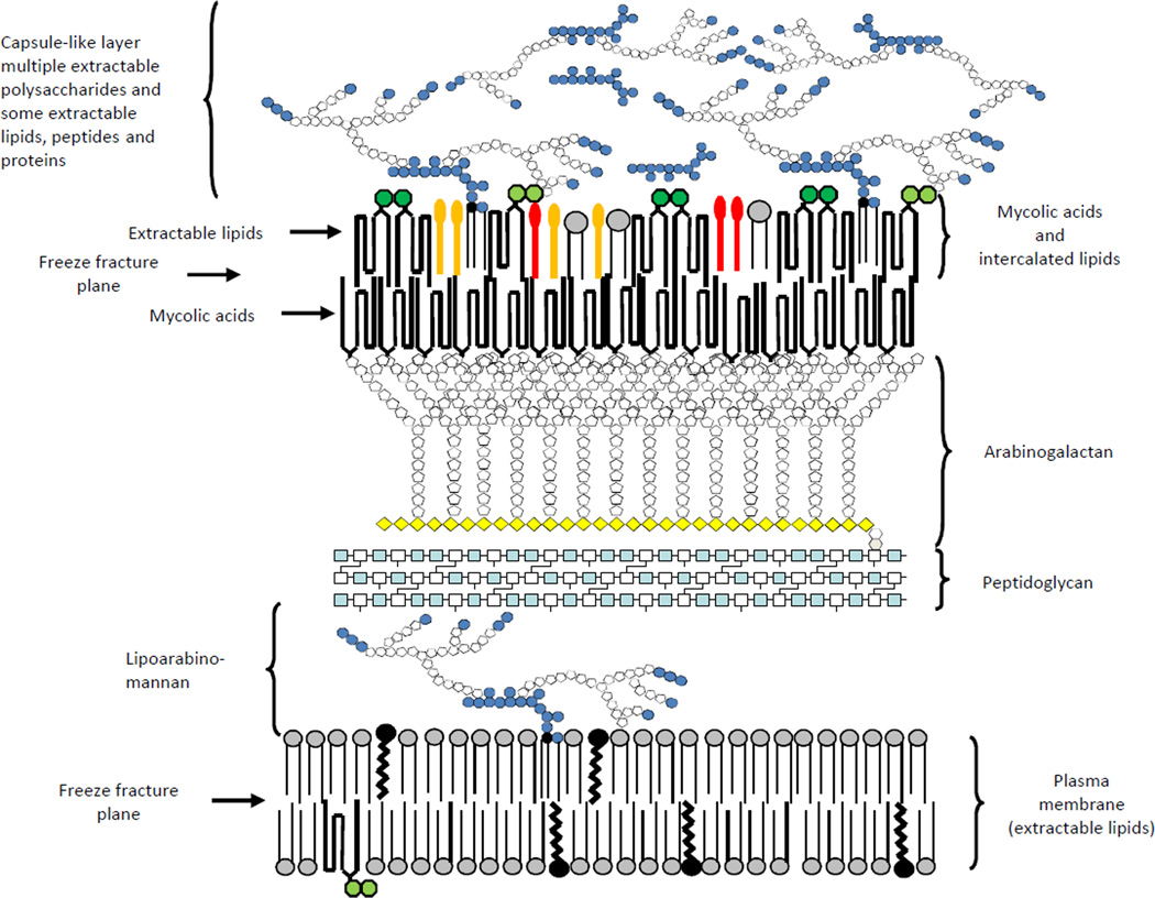
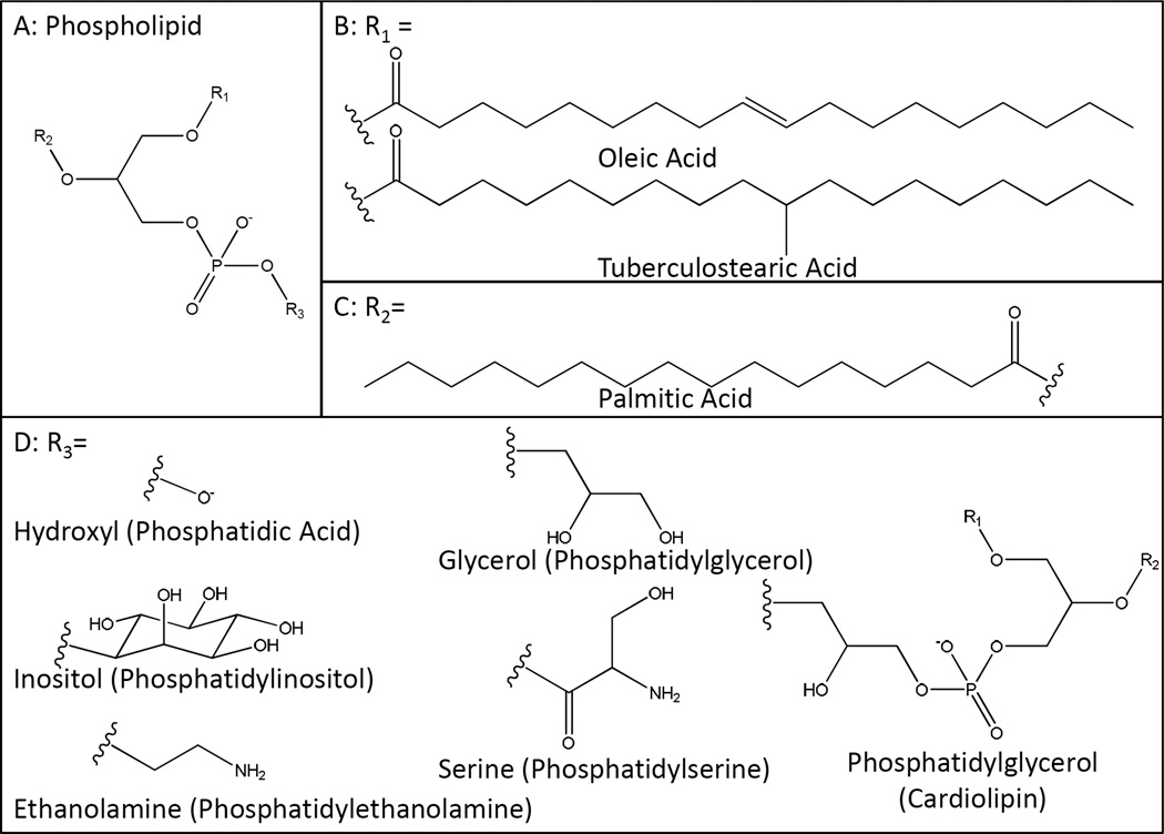
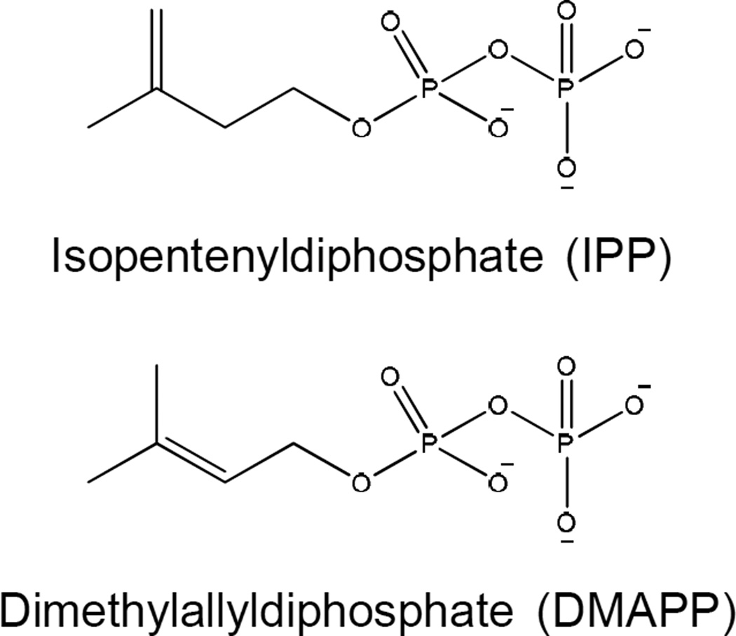
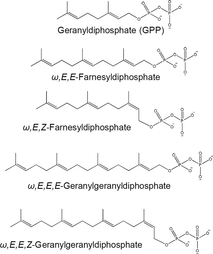
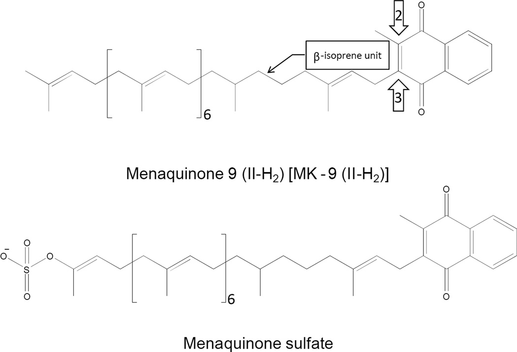
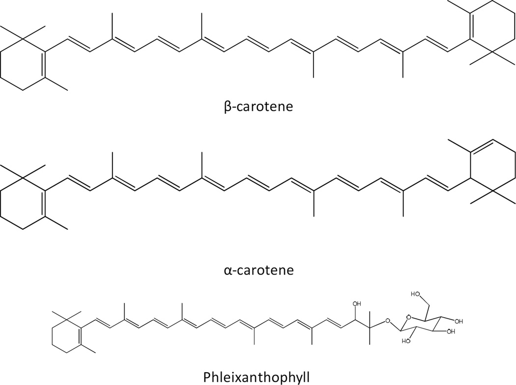
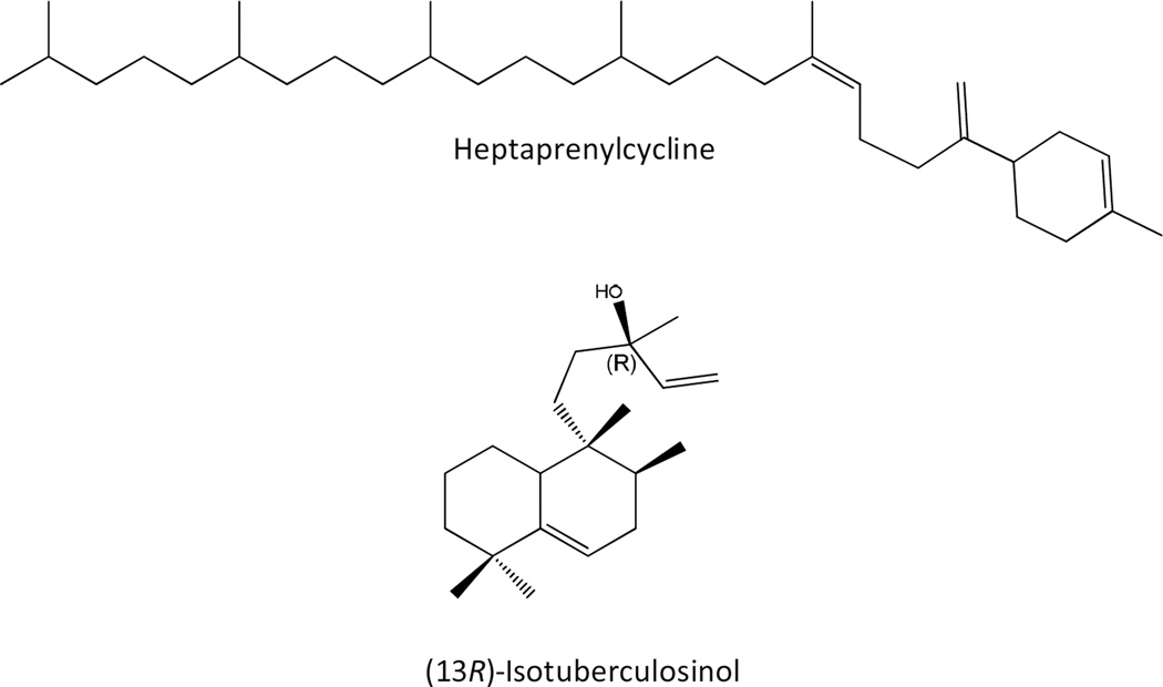
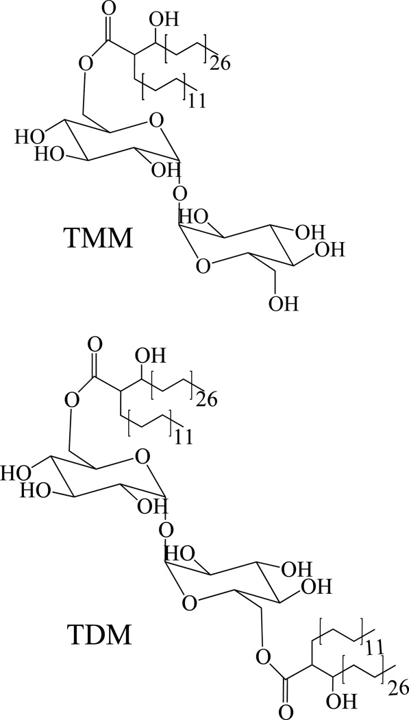
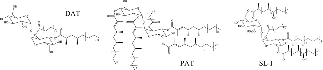
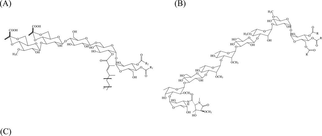
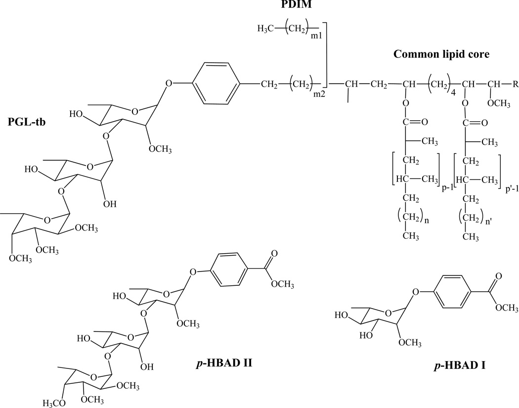
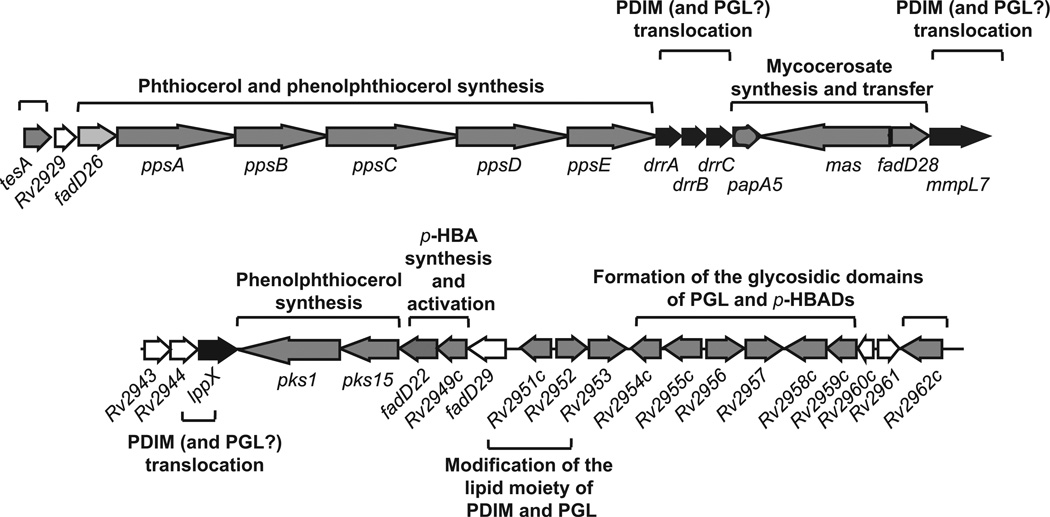
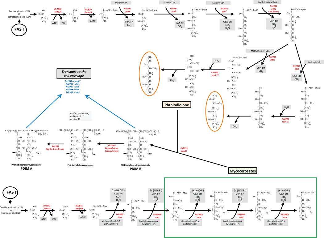
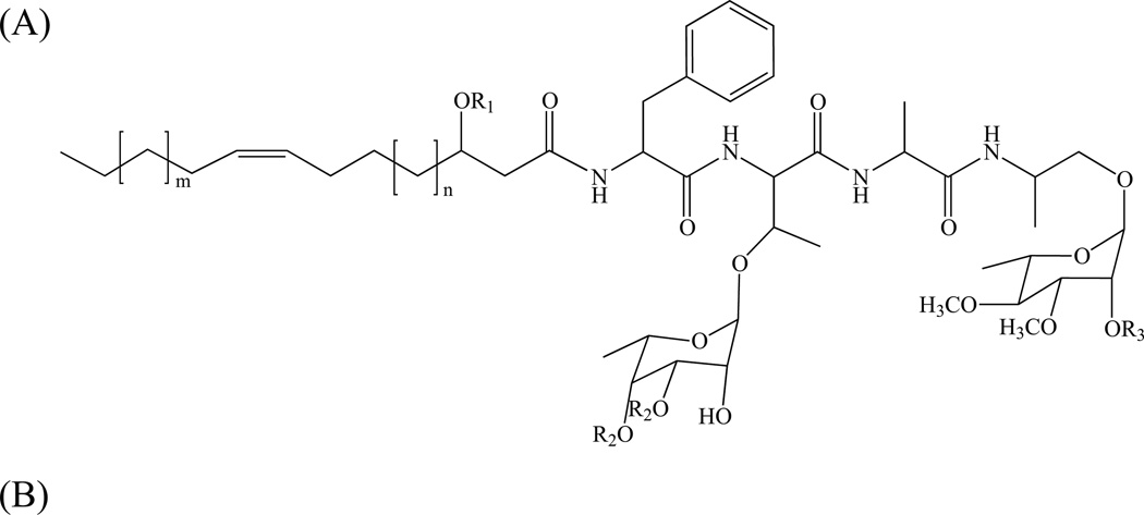
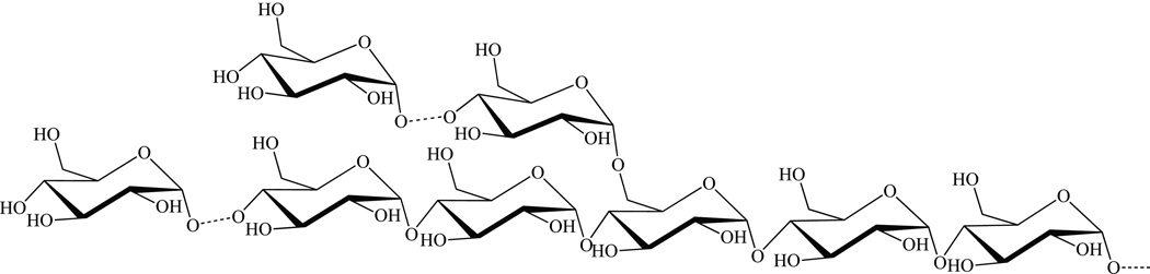
This chapter summarizes what is currently known of the structures, physiological roles, involvement in pathogenicity and biogenesis of a variety of non-covalently bound cell envelope lipids and glycoconjugates of Mycobacterium tuberculosis and other Mycobacterium species. Topics addressed in this chapter include phospholipids; phosphatidylinositol mannosides; triglycerides; isoprenoids and related compounds (polyprenyl phosphate, menaquinones, carotenoids, non-carotenoid cyclic isoprenoids); acyltrehaloses (lipooligosaccharides, trehalose mono- and di-mycolates, sulfolipids, di- and poly-acyltrehaloses); mannosyl-beta-1-phosphomycoketides; glycopeptidolipids; phthiocerol dimycocerosates, para-hydroxybenzoic acids and phenolic glycolipids; mycobactins; mycolactones; and capsular polysaccharides.


Figures

Figure 1
Figure 1. Schematic representation of the Mtb cell envelope
Many of the classes of lipids and glycolipids discussed in the text are represented schematically and are shown in probable locations in the cell envelope. The structures with light and dark green hexagons represent trehalose mono- and dimycolates, respectively, the red lollipops represent phthiocerol dimycocerosates, while the gold ones represent sulfolipids, diacyltrehaloses and polyacyltrehaloses. Grey circles represent phospholipid headgroups, black circles, isoprenoids, light blue squares GlcNAc, white squares MurNAc, white pentagons arabinofuranose, yellow diamonds galactofuranose, and blue hexagons mannose. The overall schematic and individual structures are not drawn to scale and the numbers of carbohydrate residues shown are not representative of the actual molecules. Proteins and peptides are not shown for the sake of clarity.
Figure 2
Figure 2. Structures of mycobacterial phospholipids
Figure 3
Figure 3. Structures of isopentenyldiphosphate and dimethylallyldiphosphate
These molecules are precursors of all isoprenoid compounds.
Figure 4
Figure 4. Structures of representative short-chain isopentenyldiphosphates synthesized by mycobacteria
The sterochemical conformation is shown.
Figure 5
Figure 5. Structures of isoprenylphosphates reported from Mtb
Figure 6
Figure 6. Structures of the predominant menaquinone and menaquinone sulfate reported from Mtb
Carbon positions 2, 3 and the β-isoprene unit are indicated by the arrows and call out.
Figure 7
Figure 7. Structures of representative carotenoids found in mycobacteria
Figure 8
Figure 8. Structures of representative non-carotenoid cyclic isoprenoids found in mycobacteria
Figure 9
Figure 9. Structures trehalose monomycolates (TMM) and trehalose dimycolates (TDM)
Figure 10
Figure 10. Structures sulfolipids (SL), diacyltrehaloses (DAT) and polyacyltrehaloses (PAT) and biosynthetic gene clusters
The major sulfolipid SL-I (2,3,6,6’-tetraacyl α-α’-trehalose-2’-sulfate) is represented. In SL-I, trehalose is sulfated at the 2’ position and esterified with palmitic acid and the multimethyl-branched phthioceranic and hydroxyphthioceranic acids. In DAT (2,3-di-O-acyltrehalose), trehalose is esterified with palmitic acid and the multimethylbranched mycosanoic acid. In PAT, trehalose is esterified with palmitic acid and the multimethyl-branched mycolipenic acids.
Figure 11
Figure 11. Structures of major lipooligosaccharide (LOS-A) of M. smegmatis ATCC 356
(R1 and/or R2 : octanoic acid and tetra- or hexa-decanoic acid) (A) and Mtb Canettii (B); R = Ac. LOS biosynthetic gene cluster of M. smegmatis mc2155 (C). Shown is the 25.15 kb-region spanning MSMEG_4727 (pks5) to MSMEG_4741 (mmpL). ORF are depicted as arrows. Black arrows indicate genes encoding biosynthetic enzymes; grey arrows indicate putative transporter genes; white arrows, hypothetical genes of unknown function. Pks5, Mas-like polyketide synthase; Pap, putative acyltransferase; MSMEG_4729 and MSMEG_4730, putative acyltransferases; FadD, putative acyl-CoA synthase; Gtf (MSMEG_4732), putative glycosyltransferase; Gap2, putative transmembrane protein involved in glycolipid translocation; MSMEG_4734, hypothetical PE/PPE-like protein; Gtf (MSMEG_4735), putative glycosyltransferase; MSMEG_4736 and MSMEG_4737, putative pyrruvylyl transferases; MSMEG_4738, hypothetical protein; Mtf, possible O-methyltransferase; Gtf (MSMEG_4740), putative glycosyltransferase; MmpL, putative inner membrane transporter.
Figure 12
Figure 12. Structure of the predominant mannosyl-β-1-phosphomycoketide from Mtb H37Rv
(See text for details)
Figure 13
Figure 13. Structures of the phthiocerol dimycocerosates (PDIM), phenolic glycolipids (PGL) and p-hydroxybenzoic acid derivatives (p-HBADs) of Mtb
In Mtb, p, p’=3–5; n, n’=16–18; m2=15–17; m1= 20–22; R= CH2–CH3 or CH3.
Figure 14
Figure 14. Genetic organization of the PDIM and PGL locus of Mtb H37Rv
ORF are depicted as arrows. Black arrows indicate genes encoding biosynthetic enzymes; grey arrows indicate putative transporter genes; white arrows, hypothetical genes of unknown function. More details about the function of each gene are provided in Table 3 and Fig. 15. Adapted from [260].
Figure 15
Figure 15. The PDIM biosynthetic pathway
(See text for details)
Figure 16
Figure 16
(A) Structure of the non-specific glycopeptidolipids of M. smegmatis. R1 = -H or –CH3; R2 = -H or –Ac; R3, -CH3, -succinyl, -rhamnosyl or -2-O-succinylrhamnosyl; m = 12–14; n = 6–10. (B) GPL biosynthetic gene cluster of M. smegmatis mc2155. Shown is the 64.97 kb-region spanning MSMEG_0380 (mmpS4) to MSMEG_0413. ORF are depicted as arrows. Black arrows indicate genes encoding biosynthetic enzymes; grey arrows indicate putative transporter genes; white arrows, putative regulatory genes. Chp, putative acyltransferase; FadE, putative acyl-CoA dehydrogenase; PapA, putative acyltransferase. Other genes are described in the text.
Figure 17
Figure 17. Structure and biosynthesis of α-D-glucans in Mtb
(See text for details)
Figure 18
Figure 18. Representative structures of mycobactins and carboxymycobactins from Mtb
(See text for details). Mycobactins: R1 = H; R2 = (CH2)nCH3, n = 16–19; (CH2)xCH=CH(CH2)yCH3, x+y = 14–17. Carboxymycobactins: R1 = H, CH3; R2 = (CH2)nCOOCH3/COOH, n = 1–7; (CH2)xCH=CH(CH2)yCOOCH3/COOH, x+y = 1–5.
Figure 19
Figure 19. Representative structure of a mycolactone from M. ulcerans
The genes involved in the biosynthesis of the various constituents of mycolactone are indicated on the structure.

Similar articles

Cited by

References

    1. Daffé M, Draper P. The envelope layers of mycobacteria with reference to their pathogenicity. Adv Microb Physiol. 1998;39:131–203. - PubMed
    1. Jackson M, McNeil MR, Brennan PJ. Progress in targeting cell envelope biogenesis in Mycobacterium tuberculosis. Future Microbiol. 2013 in press. - PMC - PubMed
    1. Hoffmann C, Leis A, Niederweis M, Plitzko JM, Engelhardt H. Disclosure of the mycobacterial outer membrane: cryo-electron tomography and vitreous sections reveal the lipid bilayer structure. Proc. Natl. Acad. Sci. USA. 2008;105:3963–3967. - PMC - PubMed
    1. Zuber B, Chami M, Houssin C, Dubochet J, Griffiths G, Daffe M. Direct visualization of the outer membrane of mycobacteria and corynebacteria in their native state. J. Bacteriol. 2008;190:5672–5680. - PMC - PubMed
    1. Sani M, Houben ENG, Geurtsen J, Pierson J, de Punder K, van Zon M, Wever B, Piersma SR, Jimenez CR, Daffe M, Appelmelk BJ, Bitter W, van der Wel N, Peters PJ. Direct visualization by cryo-EM of the mycobacterial capsular layer: a labile structure containing ESX-1-secreted proteins. PLoS Pathog. 2010;6:e1000794. - PMC - PubMed