

Pseudophacomorphic Glaucoma along with Pupillary Block after Visian™ Implantable Collamer Lens Implantation for High Myopia
- PMID: 25485179
- PMCID: PMC4254834
- DOI: 10.4236/ojoph.2014.44017
Pseudophacomorphic Glaucoma along with Pupillary Block after Visian™ Implantable Collamer Lens Implantation for High Myopia
Abstract
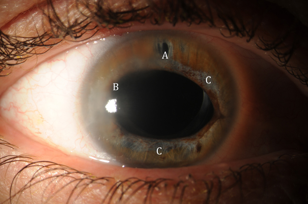
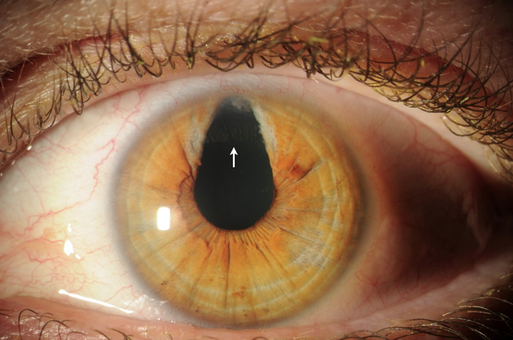
Purpose: To report a case of bilateral glaucoma related to pseudophacomorphic mechanism in one eye and pupillary block in the other eye after Visian Implantable Collamer Lens (ICL, STAAR Surgical) insertion.
Methods: A 44 year-old female with high myopia underwent bilateral ICL implantation of MICL12.6 after sulcus diameter measurements were performed by Pentacam.
Results: Pseudophacomorphic glaucoma-related angle closure occurred due to lens oversizing in the right eye. The mechanism was relieved via ICL explantation. In the left eye, pupillary block developed in a subacute manner after closure of the peripheral iridotomy (PI). The attack was ameliorated by reestablishing patency of the iridotomy.
Conclusions: ICL-related glaucomatous attacks may result from improper sizing as well as from placement of a single PI. Identification of the proper mechanism is vital as treatments differ significantly. In pseudophacomorphic glaucoma, explantation is needed. In pupillary block glaucoma, treatment involves establishment of a patent PI.
Keywords: Glaucoma; Pseudophacomorphic; Pupillary Block; Visian ICL.
Figures
References
-
- Huang D, Schallhorn SC, Sugar A, et al. Phakic intraocular lens implantation for the correction of myopia: a report by the American Academy of Ophthalmology. Ophthalmology. 2009;116:2244–2258. http://dx.doi.org/10.1016/j.ophtha.2009.08.018. - DOI - PubMed
-
- Igarashi A, Shimizu K, Kamiya K. Eight-year follow-up of posterior chamber phakic intraocular lens implantation for moderate to high myopia. Am J Ophthalmol. 2014;157:532–539. http://dx.doi.org/10.1016/j.ajo.2013.11.006. - DOI - PubMed
-
- Azar DT. Refractive Surgery. 2nd Edition. New York, New York: Elsevier Inc; 2007.
-
- Kojima T, et al. Optimization of an implantable collamer lens sizing method using high-frequency ultrasound biomicroscopy. Am J Ophthalmol. 2012;153(4):632–637. http://dx.doi.org/10.1016/j.ajo.2011.06.031. - DOI - PubMed
-
- Reinstein DZ, Lovisolo CF, Archer TJ, Gobbe M. Comparison of postoperative vault height predictability using white-to-white or sulcus diameter-based sizing for the visian implantable collamer lens. J Refract Surg. 2013;29(1):30–35. http://dx.doi.org/10.3928/1081597X-20121210-02. - DOI - PubMed
Grants and funding
LinkOut - more resources
Full Text Sources
Other Literature Sources
Research Materials
Miscellaneous