. 2014:2014:908098.
doi: 10.1155/2014/908098. Epub 2014 Nov 18.

Impact of ellagic acid in bone formation after tooth extraction: an experimental study on diabetic rats

Affiliations

Impact of ellagic acid in bone formation after tooth extraction: an experimental study on diabetic rats

Mazen M Jamil Al-Obaidi et al. ScientificWorldJournal. 2014.

Abstract

Objectives: To estimate the impact of ellagic acid (EA) towards healing tooth socket in diabetic animals, after tooth extraction.

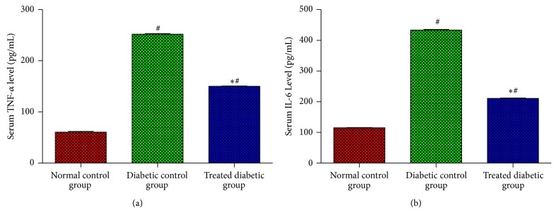
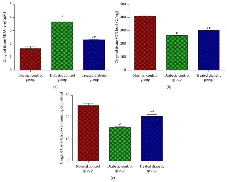
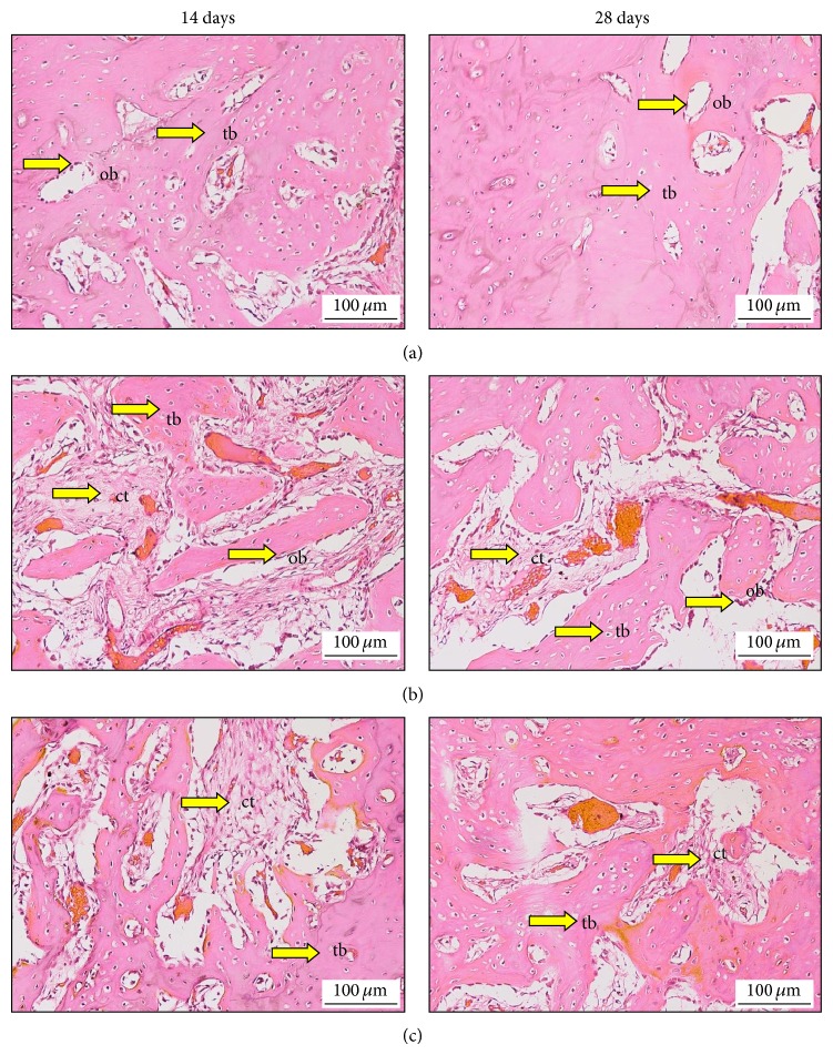
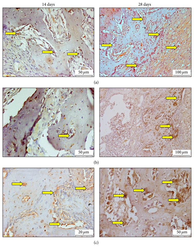
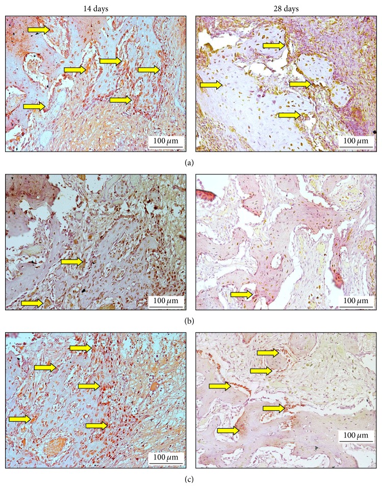
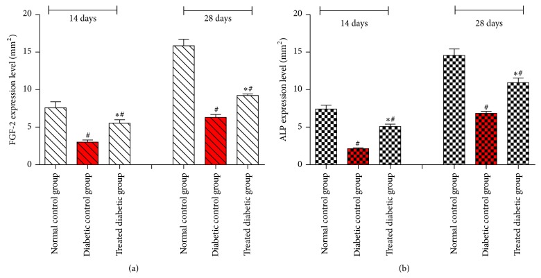
Methods: Twenty-four Sprague Dawley male rats weighing 250-300 g were selected for this study. All animals were intraperitoneally injected with 45 mg/kg (b.w.) of freshly prepared streptozotocin (STZ), to induce diabetic mellitus. Then, the animals were anesthetized, and the upper left central incisor was extracted and the whole extracted sockets were filled with Rosuvastatin (RSV). The rats were separated into three groups, comprising 8 rats each. The first group was considered as normal control group and orally treated with normal saline. The second group was regarded as diabetic control group and orally treated with normal saline, whereas the third group comprised diabetic rats, administrated with EA (50 mg/kg) orally. The maxilla tissue stained by eosin and hematoxylin (H&E) was used for histological examinations and immunohistochemical technique. Fibroblast growth factor (FGF-2) and alkaline phosphatase (ALP) were used to evaluate the healing process in the extracted tooth socket by immunohistochemistry test.

Results: The reactions of immunohistochemistry for FGF-2 and ALP presented stronger expression, predominantly in EA treated diabetic rat, than the untreated diabetic rat.

Conclusion: These findings suggest that the administration of EA combined with RSV may have accelerated the healing process of the tooth socket of diabetic rats, after tooth extraction.


Figures

Figure 1
Figure 1
Effects of EA on inflammatory cytokines TNF-α (a) and IL-6 (b) levels in serum experimental rats after tooth extraction. Each bar represents the mean ± SD. *Significant difference compared to diabetic control group (P < 0.05). #Significant difference compared to normal control group (P < 0.05).
Figure 2
Figure 2
Effects of EA on antioxidant status activities MDA (a), SOD (b), and CAT (c) levels in gingival tissue after tooth extraction in normal and diabetic rats. Each bar has been represented as mean ± SD. *Significant difference compared to diabetic control group (P < 0.05). #Significant difference compared to normal control group (P < 0.05).
Figure 3
Figure 3
The healing process in the alveolar bone in normal and diabetic rats at 14 days (left side) and 28 days (right side). (a) Normal control group treated with normal saline (5 mL/kg). (b) The untreated diabetic rat was fed with normal saline (5 mL/kg), and (c) treated diabetic rat administrated with EA (50 mg/kg/b.w.). Trabeculae bone (tb), connective tissue (ct), osteoblasts (ob). Histological sections stained with haematoxylin and eosin 200x.
Figure 4
Figure 4
Expressions of ALP immunolabeling in socket normal and diabetic animals at 14 (left side) and 28 (right side) days after tooth extraction of the right maxillary incisor. (a) Normal control group, (b) diabetic untreated rat (RSV+ normal saline), (c) diabetic rat treated with (RSV+EA). DAB with hematoxylin counterstaining, original 20x (ALP = arrows; DAB = diaminobenzidine).
Figure 5
Figure 5
Expressions of FGF-2 immunolabeling in socket normal and diabetic animals at 14 (left side) and 28 (right side) days after tooth extraction of the right maxillary incisor. (a) Normal control group, (b) diabetic untreated rat (RSV+ normal saline), (c) diabetic rat treated with (RSV+EA). DAB with hematoxylin counterstaining, original 20x (FGF-2 = arrows; DAB = diaminobenzidine).
Figure 6
Figure 6
Quantitative analysis of FGF-2 (a) and ALP (b) expression levels on 14th and 28th days in socket of experimental groups. Data were quantified by image analysis software as mean ± SD and analyzed by one-way ANOVA (SPSS version 20); (∗) indicates significant difference P < 0.05 versus untreated group. (#) indicates significant difference P < 0.05 versus normal control group.

References

    1. O'Ryan F. S., Lo J. C. Bisphosphonate-related osteonecrosis of the jaw in patients with oral bisphosphonate exposure: clinical course and outcomes. Journal of Oral and Maxillofacial Surgery. 2012;70(8):1844–1853. doi: 10.1016/j.joms.2011.08.033. - DOI - PubMed
    1. Araújo M. G., Lindhe J. Ridge alterations following tooth extraction with and without flap elevation: an experimental study in the dog. Clinical Oral Implants Research. 2009;20(6):545–549. doi: 10.1111/j.1600-0501.2008.01703.x. - DOI - PubMed
    1. Mealey B. L., Ocampo G. L. Diabetes mellitus and periodontal disease. Periodontology 2000. 2007;44(1):127–153. doi: 10.1111/j.1600-0757.2006.00193.x. - DOI - PubMed
    1. Vernillo A. T. Diabetes mellitus: relevance to dental treatment. Oral Surgery, Oral Medicine, Oral Pathology, Oral Radiology, and Endodontics. 2001;91(3):263–270. doi: 10.1067/moe.2001.114002. - DOI - PubMed
    1. Mellado-Valero A., Ferrer García J. C., Herrera Ballester A., Labaig Rueda C. Effects of diabetes on the osseointegration of dental implants. Medicina Oral, Patología Oral y Cirugía Bucal. 2007;12(1):E38–E43. - PubMed

Publication types

MeSH terms

LinkOut - more resources