

Rapid D-Affine biventricular cardiac function with polar prediction
- PMID: 25485422
- PMCID: PMC4260812
- DOI: 10.1007/978-3-319-10470-6_68
Rapid D-Affine biventricular cardiac function with polar prediction
Abstract
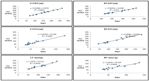
Although many solutions have been proposed for left ven-tricular functional analysis of the heart, right and left (bi-) ventricular function has been problematic due to the complex geometry and large motions. Biventricular function is particularly important in congenital heart disease, the most common type of birth defects. We describe a rapid interactive analysis tool for biventricular function which incorporates 1) a 3D+ time finite element model of biventricular geometry, 2) a fast prediction step which estimates an initial geometry in a polar coordinate system, and 3) a Cartesian update which penalizes deviations from affine transformations (D-Affine) from a prior. Solution times were very rapid, enabling interaction in real time using guide point modeling. The method was applied to 13 patients with congenital heart disease and compared with the clinical gold standard of manual tracing. Results between the methods showed good correlation (R2 > 0.9) and good precision (volume < 17 ml; mass < 11g) for both chambers.
Figures


References
-
- Bistoquet A, et al. Myocardial deformation recovery from cine mri using a nearly incompressible biventricular model. Medical Image Analysis. 2008;12(1):69–85. - PubMed
-
- Chen Y, et al. Object modelling by registration of multiple range images. Image and Vision Computing. 1992;10(3):145–155.
-
- Commowick O, et al. An efficient locally affine framework for the smooth registration of anatomical structures. Medical Image Analysis. 2008;12(4):427–441. - PubMed
-
- Grothues F, et al. Interstudy reproducibility of right ventricular volumes, function, and mass with cardiovascular magnetic resonance. American Heart Journal. 2004;147(2):218–223. - PubMed
-
- Haddad F, et al. Right ventricular function in cardiovascular disease, part II: pathophysiology, clinical importance, and management of right ventricular failure. Circulation. 2008;117(13):1717–1731. - PubMed
Publication types
MeSH terms
Grants and funding
LinkOut - more resources
Full Text Sources
Medical