Review
. 2015 Feb;122(2):448-64.
doi: 10.1097/ALN.0000000000000559.

Acute and perioperative care of the burn-injured patient

Affiliations
Review

Acute and perioperative care of the burn-injured patient

Edward A Bittner et al. Anesthesiology. 2015 Feb.

Abstract

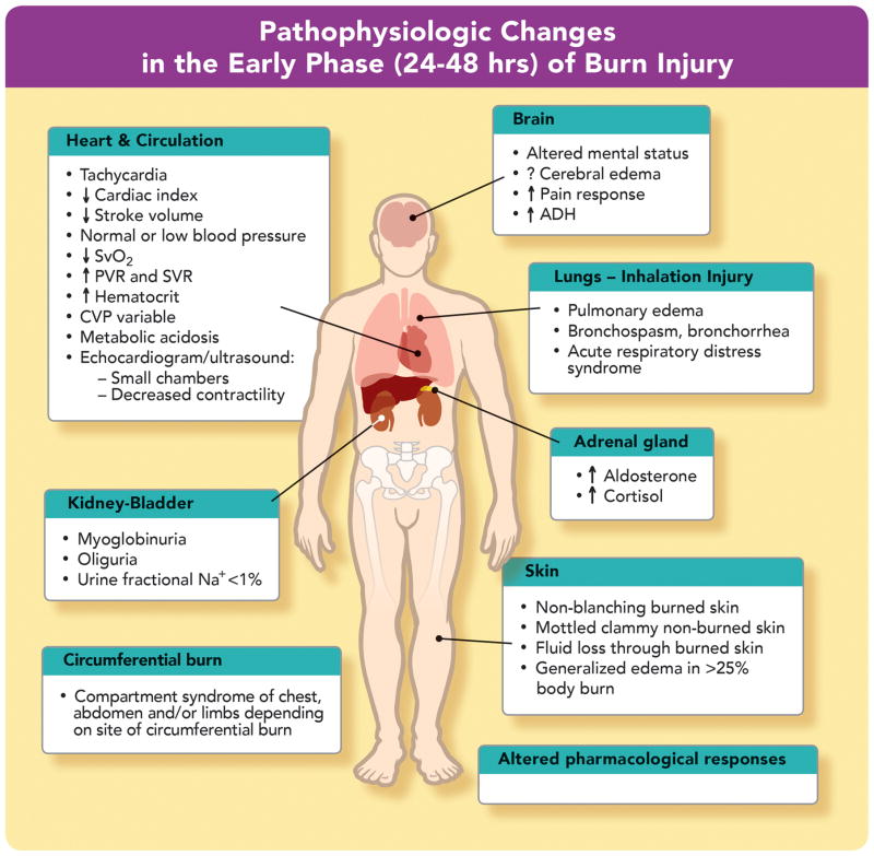
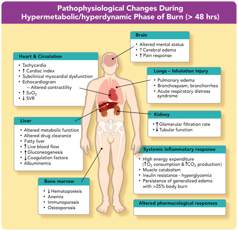
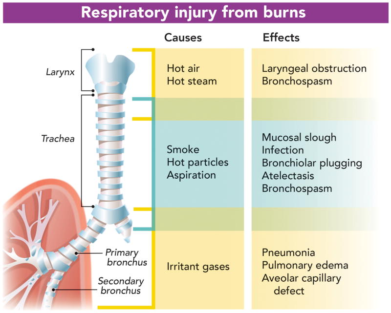
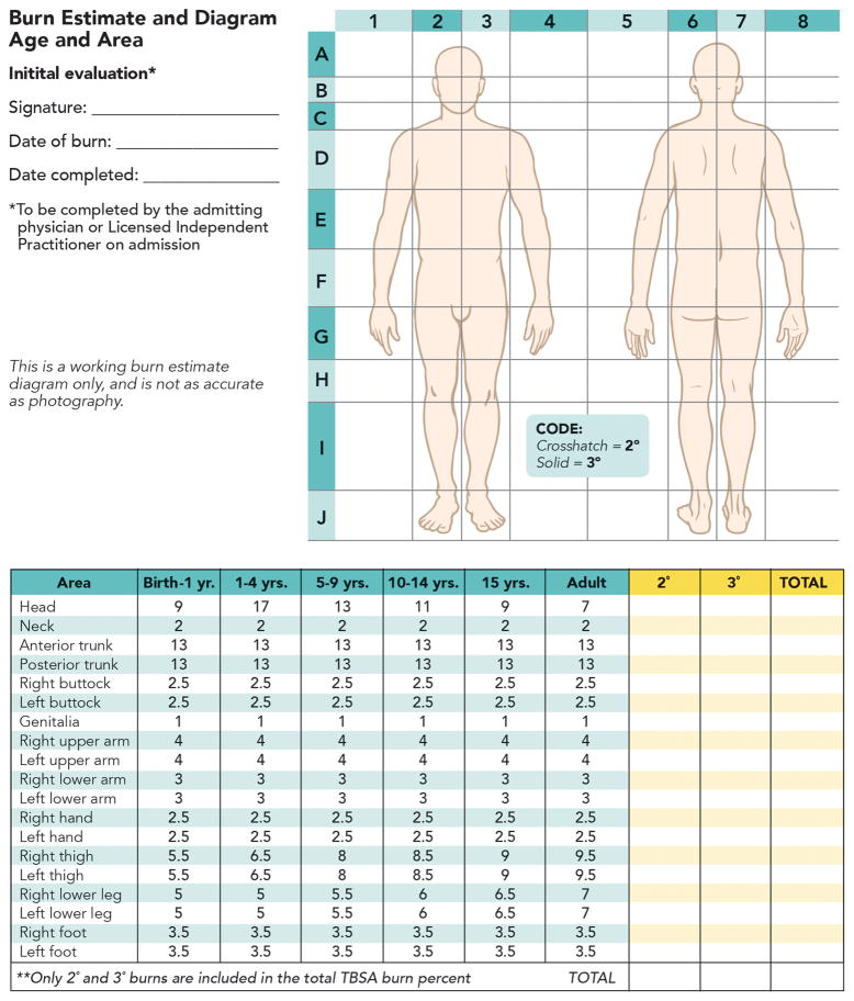
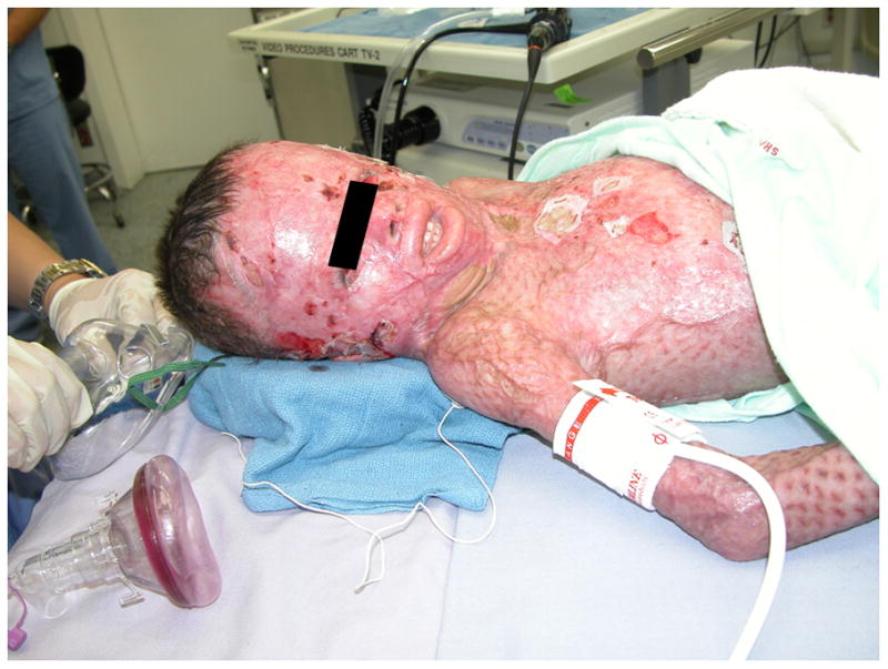
Care of burn-injured patients requires knowledge of the pathophysiologic changes affecting virtually all organs from the onset of injury until wounds are healed. Massive airway and/or lung edema can occur rapidly and unpredictably after burn and/or inhalation injury. Hemodynamics in the early phase of severe burn injury is characterized by a reduction in cardiac output and increased systemic and pulmonary vascular resistance. Approximately 2 to 5 days after major burn injury, a hyperdynamic and hypermetabolic state develops. Electrical burns result in morbidity much higher than expected based on burn size alone. Formulae for fluid resuscitation should serve only as guideline; fluids should be titrated to physiologic endpoints. Burn injury is associated basal and procedural pain requiring higher than normal opioid and sedative doses. Operating room concerns for the burn-injured patient include airway abnormalities, impaired lung function, vascular access, deceptively large and rapid blood loss, hypothermia, and altered pharmacology.


Conflict of interest statement

The authors declare no competing interests

Figures

FIGURE 1
FIGURE 1. Pathophysiologic changes in the early phase (24–48 h) of burn injury
The early (ebb) phase of burn injury is characterized by decreased cardiac output, and decreased blood flow to all organs. The decreased cardiac output is due to loss of intravascular volume, direct myocardial depression, increased pulmonary and systemic vascular resistance (PVR and SVR, respectively) and hemoconcentration, and can lead to metabolic acidosis, and venous desaturation (↓SVO2). Decreased urine flow results from decreased glomerular filtration, and elevated aldosterone and antidiuretic hormone levels (ADH) levels. Oxygenation and ventilation problems can occur due to inhalation injury and/or distant effects of burn on airways and lung. Compartment syndrome ensues if there is circumferential burn with no escharotomy performed to release the constriction. Compartment Syndrome can also occur in abdomen, extremities or orbits without local or circumferential burns. Mental status can be altered because of hypoxia, inhaled toxins and/or drugs. The reasons why heart rate, blood pressure, and central venous pressure (CVP) can be poor indicators of volume status are explained in table 3.
FIGURE 2
FIGURE 2. Pathophysiological changes during hypermetabolic/hyperdynamic phase of burn
At 48–72 h after burn, the hypermetabolic-hyperdynamic (flow) phase starts, characterized by increased oxygen consumption, carbon dioxide production, and cardiac output, with enhanced blood flow to all organs including skin, kidney (glomerular filtration rate [GFR]) and liver, and decreased systemic vascular resistance (SVR). Increased venous oxygen saturation (↑SVO2) is related to peripheral arteriovenous shunting. The markedly decreased SVR mimics sepsis. Lungs and airways may continue to be affected because of inhalation injury and/or acute respiratory distress syndrome. Pulmonary edema can occur due to distant effects of major burn and to reabsorption of edema fluid (hypervolemia). The altered mental status may be related to burn itself and/or concomitant drug therapy. Release of catabolic hormones and insulin resistance leads to muscle protein catabolism and hyperglycemia.
FIGURE 3
FIGURE 3. Respiratory inadequacy due to direct injury: putative agents and site of injury
The noxious agents released from the burning material can affect different parts of the airway. The cartoon indicates which part of the respiratory system is affected by each gas, toxin or chemical (“Cause”). The pathophysiological effects of each of these noxious agents is also indicated (“Effects”). Thermal or chemical injury can lead to edema of face, pharynx, glottis and larynx. Injury to trachea and bronchi leads to bronchospasm and bronchorrhea. Chemical and toxin injury can lead to alveolar damage and pulmonary edema.
FIGURE 4
FIGURE 4. Lund-Browder burn diagram and table
Lund-Browder burn diagram and table indicate the varying proportions in surface area in persons with different ages. A careful burn diagram should be completed at the time of initial evaluation including wound size, location, and estimated burn depth. Lund-Browder chart should be used in pediatric patients because the body surface area relationships vary with age. LIP = licensed independent practitioner; TBSA = total burn surface area.
FIGURE 5
FIGURE 5. Severe scar contracture developing before complete wound coverage
In contrast to edema affecting airways in the early phase, burn scar contraction of mouth and neck can complicate airway management during acute recovery phase. Reduced mandibular mobility and contraction around oral commissures can make it difficult or impossible to advance the jaw and open mouth. Furthermore, the airway can become obstructed by collapse of pharyngeal tissues during induction of general anesthesia. In these instances direct laryngoscopy can be difficult or impossible because the larynx also can be tethered to surrounding structures. Awake fiber optic intubation is an option. Ketamine provides analgesia and maintains respiratory drive and pharyngeal tone for pediatric patients and adults who will not tolerate awake intubation.
FIGURE 6
FIGURE 6. Dose–response curves and time to maximal effect of rocuronium in adult burned and non-burned patients
Dose versus time to percent twitch suppression for rocuronium in control subjects and burned subjects of mean 40% total body surface area (TBSA) burn and studied at least one week after burn. In normal patients dose of 0.9 mg/kg rocuronium caused 95% twitch suppression in ≤60 s. The same dose has an onset of >120 s following major burn. Increasing doses of rocuronium shifted dose-response curves to the left. However, even with 1.5 mg/kg dose, the onset was still >90 s. TOF Ratio refers to train-of-four ratio recorded in muscle during 2Hz nerve stimulation.,
FIGURE 7
FIGURE 7. Burn injury-induced tolerance to narcotics and sedatives
A 17-yr-old male sustained 90% flame burn injury requiring mechanical ventilation, multiple surgeries and anesthetics. The graph indicates the mg/kg/hr doses of morphine and midazolam administered over time after burn starting from week 1 to week 25. At one stage the intravenous morphine and midazolam doses required exceeded 55mg/hr of each. During procedures (e.g., dressing changes) additional doses of ketamine, dexmedetomidine, fentanyl, and/or propofol were administered pro re nata (PRN). Morerecently, when the doses of morphine and midazolam exceeds 0.5mg/kg/hr, we institute dexmedetomidine or ketamine infusions as sedative and change the opioid from morphine to fentanyl or vice versa (See also table 5)

References

    1. Cuthbertson DP. Postshock metabolic response. Lancet. 1942;239:433–7.
    1. Martyn JA, Aikawa N, Wilson RS, Szyfelbein SK, Burke JF. Extrapulmonary factors influencing the ration of arterial oxygen tension to inspired oxygen concentration in burn patients. Crit Care Med. 1979;7:492–6. - PubMed
    1. Wilmore DW, Goodwin CW, Aulick LH, Powanda MC, Mason AD, Jr, Pruitt BA. Effect of injury and infection on visceral metabolism and circulation. Ann Surg. 1980;192:491–504. - PMC - PubMed
    1. Aulick LH, McManus AT, Mason AD, Jr, Pruitt BA., Jr Effects of infection on oxygen consumption and core temperature in experimental thermal injury. Ann Surg. 1986;204:48–52. - PMC - PubMed
    1. Rehberg S, Maybauer MO, Enkhbaatar P, Maybauer DM, Yamamoto Y, Traber DL. Pathophysiology, management and treatment of smoke inhalation injury. Expert Rev Respir Med. 2009;3:283–97. - PMC - PubMed

Publication types