

Loss of circadian clock gene expression is associated with tumor progression in breast cancer
- PMID: 25485508
- PMCID: PMC4613905
- DOI: 10.4161/15384101.2014.954454
Loss of circadian clock gene expression is associated with tumor progression in breast cancer
Abstract
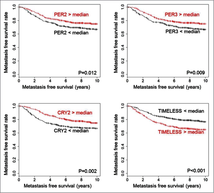
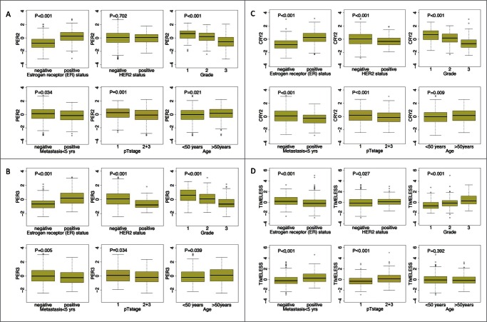
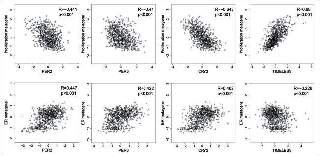
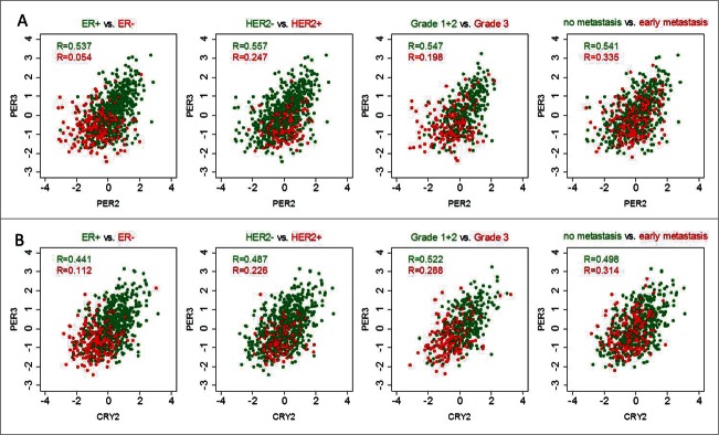
Several studies suggest a link between circadian rhythm disturbances and tumorigenesis. However, the association between circadian clock genes and prognosis in breast cancer has not been systematically studied. Therefore, we examined the expression of 17 clock components in tumors from 766 node-negative breast cancer patients that were untreated in both neoadjuvant and adjuvant settings. In addition, their association with metastasis-free survival (MFS) and correlation to clinicopathological parameters were investigated. Aiming to estimate functionality of the clockwork, we studied clock gene expression relationships by correlation analysis. Higher expression of several clock genes (e.g., CLOCK, PER1, PER2, PER3, CRY2, NPAS2 and RORC) was found to be associated with longer MFS in univariate Cox regression analyses (HR<1 and FDR-adjusted P < 0.05). Stratification according to molecular subtype revealed prognostic relevance for PER1, PER3, CRY2 and NFIL3 in the ER+/HER2- subgroup, CLOCK and NPAS2 in the ER-/HER2- subtype, and ARNTL2 in HER2+ breast cancer. In the multivariate Cox model, only PER3 (HR = 0.66; P = 0.016) and RORC (HR = 0.42; P = 0.003) were found to be associated with survival outcome independent of established clinicopathological parameters. Pairwise correlations between functionally-related clock genes (e.g., PER2-PER3 and CRY2-PER3) were stronger in ER+, HER2- and low-grade carcinomas; whereas, weaker correlation coefficients were observed in ER- and HER2+ tumors, high-grade tumors and tumors that progressed to metastatic disease. In conclusion, loss of clock genes is associated with worse prognosis in breast cancer. Coordinated co-expression of clock genes, indicative of a functional circadian clock, is maintained in ER+, HER2-, low grade and non-metastasizing tumors but is compromised in more aggressive carcinomas.
Keywords: ARNTL/2, aryl hydrocarbon receptor nuclear translocator-like/2; BHLHE40/41, basic helix-loop-helix family, member e; CLOCK, circadian locomotor output cycles kaput; CRY1/2, cryptochrome circadian clock 1/2; DBP, D site of albumin promoter (albumin D-box) binding protein; DFS, disease-free survival; ER, estrogen receptor; HER2, human epidermal growth factor receptor 2; HR, hazard ratio; MFS, metastasis-free survival; NFIL3, nuclear factor, interleukin 3 regulated; NPAS2, neuronal PAS domain protein 2; NR1D2, nuclear receptor subfamily 1, group D, member 2; PER1/2/3, period circadian clock 1/2/3; RORA/B/C, RAR-related orphan receptor alpha/beta/gamma; SCN, suprachiasmatic nucleus; breast cancer; circadian clock; clock genes; estrogen receptor; metastasis-free survival; tumor progression.
Figures
References
-
- Dibner C, Schibler U, Albrecht U. The mammalian circadian timing system: organization and coordination of central and peripheral clocks. Ann Rev physiol 2010; 72:517-49; PMID:20148687; http://dx.doi.org/10.1146/annurev-physiol-021909-135821 - DOI - PubMed
-
- Barclay JL, Tsang AH, Oster H. Interaction of central and peripheral clocks in physiological regulation. Prog Brain Res 2012; 199:163-81; PMID:22877665; http://dx.doi.org/10.1016/B978-0-444-59427-3.00030-7 - DOI - PubMed
-
- Mohawk JA, Green CB, Takahashi JS. Central and peripheral circadian clocks in mammals. Ann Rev Neurosci 2012; 35:445-62; PMID:22483041; http://dx.doi.org/10.1146/annurev-neuro-060909-153128 - DOI - PMC - PubMed
-
- Lowrey PL, Takahashi JS. Genetics of circadian rhythms in Mammalian model organisms. Adv Genet 2011; 74:175-230; PMID:21924978; http://dx.doi.org/10.1016/B978-0-12-387690-4.00006-4 - DOI - PMC - PubMed
-
- Ko CH, Takahashi JS. Molecular components of the mammalian circadian clock. Human Mol Genet 2006; 15 Spec No 2:R271-7; http://dx.doi.org/10.1093/hmg/ddl207 - DOI - PubMed
Publication types
MeSH terms
Substances
LinkOut - more resources
Full Text Sources
Other Literature Sources
Medical
Molecular Biology Databases
Research Materials
Miscellaneous