. 2015 Jan;33(1):64-72.
doi: 10.1038/nbt.3071. Epub 2014 Dec 8.

Injectable, spontaneously assembling, inorganic scaffolds modulate immune cells in vivo and increase vaccine efficacy

Affiliations

Injectable, spontaneously assembling, inorganic scaffolds modulate immune cells in vivo and increase vaccine efficacy

Jaeyun Kim et al. Nat Biotechnol. 2015 Jan.

Abstract

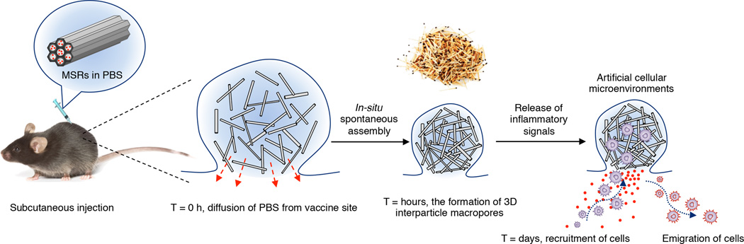
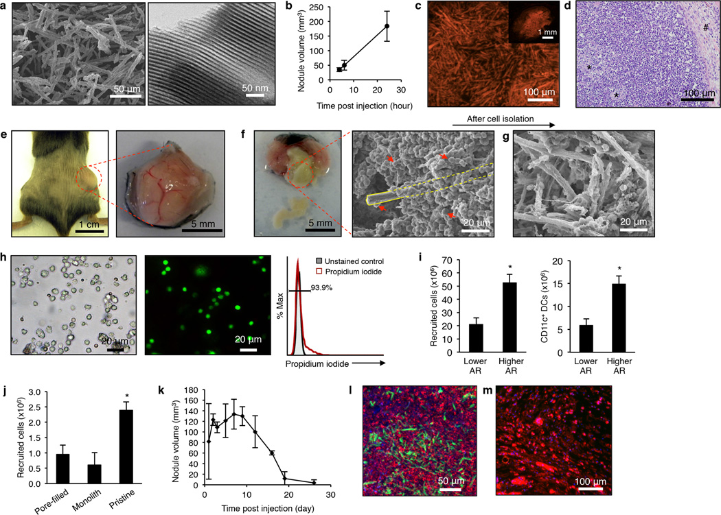
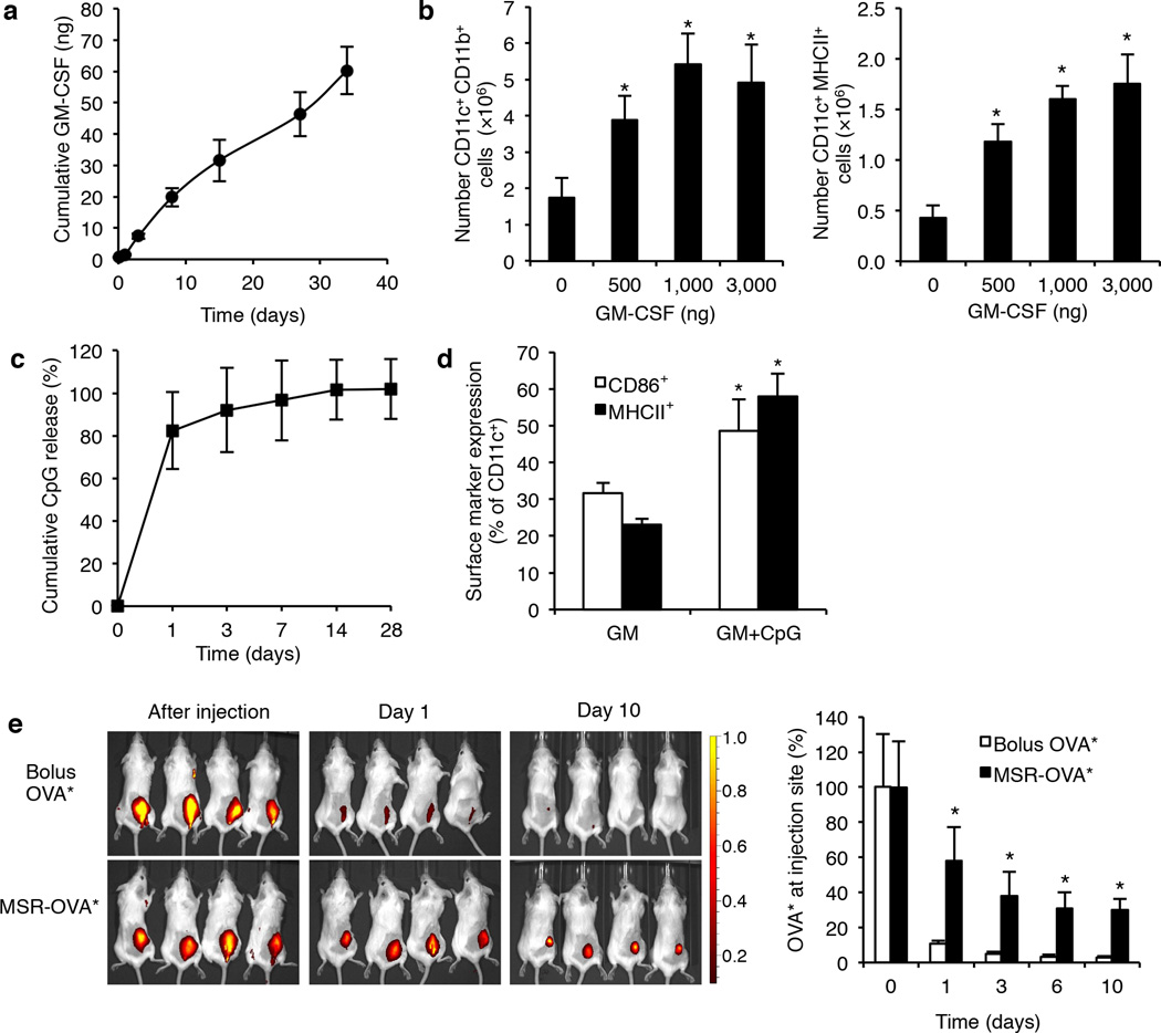
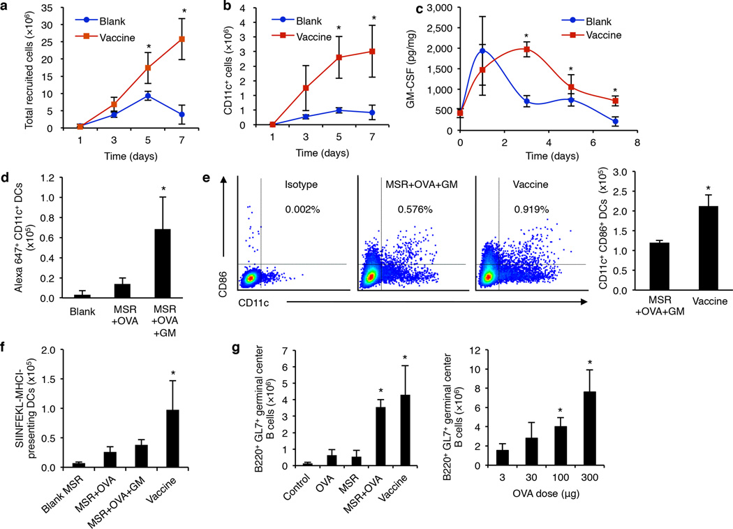
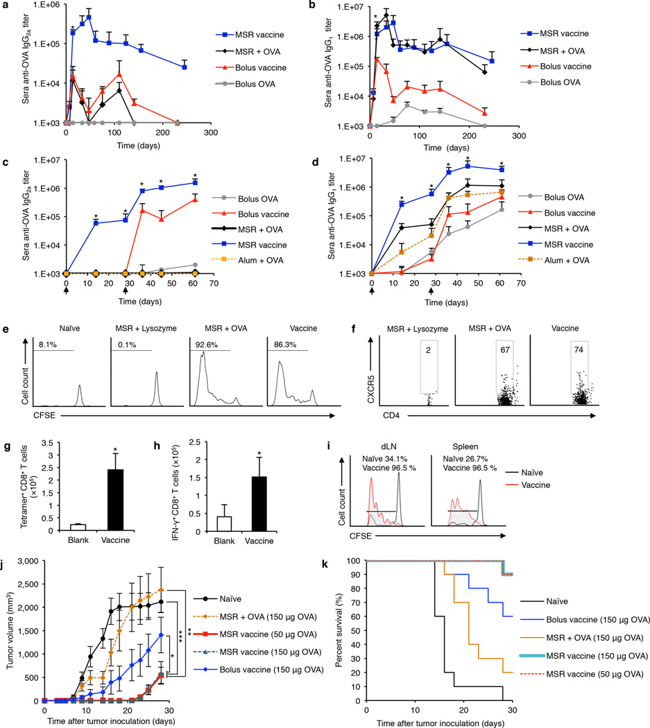
Implanting materials in the body to program host immune cells is a promising alternative to transplantation of cells manipulated ex vivo to direct an immune response, but doing so requires a surgical procedure. Here we demonstrate that high-aspect-ratio, mesoporous silica rods (MSRs) injected with a needle spontaneously assemble in vivo to form macroporous structures that provide a 3D cellular microenvironment for host immune cells. In mice, substantial numbers of dendritic cells are recruited to the pores between the scaffold rods. The recruitment of dendritic cells and their subsequent homing to lymph nodes can be modulated by sustained release of inflammatory signals and adjuvants from the scaffold. Moreover, injection of an MSR-based vaccine formulation enhances systemic helper T cells TH1 and TH2 serum antibody and cytotoxic T-cell levels compared to bolus controls. These findings suggest that injectable MSRs may serve as a multifunctional vaccine platform to modulate host immune cell function and provoke adaptive immune responses.


Figures

Figure 1
Figure 1. A schematic representation of in vivo spontaneous assembly of mesoporous silica rods (MSRs) and recruitment of host cells for maturation
A PBS dispersion of MSRs is injected into subcutaneous tissue of mice to form a pocket. After diffusion of PBS from the pocket, in situ spontaneous assembly of MSRs, analogous to the random assembly of thrown matchsticks, results in the formation of three-dimensional interparticle spaces where host cells can be recruited and educated by the payloads in MSRs. Educated cells may then emigrate from the structure to interact with other immune cells.
Figure 2
Figure 2. Subcutaneous injection of blank MSRs results in their spontaneous assembly in vivo and substantial numbers of cells are recruited into interparticle pores of assembled MSRs
(a) SEM (left) and TEM (right) images of MSRs of 88 µm in length and 4.5 µm in diameter. (b) Nodule size measurements after injection of blank MSRs in time course of hours (n=3). (c) Fluorescent image of cross-section of the retrieved nodule after injection of rhodamine B-labeled MSRs. (d) H&E staining of sectioned nodule retrieved at day 3 after subcutaneous injection. *: representative crosssection of MSRs. #: surrounding fibrotic tissue. (e) Localization of MSRs as a nodule in the dorsal flank of a mouse one day after subcutaneous injection (left) and the retrieved nodule (right). (f) Isolation of MSR-scaffold and cells from the nodule (left) and SEM photomicrograph (right) demonstrating a high number of recruited cells. Yellow outline represents a visible MSR. Red arrows indicate the representative cells. (g) SEM image of MSR-scaffold after removal of most recruited cells. (h) Representative bright-field optical microscope of the isolated cells (left), fluorescent image of the cells after live-dead staining (middle, green: live cells, red: dead cells), and propidium iodide (PI) flow cytometry analysis of retrieved cells from the nodule (right). (i) Number of total recruited cells (left) and CD11c+ DCs (right) into MSRs (20 mg) with lower and higher aspect ratio, respectively, at day 7 post injection (n=3). (j) Number of total recruited cells in 5 mg of pore-filled, pressed, and pristine MSRs, respectively, at day 3 post injection (n=4). (k) Nodule size measurement after injection of blank MSR in time course over weeks (n=3). (l,m) Confocal images of sectioned nodules retrieved at (l) day 7 and (m) day 28 post-injection, respectively. The injected MSRs were labeled with AF488 and loaded with GM-CSF (1 µg). The cryosections were stained with DAPI and rhodamine-phalloidin. Biological replicates were used and studies were repeated at least 2 times in the lab. Values represent mean and s.d. * represents p<0.05.
Figure 3
Figure 3. Cytokine, PAMP, and model antigen are released from MSR-scaffold in sustained manner in vitro and in vivo
(a)In vitro level of bioactive GM-CSF released from MSRs analyzed using ELISA. (b) Numbers of CD11c+ CD11b+ (left) and CD11c+ MHC II+ (right) DCs recruited to MSR-scaffold loaded with different amounts of GM-CSF (n=4). (c)In vitro release of CpG from MSRs, as determined using OliGreen ssDNA assay. (d) Levels of activation markers (CD86 and MHC II) of recruited CD11c+ DCs with injection of MSRs loaded with GM-CSF, or both GM-CSF and CpG, respectively. (n=4) (e) Representative NIR fluorescent images of mice injected with bolus OVA labeled with Alexafluor-647 (OVA*) or MSRs loaded with same amount of OVA* (left), and relative OVA* remaining in injection sites as a function of time, as based on fluorescent imaging (right) (n=4). Biological replicates were used and studies were repeated at least 2 times in the lab. Values represent mean and s.d. * represents p<0.05.
Figure 4
Figure 4. Vaccine formulation consisting of MSRs loaded with GM-CSF, CpG, and OVA is able to recruit DCs, program them with antigen and PAMP, and enhance their trafficking to the dLN to exert systemic effects
(a,b) Total number of recruited host cells (a) and CD11c+ DCs (b) in blank or vaccine MSR scaffolds (5 mg), respectively (n=4). (c) In vivo GM-CSF levels in tissue around the injection sites of blank or vaccine MSR scaffolds (n=4). In (a–c), the comparisons were made between two groups (vaccine versus blank). (d) Numbers of Alexafluor 647+ DCs in dLNs after injections of blank MSR scaffold, MSRs loaded with OVA*, or MSRs loaded with OVA* and GM-CSF, respectively (n=4). (e) Representative flow cytometry plots to analyze activated CD11c+ CD86+ DCs in dLNs in mice immunized with MSRs loaded with OVA/GM-CSF or MSRs loaded with OVA/GM-CSF/CpG (Vaccine), respectively (left), and numbers of CD11c+ CD86+ DCs in dLNs (right) (n=4). (f) Numbers of DCs presenting SIINKEKL-MHCI in dLNs at day 7 post immunization (n=4). (g) Numbers of B220+ GL7+ germinal center B cells at day 7 post immunization in untreated mice or mice treated with MSR containing various cargoes (left), and impact of OVA dose on number of germinal center B cells (right) (n=4). Biological replicates were used and studies were repeated at least 2 times in the lab. Values represent mean and s.d. *represents p<0.05.
Figure 5
Figure 5. MSR vaccine generates potent humoral and cellular immune responses against a model antigen, OVA
(a,b) ELISA analysis of sera OVA-specific IgG2a (a) or IgG1 (b) after immunization with bolus OVA, soluble components of the vaccine (bolus vaccine), MSRs loaded with OVA, or MSR vaccine, respectively (n=5). (c,d) ELISA analysis of sera OVA-specific (c) IgG2a and (d) IgG1 after immunization with bolus OVA, bolus vaccine formulation (GM-CSF, CpG, OVA), Imject Alum with OVA, MSR with OVA, and the MSR vaccine formulation on day 0, and boosted with the same formulations for each condition on day 30 (n=5). In (a–d), comparisons were made between vaccine and bolus formulation groups. (e,f) Flow cytometric analysis of proliferation of OVA-specific CD4+ T cells (gated on Thy1.2+ CD3+ T cells) (e) and CD4+ CXCR5+ T follicular helper (TFH) cells (gated on Thy1.2+ CD3+ T cells) (f) in naïve mice, or mice at day 3 post immunization with MSR loaded with non-specific antigen (MSR + Lysozyme), MSR loaded with OVA (MSR + OVA), or complete MSR vaccine (Vaccine) (n=4). (g,h) Number of tetramer+ CD8+ T cells (g) and IFN-γ+ CD8+ T cells (h) in spleen 7 days after vaccination with blank MSR (Blank) or complete MSR vaccine (Vaccine) (n=4). (i) Flow cytometric analysis of proliferation of OVA-specific CD8+ T cells in the draining lymph node (dLN; left) and spleen (right) of naïve mice, and mice at day 3 post immunization with complete MSR vaccine (Vaccine). (j,k) Prophylactic cancer vaccine study using injectable MSRs. (j) EG.7-OVA tumor volume and (k) survival rate after subcutaneous injection of various vaccine formulations 10 days before tumor inoculation (n=10). In (j), the tumor volumes were compared on day 21, 23 and 25, following the onset of tumor growth in the vaccine group. Biological replicates were used and studies were repeated at least 2 times in the lab. Values represent mean and S.D. with exception of Fig. 5j, which is expressed as mean ± SEM. *, **, and *** represents p<0.05, p<0.01, and p<0.001, respectively.

Similar articles

Cited by

References

    1. Jähnisch H, et al. Dendritic cell-based immunotherapy for prostate cancer. Clin. Dev. Immunol. 2010;2010:1–8. - PMC - PubMed
    1. Porter DL, Levine BL, Kalos M, Bagg A, June CH. Chimeric antigen receptor-modified T cells in chronic lymphoid leukemia. N. Engl. J. Med. 2011;365:725–733. - PMC - PubMed
    1. Ali OA, Huebsch N, Cao L, Dranoff G, Mooney DJ. Infection-mimicking materials to program dendritic cells in situ. Nat. Mater. 2009;8:151–158. - PMC - PubMed
    1. Ali OA, Emerich D, Dranoff G, Mooney DJ. In situ regulation of DC subsets and T cells mediates tumor regression in mice. Sci. Transl. Med. 2009;1 8ra19. - PMC - PubMed
    1. Stephan MT, Moon JJ, Um SH, Bershteyn A, Irvine DJ. Therapeutic cell engineering with surface-conjugated synthetic nanoparticles. Nat. Med. 2010;16:1035–1041. - PMC - PubMed

Publication types