. 2015 Jan;125(1):304-15.
doi: 10.1172/JCI76357. Epub 2014 Dec 8.

LMP1-deficient Epstein-Barr virus mutant requires T cells for lymphomagenesis

LMP1-deficient Epstein-Barr virus mutant requires T cells for lymphomagenesis

Shi-Dong Ma et al. J Clin Invest. 2015 Jan.

Abstract

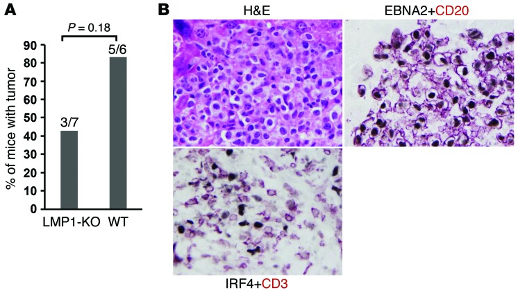
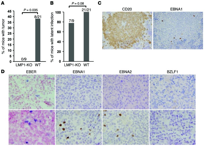
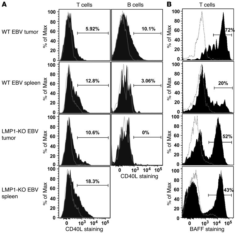
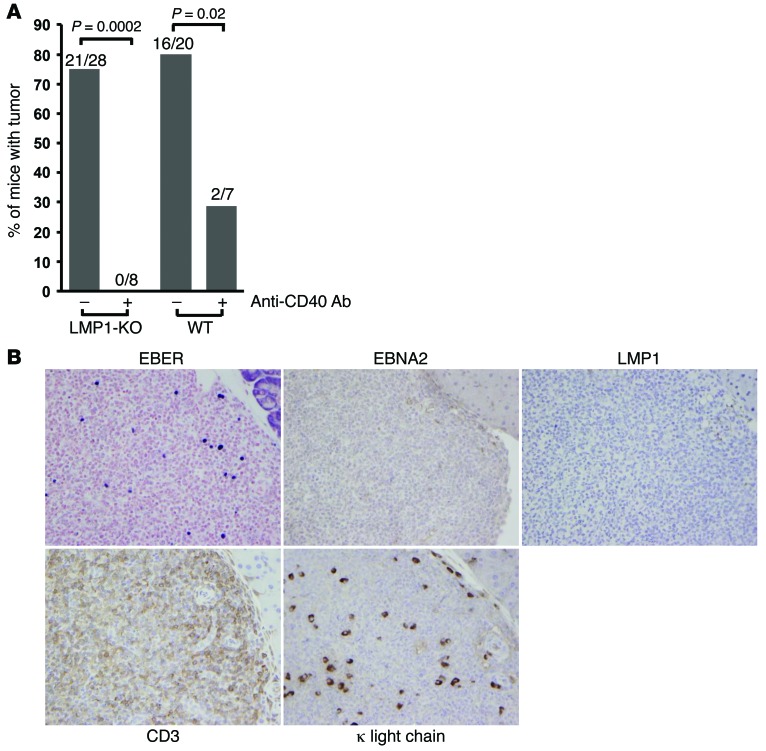
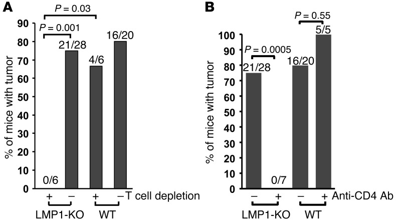
Epstein-Barr virus (EBV) infection transforms B cells in vitro and is associated with human B cell lymphomas. The major EBV oncoprotein, latent membrane protein 1 (LMP1), mimics constitutively active CD40 and is essential for outgrowth of EBV-transformed B cells in vitro; however, EBV-positive diffuse large B cell lymphomas and Burkitt lymphomas often express little or no LMP1. Thus, EBV may contribute to the development and maintenance of human lymphomas even in the absence of LMP1. Here, we found that i.p. injection of human cord blood mononuclear cells infected with a LMP1-deficient EBV into immunodeficient mice induces B cell lymphomas. In this model, lymphoma development required the presence of CD4+ T cells in cord blood and was inhibited by CD40-blocking Abs. In contrast, LMP1-deficient EBV established persistent latency but did not induce lymphomas when directly injected into mice engrafted with human fetal CD34+ cells and human thymus. WT EBV induced lymphomas in both mouse models and did not require coinjected T cells in the cord blood model. Together, these results demonstrate that LMP1 is not essential for EBV-induced lymphomas in vivo and suggest that T cells supply signals that substitute for LMP1 in EBV-positive B cell lymphomagenesis.


Figures

Figure 11
Figure 11. LMP1-KO EBV can induce lymphomas in a subset of hNSG(thy) mice treated with the T cell receptor Ab OKT3.
(A) Incidence and percentage of LMP1-KO EBV– and WT EBV–infected hNSG(thy) mice that developed lymphomas when treated with OKT3 Ab. Data were combined from 2 experiments; 2-tailed Fisher exact test was used for statistical analysis. (B) An LMP1-KO EBV–infected lymphoma invading the liver in an OKT3-treated infected hNSG(thy) mouse was stained for H&E, costained for EBNA2 (purple) and CD20 (red), or costained for CD3 (red) and IRF4 (purple). Even with OKT3 treatment, IRF4+ EBV-infected lymphoma cells were infiltrated by CD3+ T cells. Neoplastic cells coexpressed EBNA2 and CD20, which indicated that they are B lymphocytes with type III latency. Original magnification, ×100.
Figure 10
Figure 10. LMP1-KO EBV cannot induce lymphomas in highly immunocompetent hNSG(thy) mice, but can establish long-term viral latency.
(A and B) Incidence and percentage of LMP1-KO EBV– and WT EBV–infected hNSG(thy) mice that developed lymphomas (A) or latent EBV infection (B). Data were combined from 4 experiments; 2-tailed Fisher exact test was used for statistical analysis. (C) The spleen of an LMP1-KO EBV–infected hNSG(thy) mice was stained with CD20 or anti-EBNA1 Ab. A small fraction of CD20+ B lymphocytes harbored EBV infection, consistent with viral latency. This animal also had a detectable viral load in the blood at the time of euthanasia. (D) Spleens from 2 LMP1-KO EBV–infected hNSG(thy) mice subjected to EBER ISH and IHC for EBNA1, EBNA2, and BZLF1. Original magnification, ×40 (C); ×100 (D).
Figure 9
Figure 9. T cells, but not B cells, from LMP1-KO EBV tumors express CD40L, and T cells from tumors express high levels of BAFF.
Cells isolated from tumors or spleens of mice injected with LMP1-KO EBV– or WT EBV–infected cord blood were stained with Abs specific for human CD45, CD19, CD20, CD3, CD40L, BAFF, or the respective isotype-matched negative controls, then analyzed by flow cytometry. Samples were gated on single cells expressing human CD45, CD19, and CD20 (B cells) or CD45 and CD3 (T cells). Black shaded histograms show staining for CD40L (A) or BAFF (B); dashed gray histograms show staining of the same population of cells by the isotype control. Percentages denote the fraction of CD40L+ or BAFF+ cells above the isotype control background.
Figure 8
Figure 8. CD40 signaling supports the growth of both WT EBV– and LMP1-KO EBV–induced cord blood lymphomas in NSG mice.
(A) Incidence and percentage of NSG mice developing lymphomas after injection with WT EBV– or LMP1-KO EBV–infected human cord blood in the presence or absence of CD40-blocking Ab. Data were derived by combining the results of 2 experiments each with and without anti-CD40 Ab and of 4 no-Ab experiments; 2-tailed Fisher exact test was used for statistical analysis. (B) A peripancreatic granuloma-like lesion in a NSG mouse injected with WT EBV–infected cord blood and treated with CD40-blocking Ab, stained for EBER, EBNA2, LMP1, CD3, and κ light chain. EBV-infected B cells were surrounded by T cells. Original magnification, ×40.
Figure 7
Figure 7. CD4+ T cells support the growth of LMP1-KO EBV–induced cord blood lymphomas in NSG mice.
(A) Incidence and percentage of NSG mice that developed lymphomas after injection with WT EBV– or LMP1-KO EBV–infected human cord blood in the presence or absence of T cell–depleting treatments. Data were derived by combining the results of 2 experiments each with and without T cell depletion and of 4 nondepletion experiments; 2-tailed Fisher exact test was used for statistical analysis. (B) Incidence and percentage of NSG mice developing lymphomas after injection with WT EBV– or LMP1-KO EBV–infected human cord blood in the presence or absence of CD4+ T cell–depleting Ab (CD4R1). Data were derived by combining the results of 2 experiments each with and without anti-CD4 Ab and of 4 no-Ab experiments; 2-tailed Fisher exact test was used for statistical analysis.
Figure 6
Figure 6. LMP1-KO EBV–infected cord blood lymphomas can become monoclonal.
IHC staining using κ- and λ-specific light chain Abs revealed κ light chain dominance in an LMP1-KO EBV–infected pancreatic lymphoma as well as polyclonality in the spleen tissue from same animal. Original magnification, ×10.
Figure 5
Figure 5. LMP1-KO EBV–induced lymphomas do not express LMP1.
(A) EBER ISH and LMP1 IHC staining of representative WT EBV– and LMP1-KO EBV–infected cord blood lymphomas. Original magnification, ×20. (B) PCR analysis was performed on isolated tumor DNA prepared from 1 WT EBV–infected tumor, 2 different LMP1-KO EBV–infected tumors, or water control, using primers that amplify the LMP1 gene (L) or the viral BZLF1 promoter (Z).
Figure 4
Figure 4. LMP1-KO EBV–induced lymphomas are highly proliferative, activated DLBCLs that express c-MYC.
(A) LMP1-KO EBV–infected B cells infiltrating the gall bladder wall expressed EBER, CD20, Ki67, and IRF4. (B) Representative LMP1-KO EBV– and WT EBV–infected lymphomas invading the pancreas, stained for either Ki67 or c-MYC (purple) and costained with CD20 (brown). Original magnification, ×10 (A); ×100 (B).
Figure 3
Figure 3. LMP1-KO EBV–infected cord blood lymphomas have type III latency and are heavily infiltrated by T cells.
(A) An LMP1-KO EBV–infected cord blood lymphoma invading the pancreas was stained for H&E, EBV EBNA2 protein, CD20, and CD3. The lymphoma was EBNA2+ and CD20+ and infiltrated by numerous CD3+ T cells. Original magnification, ×40. (B) RT-PCR was performed on RNA isolated from 2 different LMP1-KO EBV–infected tumors to detect Qp- and Cp-derived EBNA transcripts. Mutu I (type I latency) and Kem III (type III latency) cells were used as controls.
Figure 2
Figure 2. WT and LMP1-KO EBV–infected cord blood cells, but not mock-infected cells, promote lymphomas in NSG mice.
(A) Incidence and percentage of NSG mice developing lymphomas after injection with mock-infected, WT EBV–infected, or LMP1-KO EBV–infected human cord blood. 6 experiments were performed; 2-tailed Fisher exact test was used for statistical analysis. (B) EBV plasma viral loads (obtained at the time of euthanasia) in mice injected with WT EBV– or LMP1-KO EBV–infected cord blood. Animal plasma from 3 experiments were used for testing viral loads; Wilcoxon rank-sum test was used for statistical analysis. For the animals with very high plasma viral loads, exact values are shown.
Figure 1
Figure 1. LMP1-KO EBV–infected human cord blood induces invasive lymphomas in NSG mice.
Representative H&E-stained LMP1-KO EBV– and WT EBV–infected lymphomas invading the pancreas. EBER was detected by ISH, and cellular proteins IRF4 and CD20 were detected by IHC. Original magnification, ×10 (top row), ×100 (bottom rows).

Similar articles

Cited by

References

    1. Vereide D, Sugden B. Insights into the evolution of lymphomas induced by Epstein-Barr virus. Adv Cancer Res. 2010;108:1–19. - PubMed
    1. Vereide DT, Sugden B. Lymphomas differ in their dependence on Epstein-Barr virus. Blood. 2011;117(6):1977–1985. doi: 10.1182/blood-2010-05-285791. - DOI - PMC - PubMed
    1. Kieff ED, Rickinson AB. Epstein-Barr Virus and its replication. In: Fields BN, Knipe DM, Howley PM, Griffin DE eds. Fields’ Virology. 5th ed. Philadelphia, Pennsylvania, USA: Wolters Kluwer Health/Lippincott Williams & Wilkins; 2007:2603–2654.
    1. Rickinson AB, Kieff ED. Epstein-Barr Virus. In: Fields BN, Knipe DM, Howley PM, Griffin DE eds. Fields’ Virology. 5th ed. Philadelphia, Pennsylvania, USA: Wolters Kluwer Health/Lippincott Williams & Wilkins; 2007:2655–2700.
    1. Rastelli J, et al. LMP1 signaling can replace CD40 signaling in B cells in vivo and has unique features of inducing class-switch recombination to IgG1. Blood. 2008;111(3):1448–1455. - PubMed

Publication types

MeSH terms

Substances