. 2014 Dec 8;9(12):e114651.
doi: 10.1371/journal.pone.0114651. eCollection 2014.

Identification of the interactors of human nibrin (NBN) and of its 26 kDa and 70 kDa fragments arising from the NBN 657del5 founder mutation

Affiliations

Identification of the interactors of human nibrin (NBN) and of its 26 kDa and 70 kDa fragments arising from the NBN 657del5 founder mutation

Domenica Cilli et al. PLoS One. .

Abstract

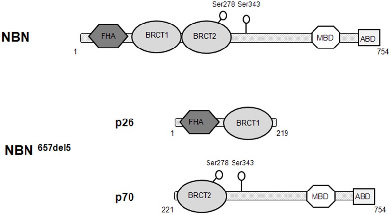
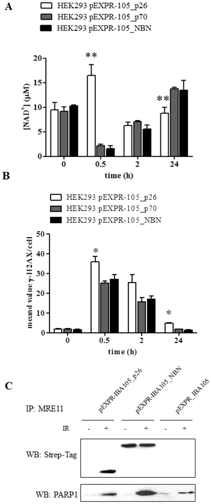
Nibrin (also named NBN or NBS1) is a component of the MRE11/RAD50/NBN complex, which is involved in early steps of DNA double strand breaks sensing and repair. Mutations within the NBN gene are responsible for the Nijmegen breakage syndrome (NBS). The 90% of NBS patients are homozygous for the 657del5 mutation, which determines the synthesis of two truncated proteins of 26 kDa (p26) and 70 kDa (p70). Here, HEK293 cells have been exploited to transiently express either the full-length NBN protein or the p26 or p70 fragments, followed by affinity chromatography enrichment of the eluates. The application of an unsupervised proteomics approach, based upon SDS-PAGE separation and shotgun digestion of protein bands followed by MS/MS protein identification, indicates the occurrence of previously unreported protein interacting partners of the full-length NBN protein and the p26 fragment containing the FHA/BRCT1 domains, especially after cell irradiation. In particular, results obtained shed light on new possible roles of NBN and of the p26 fragment in ROS scavenging, in the DNA damage response, and in protein folding and degradation. In particular, here we show that p26 interacts with PARP1 after irradiation, and this interaction exerts an inhibitory effect on PARP1 activity as measured by NAD+ levels. Furthermore, the p26-PARP1 interaction seems to be responsible for the persistence of ROS, and in turn of DSBs, at 24 h from IR. Since some of the newly identified interactors of the p26 and p70 fragments have not been found to interact with the full-length NBN, these interactions may somehow contribute to the key biological phenomena underpinning NBS.


Conflict of interest statement

Competing Interests: The authors have declared that no competing interests exist.

Figures

Figure 1
Figure 1. Schematic representation of the wild type and mutated NBN proteins.
(A) Structure of the NBN wild type protein, having a molecular weight of approximately 85 kDa. (B) The 657del5 mutation, which splits up the tandem BRCT domains, determines the expression of two truncated proteins of 26 and 70kDa. The Ser278 (S278) and Ser343 (S343) residues are phosphorylated by ATM in response to the DNA damage induction. (FHA: fork-head associated domain; BRCT: breast cancer 1 (BRCA1) carboxy-terminal domain; MBD: MRE11 binding domain; ABM: ATM binding motif). For details, see the text.
Figure 2
Figure 2. Evaluation of the Strep-tag recombinant proteins expression and Western blot analysis of NBN, p26 and p70 interactors.
(A) Protein lysates, obtained after 24 and 48 h from HEK293 transient tranfections, were analyzed by Western blot using a StrepMAB-Classic horse radish peroxidase conjugated antibody. The highest level of expression was observed after 48 h from transfection, for all the recombinant proteins. Protein extracts obtained from HEK293 cells transfected with the empty vector were used as negative control. (B) Check of the expression of the Strep-tag recombinant proteins. Immunoblots were performed using the protein eluates obtained from HEK293 transfected cells either untreated or exposed to 2 Gy of X-rays, lysed after 30 minutes and purified by Strep-tag chromatography. Filters were probed with the anti-NBN antibody directed against the full-length protein. (C) Protein eluates obtained from HEK293 transfected cells exposed to 2 Gy of X-rays were lysed after 30 minutes, purified by Strep-tag chromatography, and analysed by Western blot using the following antibodies: anti-53BP1 rabbit polyclonal, anti-ATM mouse monoclonal, anti-ATM pSer1981 mouse monoclonal, anti-BRCA1 mouse monoclonal, anti-CHK2 pThr68 rabbit polyclonal, anti-CHK2 mouse monoclonal, anti-CtIP rabbit polyclonal, γ-H2AX rabbit polyclonal, anti-Hsp90 rabbit polyclonal, anti-MRE11 mouse monoclonal, anti-NBN mouse monoclonal, anti-PARP1 mouse monoclonal, anti-PP2A rabbit polyclonal, anti-PML rabbit polyclonal, anti-SMC1 rabbit polyclonal.
Figure 3
Figure 3. Interactome of the NBN full-length protein.
The interactome was gleaned from SDS-PAGE-MS/MS analysis of the streptavidin chromatography eluates obtained from cells expressing the full-length NBN protein (A) in control conditions or (B) following 2 Gy of X-rays. Colored nodes indicate those proteins that have been identified experimentally in the present study, while grey nodes indicate likely additional interactors that are predicted on the basis of evidences available from the literature. For details, see the text.
Figure 4
Figure 4. Interactome of the p26 fragment.
The interactome was gleaned by merging the results obtained through SDS-PAGE-MS/MS analysis of the streptavidin chromatography eluates obtained from cells expressing the p26 protein fragment (A) in control conditions or (B) following 2 Gy of X-rays. For details, see the text.
Figure 5
Figure 5. (A) Measurement of NAD+ levels in HEK293 transfected cells.
Cells transfected with either the full length or the p26 NBN fragment were exposed to 2 Gy of X-rays and harvested after 0.5, 2, and 24 h. The concentration of NAD+ was determined using 2×10⁵ transfected cells. Error bars represent the standard deviation from two independent experiments. (**p<0.01; *p<0.05). (B) DSBs repair analysis evaluated by γ-H2AX foci. HEK293 cells transfected with either the full length or the p26 NBN fragment were exposed to 2 Gy of X-rays and harvested after 0.5, 2, and 24 h. Fixed cells were stained with anti-γ-H2AX, and the number of γ-H2AX foci was counted in 50 cells/experiment, in two repeated experiments. (**p<0.01; *p<0.05). (C) Co-immunoprecipitation experiments aimed at evaluating the interaction of MRE11 with the Strep-tagged proteins and PARP1. HEK293 cells transfected with either the full length or the p26 NBN fragment were exposed to 2 Gy of X-rays and harvested after 0.5 h. Protein eluates were immunoprecipitated with MRE11 and blotted with anti-Strep-tag and anti-PARP1 mouse monoclonal antibodies.
Figure 6
Figure 6. Interactome of the p70 fragment.
The interactome was gleaned by merging the results obtained through SDS-PAGE-MS/MS analysis of the streptavidin chromatography eluates obtained from cells expressing the p70 protein fragment (A) in control conditions or (B) following 2 Gy of X-rays. For details, see the text.

Similar articles

Cited by

References

    1. D'Amours D, Jackson SP (2002) The Mre11 complex: at the crossroads of dna repair and checkpoint signalling. Nat Rev Mol Cell Biol 3:317–327. - PubMed
    1. Demuth I, Frappart PO, Hildebrand G, Melchers A, Lobitz S, et al. (2004) An inducible null mutant murine model of Nijmegen breakage syndrome proves the essential function of NBS1 in chromosomal stability and cell viability. Hum Mol Genet 13:2385–2397. - PubMed
    1. Digweed M, Sperling K (2004) Nijmegen breakage syndrome: clinical manifestation of defective response to DNA double-strand breaks. DNA Repair (Amst) 3:1207–1217. - PubMed
    1. Kobayashi J, Antoccia A, Tauchi H, Matsuura S, Komatsu K (2004) NBS1 and its functional role in the DNA damage response. DNA Repair(Amst) 3:855–861. - PubMed
    1. Iijima K, Ohara M, Seki R, Tauchi H (2008) Dancing on damaged chromatin: functions of ATM and the RAD50/MRE11/NBS1 complex in cellular responses to DNA damage. J Radiat Res 49:451–464. - PubMed

Publication types

MeSH terms