Elevated pressure causes endothelial dysfunction in mouse carotid arteries by increasing local angiotensin signaling
- PMID: 25485905
- PMCID: PMC4329479
- DOI: 10.1152/ajpheart.00775.2014
Elevated pressure causes endothelial dysfunction in mouse carotid arteries by increasing local angiotensin signaling
Abstract
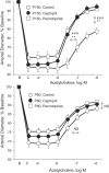
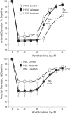
Experiments were performed to determine whether or not acute exposure to elevated pressure would disrupt endothelium-dependent dilatation by increasing local angiotensin II (ANG II) signaling. Vasomotor responses of mouse-isolated carotid arteries were analyzed in a pressure myograph at a control transmural pressure (PTM) of 80 mmHg. Acetylcholine-induced dilatation was reduced by endothelial denudation or by inhibition of nitric oxide synthase (NG-nitro-L-arginine methyl ester, 100 μM). Transient exposure to elevated PTM (150 mmHg, 180 min) inhibited dilatation to acetylcholine but did not affect responses to the nitric oxide donor diethylamine NONOate. Elevated PTM also increased endothelial reactive oxygen species, and the pressure-induced endothelial dysfunction was prevented by the direct antioxidant and NADPH oxidase inhibitor apocynin (100 μM). The increase in endothelial reactive oxygen species in response to elevated PTM was reduced by the ANG II type 1 receptor (AT1R) antagonists losartan (3 μM) or valsartan (1 μM). Indeed, elevated PTM caused marked expression of angiotensinogen, the precursor of ANG II. Inhibition of ANG II signaling, by blocking angiotensin-converting enzyme (1 μM perindoprilat or 10 μM captopril) or blocking AT1Rs prevented the impaired response to acetylcholine in arteries exposed to 150 mmHg but did not affect dilatation to the muscarinic agonist in arteries maintained at 80 mmHg. After the inhibition of ANG II, elevated pressure no longer impaired endothelial dilatation. In arteries treated with perindoprilat to inhibit endogenous formation of the peptide, exogenous ANG II (0.3 μM, 180 min) inhibited dilatation to acetylcholine. Therefore, elevated pressure rapidly impairs endothelium-dependent dilatation by causing ANG expression and enabling ANG II-dependent activation of AT1Rs. These processes may contribute to the pathogenesis of hypertension-induced vascular dysfunction and organ injury.
Keywords: ANG II type 1 receptors; angiotensin II; endothelium; hypertension.
Copyright © 2015 the American Physiological Society.
Figures


Similar articles
-
Local renin-angiotensin system mediates endothelial dilator dysfunction in aging arteries.Am J Physiol Heart Circ Physiol. 2016 Sep 1;311(3):H849-54. doi: 10.1152/ajpheart.00422.2016. Epub 2016 Jul 15. Am J Physiol Heart Circ Physiol. 2016. PMID: 27422988 Free PMC article.
-
Effect of hyperhomocystinemia and hypertension on endothelial function in methylenetetrahydrofolate reductase-deficient mice.Arterioscler Thromb Vasc Biol. 2003 Aug 1;23(8):1352-7. doi: 10.1161/01.ATV.0000083297.47245.DA. Epub 2003 Jun 26. Arterioscler Thromb Vasc Biol. 2003. PMID: 12829522
-
Angiotensin II type 2 receptors contribute to vascular responses in spontaneously hypertensive rats treated with angiotensin II type 1 receptor antagonists.Am J Hypertens. 2005 Apr;18(4 Pt 1):493-9. doi: 10.1016/j.amjhyper.2004.11.007. Am J Hypertens. 2005. PMID: 15831358
-
Vascular-brain signaling in hypertension: role of angiotensin II and nitric oxide.Curr Hypertens Rep. 2007 Jun;9(3):242-7. doi: 10.1007/s11906-007-0043-1. Curr Hypertens Rep. 2007. PMID: 17519132 Review.
-
Interaction of endothelial nitric oxide and angiotensin in the circulation.Pharmacol Rev. 2007 Mar;59(1):54-87. doi: 10.1124/pr.59.1.2. Pharmacol Rev. 2007. PMID: 17329548 Review.
Cited by
-
Local renin-angiotensin system mediates endothelial dilator dysfunction in aging arteries.Am J Physiol Heart Circ Physiol. 2016 Sep 1;311(3):H849-54. doi: 10.1152/ajpheart.00422.2016. Epub 2016 Jul 15. Am J Physiol Heart Circ Physiol. 2016. PMID: 27422988 Free PMC article.
-
Pressure-dependent regulation of Ca2+ signalling in the vascular endothelium.J Physiol. 2015 Dec 15;593(24):5231-53. doi: 10.1113/JP271157. Epub 2015 Dec 7. J Physiol. 2015. PMID: 26507455 Free PMC article.
-
Herbal medicine Oryeongsan (Wulingsan): Cardio-renal effects via modulation of renin-angiotensin system and atrial natriuretic peptide system.Integr Med Res. 2024 Sep;13(3):101066. doi: 10.1016/j.imr.2024.101066. Epub 2024 Jun 22. Integr Med Res. 2024. PMID: 39247397 Free PMC article. Review.
-
Impaired activity of adherens junctions contributes to endothelial dilator dysfunction in ageing rat arteries.J Physiol. 2017 Aug 1;595(15):5143-5158. doi: 10.1113/JP274189. Epub 2017 Jun 30. J Physiol. 2017. PMID: 28561330 Free PMC article.
-
Mechanical activation of angiotensin II type 1 receptors causes actin remodelling and myogenic responsiveness in skeletal muscle arterioles.J Physiol. 2016 Dec 1;594(23):7027-7047. doi: 10.1113/JP272834. Epub 2016 Oct 13. J Physiol. 2016. PMID: 27531064 Free PMC article.
References
-
- Akazawa H, Yasuda N, Komuro I. Mechanisms and functions of agonist-independent activation in the angiotensin II type 1 receptor. Mol Cell Endocrinol 302: 140–147, 2009. - PubMed
-
- Bader M. Tissue renin-angiotensin-aldosterone systems: Targets for pharmacological therapy. Annu Rev Pharmacol Toxicol 50: 439–465, 2010. - PubMed
-
- Bardy N, Merval R, Benessiano J, Samuel JL, Tedgui A. Pressure and angiotensin II synergistically induce aortic fibronectin expression in organ culture model of rabbit aorta. Evidence for a pressure-induced tissue renin-angiotensin system. Circ Res 79: 70–78, 1996. - PubMed
-
- Brasier AR, Li J. Mechanisms for inducible control of angiotensinogen gene transcription. Hypertension 27: 465–475, 1996. - PubMed
Publication types
MeSH terms
Substances
Grants and funding
LinkOut - more resources
Full Text Sources
Other Literature Sources
Medical
Miscellaneous

