Review
. 2015 Mar:45:1-29.
doi: 10.1016/j.preteyeres.2014.11.005. Epub 2014 Dec 5.

Complement activation and choriocapillaris loss in early AMD: implications for pathophysiology and therapy

Affiliations
Review

Complement activation and choriocapillaris loss in early AMD: implications for pathophysiology and therapy

S Scott Whitmore et al. Prog Retin Eye Res. 2015 Mar.

Abstract

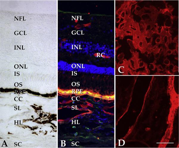
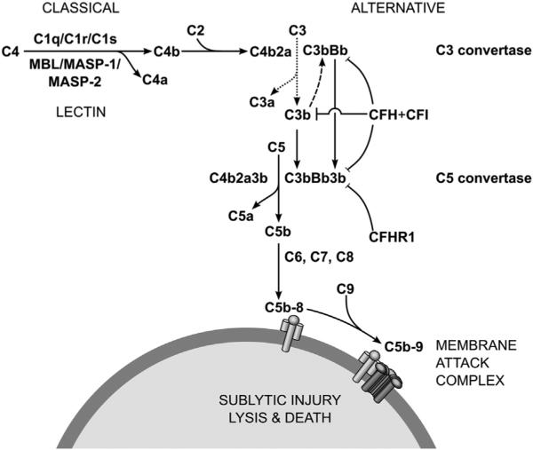
Age-related macular degeneration (AMD) is a common and devastating disease that can result in severe visual dysfunction. Over the last decade, great progress has been made in identifying genetic variants that contribute to AMD, many of which lie in genes involved in the complement cascade. In this review we discuss the significance of complement activation in AMD, particularly with respect to the formation of the membrane attack complex in the aging choriocapillaris. We review the clinical, histological and biochemical data that indicate that vascular loss in the choroid occurs very early in the pathogenesis of AMD, and discuss the potential impact of vascular dropout on the retinal pigment epithelium, Bruch's membrane and the photoreceptor cells. Finally, we present a hypothesis for the pathogenesis of early AMD and consider the implications of this model on the development of new therapies.

Keywords: Age-related macular degeneration; Choriocapillaris; Choroidmacula; Complement system; Endothelial cells; Pathophysiology.


Figures

Fig. 1
Fig. 1
Clinical appearance of drusen. Images of the right fundus of a 78 year old Caucasian female. Left panel, photograph demonstrating numerous macular drusen of various sizes; middle panel, infrared image showing location of optical coherence tomography scan in right panel indicating the sub-RPE location corresponding to drusen.
Fig. 2
Fig. 2
Clinical appearance of geographic atrophy. Fundus of an 88 year old Caucasian female. Left panel shows the right fundus (VA 20/100) with geography atrophy in the center of the macula with surrounding drusen, the largest of which are prominent in the temporal macula. Notice the confluence of the GA in this eye compared to the Right panel where the GA rings the fovea of the left eye (VA 20/40) in nummular fashion with similarly prominent drusen in peripheral macula.
Fig. 3
Fig. 3
Choroidal neovascularization. Images of 87 year old male with neovascular AMD of the right eye (VA 20/150). Upon presentation (left panel) there was prominent blood in the macula that was already dehemoglobinizing in the fovea and inferior macula corresponding to hyperreflective material on OCT (right upper panel). 3 months after anti-VEGF injections the blood has largely resolved (right lower panel) but VA is 20/100 due to atrophy of the outer retina.
Fig. 4
Fig. 4
Structure of the human macula. (A) Brightfield image of the extrafoveal macula; in normal eyes, the neural retina, RPE and choroid exist as an interdependent unit. Light enters the retina from the top of the panel, penetrates the inner retina and excites photoreceptor cell outer segments (OS). Stray photons are absorbed by melanosomes in the RPE and choroidal melanocytes (m). The phototransduction cascade results in arrest of glutamate release from photoreceptor cells and the excitation of neurons in the inner nuclear layer (INL), which in turn excite the ganglion cells (GC) that elaborate axons to the brain. The choroid itself is divided into the choriocapillaris (CC), Sattler's layer (SL), Haller's layer (HL), and the suprachoroidea, adjacent to the sclera (SC). Whereas the choriocapillaris is the vascular supply for the photoreceptor cells and RPE, the inner retina has its own vascular network (retinal capillaries, RC). B, same field as A shown with UEA-I (red), anti-CD45 antibody (green) and the nuclear stain DAPI (blue). Note the labeling of retinal and choroidal endothelial cells. The intense fluorescence at the level of the RPE is due to lipofuscin autofluorescence. C, flat section through the choriocapillaris layer shows dense, anastomosing network of large caliber capillaries (UEA-I, red); D, deeper section through the outer choroid. Scalebar, 50 μm.
Fig. 5
Fig. 5
Accumulation of the membrane attack complex in aging. Anti-MAC antibody is shown in green, UEA-I lectin is depicted in red. A, section of newborn donor without MAC immunoreactivity; B, section of 79 year old donor with extensive MAC immunoreactivity in the choriocapillaris. ELISA analyses show increased MAC in aging and additional increased MAC in AMD (see e.g. Mullins et al., in press AJP). Scalebar, 50 μm.
Fig. 6
Fig. 6
Diagram showing some of the key elements of the complement cascade. In the classical pathway, initiation occurs when C1q binds to IgG or IgM immune complexes. The lectin pathway engages when mannose-binding lectin (MBL) binds pathogen associated carbohydrate moieties, such as mannose or glucose. In the alternative pathway, C3 spontaneously hydrolyzes (dotted line) but negative regulators (e.g., CFH and CFI) bound to the extracellular matrix of host cells prevent amplifying cleavage of C3 by C3b (dashed line). Without binding opportunities for CFH, as on bacterial cell walls, positive feedback occurs ultimately leading to MAC deposition. Figure adapted from Thurman and Holers (2006).
Fig. 7
Fig. 7
Quantitative measurements of subRPE deposits and vascular density in human eyes. Linear regression analysis performed on a series of human donors shows that increasing size and/or number of drusen was negatively correlated with vascular density (filled circles/solid trendline; r2 = 0.22, P < 0.01). In contrast, the height of the RPE (open squares, dashed line) was not correlated with drusen density. Adapted from Mullins et al. (2011b).
Fig. 8
Fig. 8
Ghost vessels in the choriocapillaris are associated with pathologic subRPE deposits in AMD. A, Section of human macula labeled with UEA-I lectin shows two healthy vessels (asterisks) surrounding a UEA-I negative ghost vessel (asterisk). B, Linear regression analysis of numbers of ghost vessels versus the square root of drusen density. Drusen and ghost vessels showed a strong positive correlation (r2 = 0.57, corrected P < 0.001). Adapted from Mullins et al. (2011b).
Fig. 9
Fig. 9
In addition to being more abundant in eyes in association with an increasing volume of drusen, ghost vessels appear to be frequently spatially associated with drusen as well. Section of human macula labeled with UEA-I lectin (red) and anti-CD45 antibody (green). Druse (asterisk) appears centered over choriocapillaris ghost (arrow). See also Sohn and Mullins, 2012, “Age Related Macular Degeneration – The Recent Advances in Basic Research and Clinical Care”, Gui-Shuang Ying, editor.
Fig. 10
Fig. 10
Effects of age and AMD on choroidal thickness. (A) Scatter plot of age (x-axis) and choroidal thickness (y-axis) of 22 eyes without AMD. Among the non-AMD samples included in this study, age showed a statistically significant negative relationship to thickness (slope P < 0.05), although the correlation was weak (r2 = 0.19). (B) Donor maculae were categorized as controls (CTL), early/dry AMD (ARM), neovascular AMD (CNV), or geographic atrophy (GA). Eyes with geographic atrophy showed significant choroidal thinning compared to the other categories (P < 0.05). Diamonds indicate individuals, and filled squares indicate averages for each category. From Sohn et al. (2014b).
Fig. 11
Fig. 11
Ultrastructural appearance of basal laminar deposits in a human eye with geographic atrophy. Note the characteristic banded pattern with reproducible periodicity. These deposits typically form beneath the RPE, between the plasma membrane and basal lamina. Scalebar = 500 nm.
Fig. 12
Fig. 12
The human choriocapillaris has functional binding sites for LDL. Top, While the major site of LDL uptake is in choroidal macrophages, organ cultures of RPE/choroid exposed to fluorescent LDL show uptake in the choriocapillaris (CC), arrows. Bottom, Immunohistochemistry with anti-LDL receptor antibodies show labeling in choriocapillaris endothelial cells (CC, arrows), Dr, druse. See also Tserentsoodol et al. (2006).
Fig. 13
Fig. 13
Choriocapillaris endothelial cells elaborate processes (arrow) that project into Bruch's membrane, where they presumably serve synthetic and/or degradative functions. Loss of choriocapillaris vasculature inevitably results in loss of this function. Figure shows a transmission electron micrograph of the extramacular region of a 77 year old human donor eye. BrM, Bruch's membrane; CCEC, choriocapillaris endothelial cell; CCL, choriocapillaris lumen. Asterisk, choriocapillaris basal lamina; scalebar = 2 μm.
Fig. 14
Fig. 14
Ultrastructural comparison of endothelial cells (EC) of the retinal vasculature (top) and choriocapillaris (bottom) in mouse. In the retina, note the small diameter of the vessel, that narrowly permits the passage of a red blood cell (RBC). The retinal endothelium has relatively abundant cytosol, tight junctions (arrowhead), and a basal lamina. These capillaries are surrounded by pericytes (PC) which also have their own basal lamina and dark-staining glial processes. In contrast, choriocapillary endothelial cells (bottom) show a very small distance between their lumen and the extravascular space, especially on the surface facing the RPE basal infoldings. The inner surface of these cells is also typically fenestrated, giving the appearance of beads on a strong (arrows). A choroidal melanocyte (MC) is also depicted. Scalebar = 2 μm.
Fig. 15
Fig. 15
Schematic diagram depicting the proposed pathological processes involved in the development and progression of AMD. In a young macula (A), photoreceptor cells are supported by the RPE and choriocapillaris. During normal aging (B), MAC (green profiles) accumulates in Bruch's membrane and the choriocapillaris. The continuous exposure to MAC, especially in eyes with high-risk CFH genotypes, leads to loss of choriocapillaris endothelial cells and formation of ghost vessels (C). Impaired clearance, increased hypoxia or other events lead to increased deposition of drusen (D), which can lead to further atrophy (E) or choroidal neovascularization (F).

Similar articles

Cited by

References

    1. Abi-Hanna D, Wakefield D, Watkins S. HLA antigens in ocular tissues. I. In vivo expression in human eyes. Transplantation. 1988;45:610–613. - PubMed
    1. Ablonczy Z, Dahrouj M, Marneros AG. Progressive dysfunction of the retinal pigment epithelium and retina due to increased VEGF-A levels. FASEB J. 2014;28:2369–2379. http://dx.doi.org/10.1096/fj.13-248021. - DOI - PMC - PubMed
    1. Age-Related Eye Disease Study 2 Research Group Lutein + zeaxanthin and omega-3 fatty acids for age-related macular degeneration: the Age-Related Eye Disease Study 2 (AREDS2) randomized clinical trial. JAMA. 2013;309:2005–2015. http://dx.doi.org/10.1001/jama.2013.4997. - DOI - PubMed
    1. Age-Related Eye Disease Study Research Group A randomized, placebo-controlled, clinical trial of high-dose supplementation with vitamins C and E, beta carotene, and zinc for age-related macular degeneration and vision loss: AREDS report no. 8. Arch. Ophthalmol. 2001;119:1417–1436. - PMC - PubMed
    1. Allende A, Madigan MC, Provis JM. Endothelial cell proliferation in the choriocapillaris during human retinal differentiation. Br. J. Ophthalmol. 2006;90:1046–1051. http://dx.doi.org/10.1136/bjo.2006.092080. - DOI - PMC - PubMed

Publication types

MeSH terms

Substances