

Suppression of the PI3K pathway in vivo reduces cystitis-induced bladder hypertrophy and restores bladder capacity examined by magnetic resonance imaging
- PMID: 25486122
- PMCID: PMC4259345
- DOI: 10.1371/journal.pone.0114536
Suppression of the PI3K pathway in vivo reduces cystitis-induced bladder hypertrophy and restores bladder capacity examined by magnetic resonance imaging
Abstract
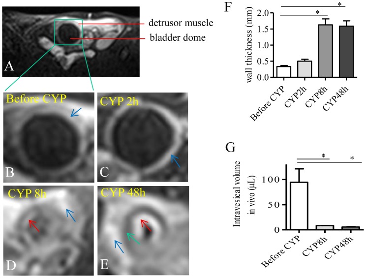
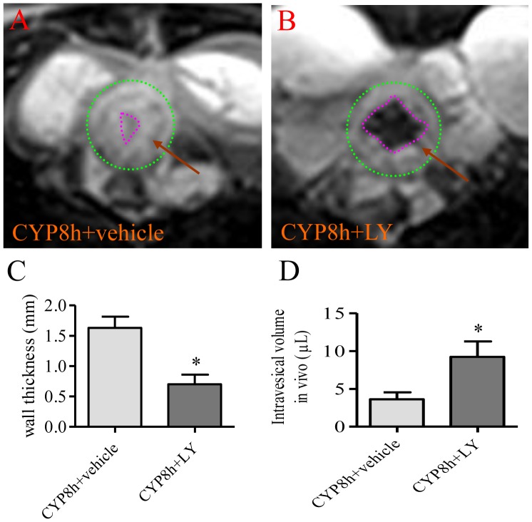
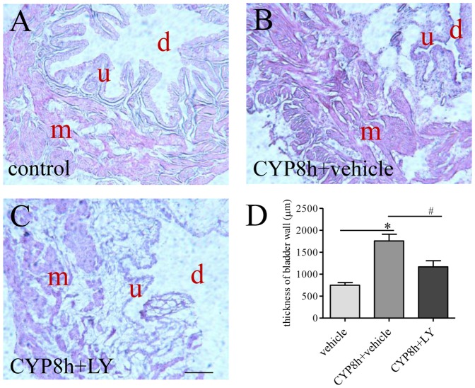
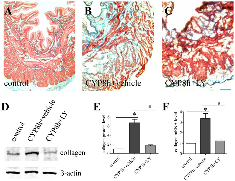
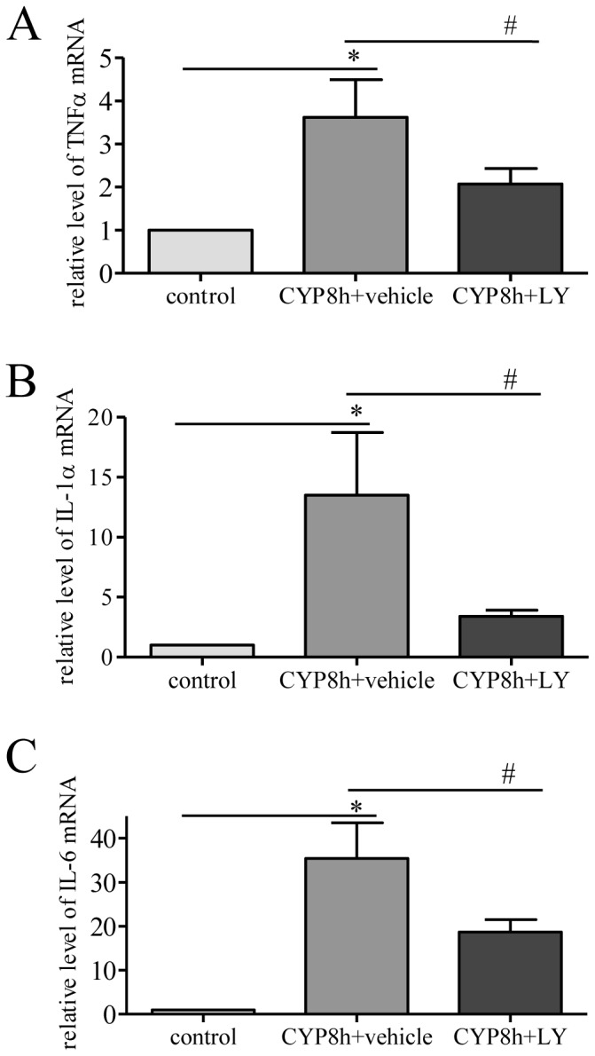
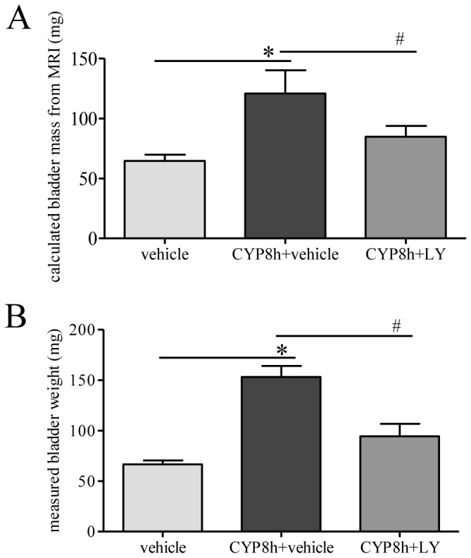
This study utilized magnetic resonance imaging (MRI) to monitor the real-time status of the urinary bladder in normal and diseased states following cyclophosphamide (CYP)-induced cystitis, and also examined the role of the phosphoinositide 3-kinase (PI3K) pathway in the regulation of urinary bladder hypertrophy in vivo. Our results showed that under MRI visualization the urinary bladder wall was significantly thickened at 8 h and 48 h post CYP injection. The intravesical volume of the urinary bladder was also markedly reduced. Treatment of the cystitis animals with a specific PI3K inhibitor LY294002 reduced cystitis-induced bladder wall thickening and enlarged the intravesical volumes. To confirm the MRI results, we performed H&E stain postmortem and examined the levels of type I collagen by real-time PCR and western blot. Inhibition of the PI3K in vivo reduced the levels of type I collagen mRNA and protein in the urinary bladder ultimately attenuating cystitis-induced bladder hypertrophy. The bladder mass calculated according to MRI data was consistent to the bladder weight measured ex vivo under each drug treatment. MRI results also showed that the urinary bladder from animals with cystitis demonstrated high magnetic signal intensity indicating considerable inflammation of the urinary bladder when compared to normal animals. This was confirmed by examination of the pro-inflammatory factors showing that interleukin (IL)-1α, IL-6 and tumor necrosis factor (TNF)α levels in the urinary bladder were increased with cystitis. Our results suggest that MRI can be a useful technique in tracing bladder anatomy and examining bladder hypertrophy in vivo during disease development and the PI3K pathway has a critical role in regulating bladder hypertrophy during cystitis.
Conflict of interest statement
Figures
Similar articles
-
Endogenous nerve growth factor regulates collagen expression and bladder hypertrophy through Akt and MAPK pathways during cystitis.J Biol Chem. 2010 Feb 5;285(6):4206-4212. doi: 10.1074/jbc.M109.040444. Epub 2009 Dec 7. J Biol Chem. 2010. PMID: 19996110 Free PMC article.
-
Urinary bladder organ hypertrophy is partially regulated by Akt1-mediated protein synthesis pathway.Life Sci. 2018 May 15;201:63-71. doi: 10.1016/j.lfs.2018.03.041. Epub 2018 Mar 21. Life Sci. 2018. PMID: 29572181 Free PMC article.
-
Blocking mammalian target of rapamycin alleviates bladder hyperactivity and pain in rats with cystitis.Mol Pain. 2016 Oct 25;12:1744806916668868. doi: 10.1177/1744806916668868. Print 2016. Mol Pain. 2016. PMID: 27780878 Free PMC article.
-
Inhibition of the PI3K/AKT pathway reduces tumor necrosis factor-alpha production in the cellular response to wear particles in vitro.Artif Organs. 2013 Mar;37(3):298-307. doi: 10.1111/j.1525-1594.2012.01568.x. Epub 2013 Jan 18. Artif Organs. 2013. PMID: 23330804
-
Inhibition of NMDAR reduces bladder hypertrophy and improves bladder function in cyclophosphamide induced cystitis.J Urol. 2015 May;193(5):1676-83. doi: 10.1016/j.juro.2014.12.092. Epub 2015 Jan 6. J Urol. 2015. PMID: 25572034 Free PMC article.
Cited by
-
Organoid modeling meets cancers of female reproductive tract.Cell Death Discov. 2024 Sep 27;10(1):410. doi: 10.1038/s41420-024-02186-x. Cell Death Discov. 2024. PMID: 39333482 Free PMC article. Review.
-
A review of recent advances on single use of antibody-drug conjugates or combination with tumor immunology therapy for gynecologic cancer.Front Pharmacol. 2022 Dec 22;13:1093666. doi: 10.3389/fphar.2022.1093666. eCollection 2022. Front Pharmacol. 2022. PMID: 36618922 Free PMC article. Review.
-
PROTACs in Epigenetic Cancer Therapy: Current Status and Future Opportunities.Molecules. 2023 Jan 26;28(3):1217. doi: 10.3390/molecules28031217. Molecules. 2023. PMID: 36770884 Free PMC article. Review.
-
Detrusor contractility to parasympathetic mediators is differentially altered in the compensated and decompensated states of diabetic bladder dysfunction.Am J Physiol Renal Physiol. 2019 Aug 1;317(2):F388-F398. doi: 10.1152/ajprenal.00178.2019. Epub 2019 May 29. Am J Physiol Renal Physiol. 2019. PMID: 31141399 Free PMC article.
-
Divergent histopathological and molecular patterns in chemically induced interstitial cystitis/bladder pain syndrome rat models.Sci Rep. 2024 Jul 12;14(1):16134. doi: 10.1038/s41598-024-67162-y. Sci Rep. 2024. PMID: 38997336 Free PMC article.
References
-
- Metcalfe PD, Wang JF, Jiao HY, Huang Y, Hori K, et al. (2010) Bladder outlet obstruction: progression from inflammation to fibrosis. Bju International 106:1686–1694. - PubMed
-
- Tyagi P, Barclay D, Zamora R, Yoshimura N, Peters K, et al. (2010) Urine cytokines suggest an inflammatory response in the overactive bladder: a pilot study. Int Urol Nephrol 42:629–635. - PubMed
-
- Jerde TJ, Bjorling DE, Steinberg H, Warner T, Saban R (2000) Determination of mouse bladder inflammatory response to E. coli lipopolysaccharide. Urol Res 28:269–273. - PubMed
-
- Malley SE, Vizzard MA (2002) Changes in urinary bladder cytokine mRNA and protein after cyclophosphamide-induced cystitis. Physiol Genomics 9:5–13. - PubMed
Publication types
MeSH terms
Substances
Grants and funding
LinkOut - more resources
Full Text Sources
Other Literature Sources
Medical
