. 2014;13(18):2859-68.
doi: 10.4161/15384101.2014.946830.

Lack of centrioles and primary cilia in STIL(-/-) mouse embryos

Affiliations

Lack of centrioles and primary cilia in STIL(-/-) mouse embryos

Ahuvit David et al. Cell Cycle. 2014.

Abstract

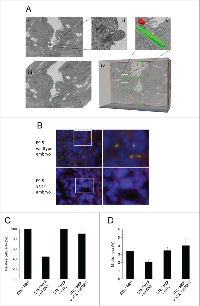
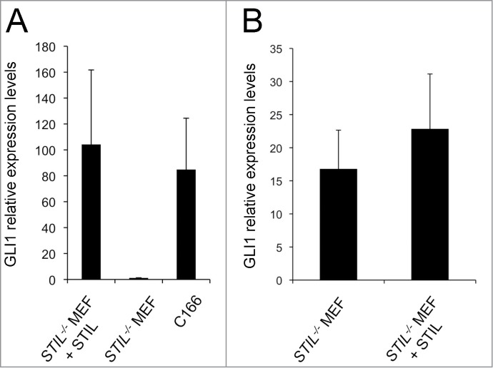
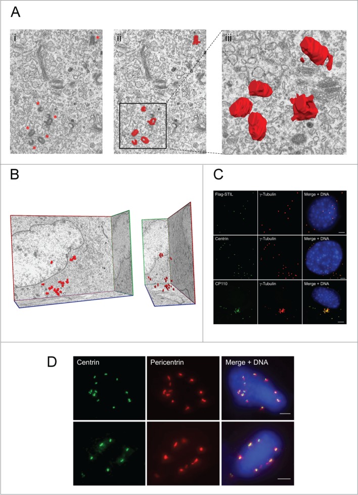
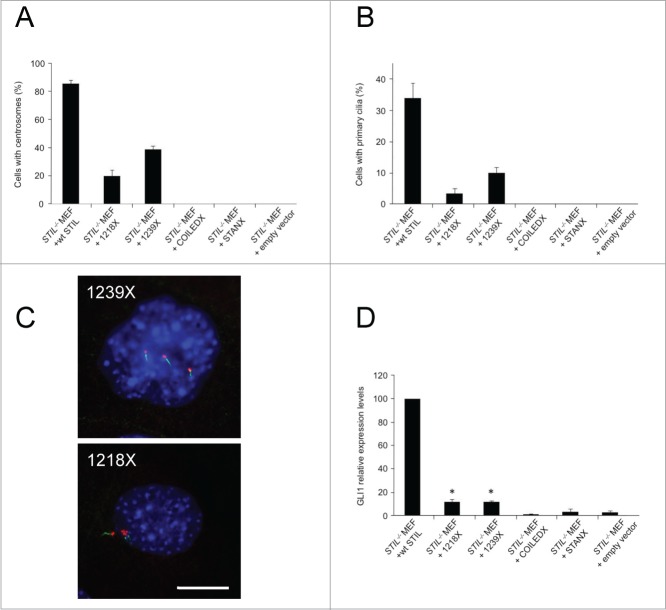
Although most animal cells contain centrosomes, consisting of a pair of centrioles, their precise contribution to cell division and embryonic development is unclear. Genetic ablation of STIL, an essential component of the centriole replication machinery in mammalian cells, causes embryonic lethality in mice around mid gestation associated with defective Hedgehog signaling. Here, we describe, by focused ion beam scanning electron microscopy, that STIL(-/-) mouse embryos do not contain centrioles or primary cilia, suggesting that these organelles are not essential for mammalian development until mid gestation. We further show that the lack of primary cilia explains the absence of Hedgehog signaling in STIL(-/-) cells. Exogenous re-expression of STIL or STIL microcephaly mutants compatible with human survival, induced non-templated, de novo generation of centrioles in STIL(-/-) cells. Thus, while the abscence of centrioles is compatible with mammalian gastrulation, lack of centrioles and primary cilia impairs Hedgehog signaling and further embryonic development.

Keywords: CDK6, cyclin-dependent kinase 6; CEP, centrosomal protein; COILEDX, coiled-coil domain deletion; E, embryonic day; FIB/SEM, focused ion beam scanning electron microscopy; MCPH, autosomal recessive primary microcephaly; MEFs, mouse embryonic fibroblasts; MTOC, microtubule organizing center; PLK4, polo kinase 4; SHH, sonic hedgehog; STAN, STIL/ANA2; STANX, STAN domain deletion; STIL; STIL, SCL/TAL1 interrupting locus; centriole; centrosome; electron microscopy; embryo; microcephaly; nm, nanometer; siRNA, small interfering RNA.


Figures

Figure 1.
Figure 1.
STIL−/− mouse embryos lack centrioles and primary cilia. (A) Focused ion beam scanning electron microscopy of an E8.5 wildtype mouse embryo. (i) Single image captured by FIB/SEM showing centrioles and a primary cilium (magnification 5700 x, scale bar 1 μm) which are depicted enlarged in (ii). (iii) Stack of serial sections with centrioles and cilia manually segmented and centrioles marked in red and cilia marked in green. Only 10 sections are shown out of the 361 serial images that compose the complete stack. (iv) Three-dimensional reconstruction of the image stack shown in (iii) after manual segmentation of centrioles and primary cilia in each section of the stack. As a representative example a 3-dimensionally reconstructed centriole/cilium pair is shown enlarged in (v). (B) Wildtype and STIL−/− E9.5 mouse embryos stained for acetylated tubulin (green), pericentrin (red) and DNA (DAPI, blue). Scale bar, 23 μm. Pictures on the right represent fold6- magnifications of the framed regions in the pictures on the left. (C) Proliferation of STIL−/− MEFs and STIL−/− MEFs reconstituted with Flag-STIL, 96 hrs after transfection with pericentrin-specific siRNA. Cellularity has been calculated as the ratio of living cells after transfection with pericentrin-specific siRNA versus a non-specific control siRNA. Error bars represent mean ± SE from 3 independent experiments. (D) Mitotic index of STIL−/− MEFs and STIL−/− MEFs reconstituted with Flag-STIL, 72 hrs after transfection with pericentrin-specific siRNA. The frequency of mitotic cells has been determined by counting at least 300 cells after transfection with pericentrin-specific siRNA vs. a non-specific control siRNA. Error bars represent mean ± SE from 3 independent experiments.
Figure 2.
Figure 2.
Primary cilia mediate STIL dependence of SHH signaling. (A) mRNA expression of GLI1 in STIL−/− MEFs and STIL−/− MEFs reconstituted with Flag-STIL 3 d after exposure to SHH-conditioned medium. C166 endothelial cells were used as a positive control. (B) mRNA expression of GLI1 in STIL−/− MEFs and STIL−/− MEFs reconstituted with Flag-STIL which have been transiently transfected with a vector expressing constitutively active GLI2 (GLI2ΔN); p=0.71. Error bars represent mean ± SE from 4 (A) and 3 (B) independent experiments. Differences in Y axis scaling in (A) and (B) are due to a lack of exposure of cells to SHH-conditioned medium in (B).
Figure 3.
Figure 3.
Exogenous STIL causes de novo formation of supernumerary centrioles in STIL−/− MEFs. (A) Single image captured by focused ion beam scanning electron microscopy (FIB/SEM) from STIL−/− MEFs reconstituted with Flag-STIL (i) before (centrioles marked with asterics) and (ii) after manual segmentation of centrioles (red, magnification 5700 x). A 3-dimensional reconstruction of an image stack consisting out of 16 single pictures showing a group of centrioles is depicted enlarged in (iii). (B) Front (left) and side view (right) of a 3-dimensional reconstruction of 301 serial sections from STIL−/− MEFs reconstituted with Flag-STIL after manual segmentation of centrioles in each section of the stack; Magnification 5700×. (C, D) STIL−/− MEFs reconstituted with Flag-STIL by lentiviral transduction were co-immunostained with antibodies to Flag (green) and γ-tubulin (red, upper panel), centrin (green) and γ-tubulin (red, middle panel), or CP110 (green) and γ-tubulin (red, lower panel) (C) as well as centrin (green) and pericentrin (red) (D) and analyzed by immunofluorescence microscopy. DNA was stained with DAPI (blue). Scale bars: 5 μm.
Figure 4.
Figure 4.
Microcephaly mutant versions of STIL partially rescue de novo centrosome formation, primary cilia formation and SHH signaling in STIL−/− MEFs. STIL mutant versions were exogenously expressed in STIL−/− MEFs and subsequently analyzed for centrosome and primary cilium formation as well as SHH signaling as a readout for cilia function. STIL−/− MEFs reconstituted with wildtype Flag-STIL (wt STIL), Flag-STIL-1218× (1218×), Flag-STIL-1239× (1239×), Flag-STIL-COILEDX (COILEDX) and Flag-STIL-STANX (STANX) by lentiviral transduction were immunostained with antibodies to γ-tubulin and poly-glutamylated tubulin. Subsequently, cells containing centrosomes (A) and primary cilia (B) were counted. Error bars represent mean ± SE from 3 independent experiments. (C) Representative examples of STIL−/− MEFs reconstituted with Flag-STIL-1239× (1239×; upper panel) or Flag-STIL-1218× (1218×; lower panel) by lentiviral transduction, co-immunostained with antibodies to γ-tubulin (red) and poly-glutamylated tubulin (green). DNA was stained with DAPI (blue). Scale bar: 14 μm. (D) mRNA expression levels of GLI1 in STIL−/− MEFs reconstituted with Flag-STIL-1218× (1218×), Flag-STIL-1239× (1239×), Flag-STIL-COILEDX (COILEDX) and Flag-STIL-STANX (STANX) by lentiviral transduction 3 d after exposure to SHH-conditioned medium, normalized to Gli1 levels in STIL−/− MEFs reconstituted with wildtype Flag-STIL (wt STIL). *, Gli1 levels in Flag-STIL-1218× (1218×) and Flag-STIL-1239× (1239×) reconstituted STIL−/− MEFs are statistically significantly different from levels of Gli1 in STIL−/− MEFs reconstituted with empty vector. Error bars represent mean ± SE from 3 independent experiments.

Comment in

  • Centrioles in the mouse: cilia and beyond.
    Bazzi H, Anderson KV. Bazzi H, et al. Cell Cycle. 2014;13(18):2809. doi: 10.4161/15384101.2014.954450. Cell Cycle. 2014. PMID: 25486466 Free PMC article. No abstract available.

References

    1. Azimzadeh J, Marshall WF. Building the centriole. Curr Biol 2010; 20:R816-25; PMID:20869612; http://dx.doi.org/ 10.1016/j.cub.2010.08.010 - DOI - PMC - PubMed
    1. Nigg EA, Stearns T. The centrosome cycle: centriole biogenesis, duplication and inherent asymmetries. Nat Cell Biol 2011; 13:1154-60; PMID:21968988; http://dx.doi.org/ 10.1038/ncb2345 - DOI - PMC - PubMed
    1. Dammermann A, Müller-Reichert T, Pelletier L, Habermann B, Desai A, Oegema K. Centriole assembly requires both centriolar and pericentriolar material proteins. Dev Cell 2004; 7:815-29; PMID:15572125; http://dx.doi.org/ 10.1016/j.devcel.2004.10.015 - DOI - PubMed
    1. Delattre M, Leidel S, Wani K, Baumer K, Bamat J, Schnabel H, Feichtinger R, Schnabel R, Gönczy P. Centriolar SAS-5 is required for centrosome duplication in C. elegans. Nat Cell Biol 2004; 6:656-64; PMID:15232593; http://dx.doi.org/ 10.1038/ncb1146 - DOI - PubMed
    1. Leidel S, Gönczy P. SAS-4 is essential for centrosome duplication in C elegans and is recruited to daughter centrioles once per cell cycle. Dev Cell 2003; 4:431-9; PMID:12636923; http://dx.doi.org/ 10.1016/S1534-5807(03)00062-5 - DOI - PubMed

Publication types

MeSH terms

Substances