

γH2AX foci on apparently intact mitotic chromosomes: not signatures of misrejoining events but signals of unresolved DNA damage
- PMID: 25486563
- PMCID: PMC4614418
- DOI: 10.4161/15384101.2014.947786
γH2AX foci on apparently intact mitotic chromosomes: not signatures of misrejoining events but signals of unresolved DNA damage
Abstract
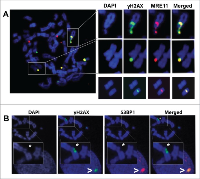
The presence of γH2AX foci on apparently intact mitotic chromosomes is controversial because they challenge the assumed relationship between γH2AX foci and DNA double-strand breaks (DSBs). In this work, we show that after irradiation during interphase, a variety of γH2AX foci are scored in mitotic cells. Surprisingly, approximately 80% of the γH2AX foci spread over apparently undamaged chromatin at Terminal or Interstitial positions and they can display variable sizes, thus being classified as Small, Medium and Big foci. Chromosome and chromatid breaks that reach mitosis are spotted with Big (60%) and Medium (30%) Terminal γH2AX foci, but very rarely are they signaled with Small γH2AX foci. To evaluate if Interstitial γH2AX foci might be signatures of misrejoining, an mFISH analysis was performed on the same slides. The results show that Interstitial γH2AX foci lying on apparently intact chromatin do not mark sites of misrejoining, and that misrejoined events were never signaled by a γH2AX foci during mitosis. Finally, when analyzing the presence of other DNA-damage response (DDR) factors we found that all γH2AX foci-regardless their coincidence with a visible break-always colocalized with MRE11, but not with 53BP1. This pattern suggests that these γH2AX foci may be hallmarks of both microscopically visible and invisible DNA damage, in which an active, although incomplete or halted DDR is taking place.
Keywords: AU, arbitrary units; DSB, DNA double-strand break; FI, fluorescence intensity; FISH, fluorescence in situ hybridization; IRIF, ionizing radiation induced foci; M phase; MRE11; MRN complex, MRE11-Rad50-Nbs1 complex; SD, standard deviation; TIF, telomere-dysfunction induced foci; illegitimate joining; mFISH, multicolor fluorescence in situ hybridization; misrepair; residual DSBs; telomeric foci; γH2AX.
Figures


Comment in
-
Spots, damn'd spots and γH2AX foci.Cell Cycle. 2015;14(7):947. doi: 10.1080/15384101.2015.1006554. Cell Cycle. 2015. PMID: 25803282 Free PMC article. No abstract available.
Similar articles
-
Breaks invisible to the DNA damage response machinery accumulate in ATM-deficient cells.Genes Chromosomes Cancer. 2009 Sep;48(9):745-59. doi: 10.1002/gcc.20679. Genes Chromosomes Cancer. 2009. PMID: 19455703
-
Identification of DNA double strand breaks at chromosome boundaries along the track of particle irradiation.Genes Chromosomes Cancer. 2016 Aug;55(8):650-60. doi: 10.1002/gcc.22367. Epub 2016 May 16. Genes Chromosomes Cancer. 2016. PMID: 27113385
-
Biodosimetry of Low Dose Ionizing Radiation Using DNA Repair Foci in Human Lymphocytes.Genes (Basel). 2020 Jan 4;11(1):58. doi: 10.3390/genes11010058. Genes (Basel). 2020. PMID: 31947954 Free PMC article.
-
DNA damage foci: Meaning and significance.Environ Mol Mutagen. 2015 Jul;56(6):491-504. doi: 10.1002/em.21944. Epub 2015 Mar 12. Environ Mol Mutagen. 2015. PMID: 25773265 Review.
-
Radiation-induced DNA repair foci: spatio-temporal aspects of formation, application for assessment of radiosensitivity and biological dosimetry.Mutat Res. 2010 Apr-Jun;704(1-3):132-41. doi: 10.1016/j.mrrev.2010.01.011. Epub 2010 Jan 22. Mutat Res. 2010. PMID: 20096808 Review.
Cited by
-
Spots, damn'd spots and γH2AX foci.Cell Cycle. 2015;14(7):947. doi: 10.1080/15384101.2015.1006554. Cell Cycle. 2015. PMID: 25803282 Free PMC article. No abstract available.
-
Incorporation of 53BP1 into phase-separated bodies in cancer cells during aberrant mitosis.J Cell Sci. 2023 Jan 1;136(1):jcs260027. doi: 10.1242/jcs.260027. Epub 2023 Jan 6. J Cell Sci. 2023. PMID: 36606487 Free PMC article.
-
Development of a multiparameter flow cytometric assay as a potential biomarker for homologous recombination deficiency in women with high-grade serous ovarian cancer.J Transl Med. 2015 Jul 22;13:239. doi: 10.1186/s12967-015-0604-z. J Transl Med. 2015. PMID: 26198537 Free PMC article.
-
Histone modifications and the DNA double-strand break response.Cell Cycle. 2018;17(21-22):2399-2410. doi: 10.1080/15384101.2018.1542899. Epub 2018 Nov 14. Cell Cycle. 2018. PMID: 30394812 Free PMC article. Review.
-
DNA double-strand break repair is impaired in presenescent Syrian hamster fibroblasts.BMC Mol Biol. 2015 Oct 12;16:18. doi: 10.1186/s12867-015-0046-4. BMC Mol Biol. 2015. PMID: 26458748 Free PMC article.
References
-
- Burma S, Chen BP, Murphy M, Kurimasa A, Chen DJ. ATM Phosphorylates Histone H2AX in response to DNA double-strand breaks. J Biol Chem 2001; 276:42462-7; PMID:11571274; http://dx.doi.org/10.1074/jbc.C100466200 - DOI - PubMed
-
- Stucki M, Clapperton JA, Mohammad D, Yaffe MB, Smerdon SJ, Jackson S. MDC1 directly binds phosphorylated histone H2AX to regulate cellular responses to DNA double-strand breaks. Cell 2005; 123:1213-26; PMID:16377563; http://dx.doi.org/10.1016/j.cell.2005.09.038 - DOI - PubMed
-
- Rogakou EP, Pilch DR, Orr AH, Ivanova VS, Bonner WM. DNA double-stranded breaks induce histone H2AX phosphorylation on Serine 139. J Biol Chem 1998; 273:5858-68; PMID:9488723; http://dx.doi.org/10.1074/jbc.273.10.5858 - DOI - PubMed
-
- Cleaver JE. γH2Ax: Biomarker of damage or functional participant in DNA repair “All that Glitters is not Gold!” Photochem Photobiol 2011; 87:1230-9; PMID:21883247; http://dx.doi.org/10.1111/j.1751-1097.2011.00995.x - DOI - PubMed
-
- Chen HT, Bhandoola A, Difilippantonio MJ, Zhu J, Brown MJ, Tai X, Rogakou EP, Brotz TM, Bonner WM, Ried T, et al. . Response to RAG-mediated V(D)J cleavage by NBS1 and g-H2AX. Science 2000; 290:1962-4; PMID:11110662; http://dx.doi.org/10.1126/science.290.5498.1962 - DOI - PMC - PubMed
Publication types
MeSH terms
Substances
LinkOut - more resources
Full Text Sources
Other Literature Sources
Research Materials
Miscellaneous