. 2015 Feb;83(2):792-801.
doi: 10.1128/IAI.02444-14. Epub 2014 Dec 8.

Impact of resolvin E1 on murine neutrophil phagocytosis in type 2 diabetes

Affiliations

Impact of resolvin E1 on murine neutrophil phagocytosis in type 2 diabetes

Bruno S Herrera et al. Infect Immun. 2015 Feb.

Abstract

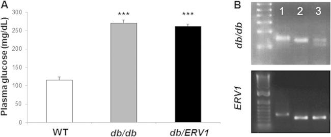
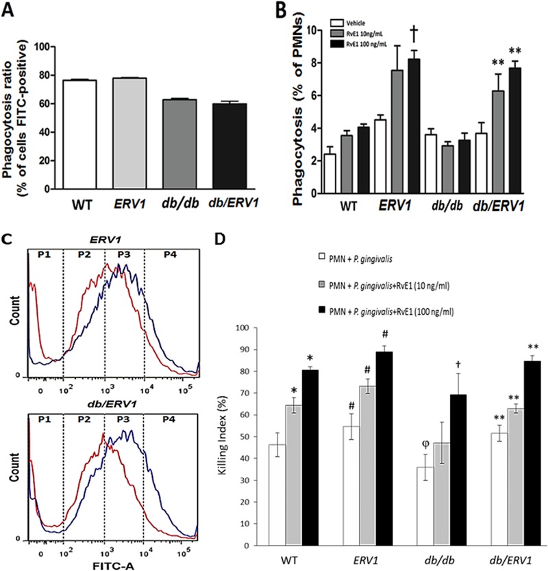
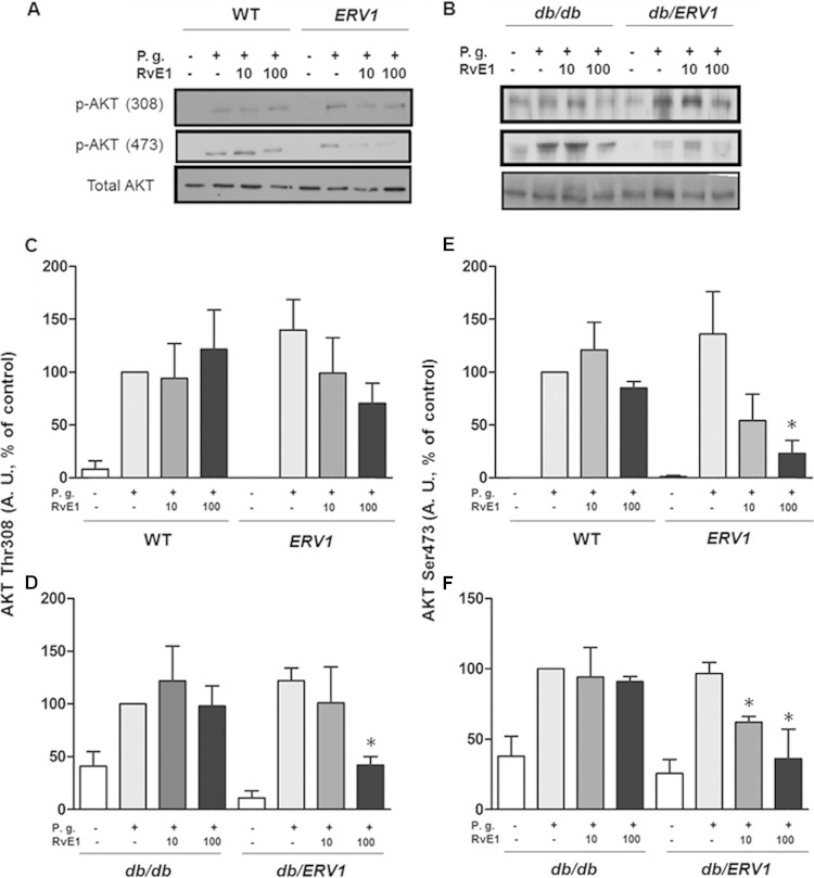
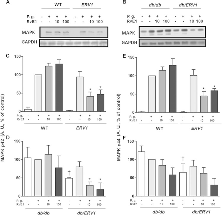
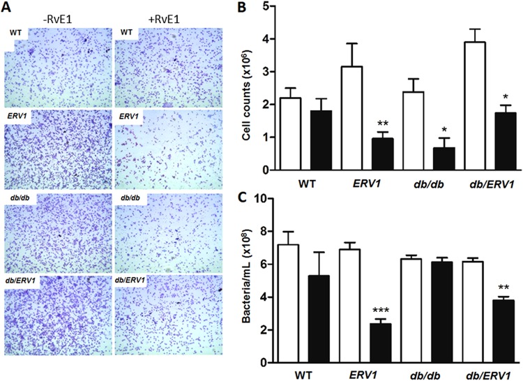
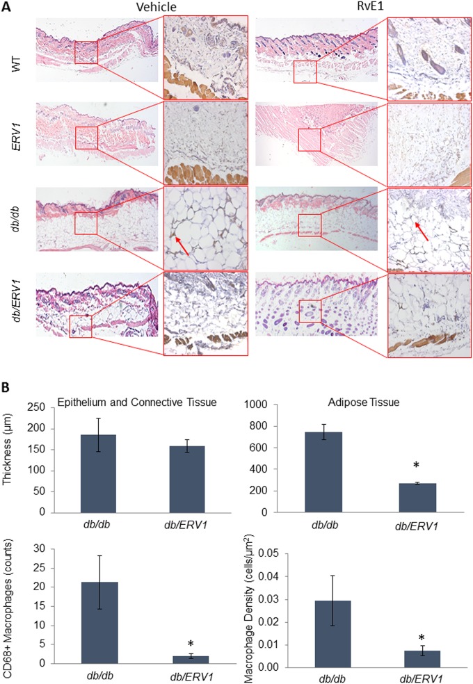
Diabetic complications involve inflammation-mediated microvascular and macrovascular damage, disruption of lipid metabolism, glycosylation of proteins, and abnormalities of neutrophil-mediated events. Resolution of inflamed tissues to health and homeostasis is an active process mediated by endogenous lipid agonists, including lipoxins and resolvins. This proresolution system appears to be compromised in type 2 diabetes (T2D). The goal of this study was to investigate unresolved inflammation in T2D. Wild-type (WT) and genetically engineered mice, including T2D mice (db/db), transgenic mice overexpressing the human resolvin E1 (RvE1) receptor (ERV1), and a newly bred strain of db/ERV1 mice, were used to determine the impact of RvE1 on the phagocytosis of Porphyromonas gingivalis in T2D. Neutrophils were isolated and incubated with fluorescein isothiocyanate-labeled P. gingivalis, and phagocytosis was measured in a fluorochrome-based assay by flow cytometry. Mitogen-activated protein kinase (MAPK) (p42 and p44) and Akt (Thr308 and Ser473) phosphorylation was analyzed by Western blotting. The mouse dorsal air pouch model was used to evaluate the in vivo impact of RvE1. Results revealed that RvE1 increased the neutrophil phagocytosis of P. gingivalis in WT animals but had no impact in db/db animals. In ERV1-transgenic and ERV1-transgenic diabetic mice, phagocytosis was significantly increased. RvE1 decreased Akt and MAPK phosphorylation in the transgenic animals. In vivo dorsal air pouch studies revealed that RvE1 decreases neutrophil influx into the pouch and increases neutrophil phagocytosis of P. gingivalis in the transgenic animals; cutaneous fat deposition was reduced, as was macrophage infiltration. The results suggest that RvE1 rescues impaired neutrophil phagocytosis in obese T2D mice overexpressing ERV1.


Figures

FIG 1
FIG 1
Mouse genotypes and phenotypes. (A) Baseline plasma glucose levels (mg/dl) show that db/db mouse plasma glucose levels are out of the normal range and that overexpression of ERV1 does not impact glycemic control. ***: db/db, P < 0.001 compared to WT; db/ERV1, P < 0.0001 compared to WT (n = 6). (B) Genotyping results. Lanes: 1, WT, 2, ERV1 transgenic (ERV1), 3, db/db-ERV1 transgenic (db/ERV1).
FIG 2
FIG 2
RvE1 increases P. gingivalis phagocytosis by neutrophils overexpressing ERV1. P. gingivalis phagocytosis by neutrophils was quantified by flow cytometry as the percentage of neutrophils containing bacteria (FITC positive) and the number of bacteria per neutrophil. (A) Percentages of FITC-positive neutrophils. Diabetic mice exhibit significantly fewer phagocytizing neutrophils (n = 9). (B) Percentages of FITC-positive neutrophils with more than 10,000 events (†, P < 0.05 compared to ERV1 with vehicle; **, P <0.01 compared to db/ERV1 with vehicle. (C) Distribution of bacteria per neutrophil by quartile. P4 = >10,000 events per cell. Note the shift to more bacteria per neutrophil with RvE1 treatment (red, no treatment; blue, 10 ng/ml RvE1; n = 8 nondiabetic and 6 diabetic mice). (D) Neutrophil bactericidal activity is enhanced by RvE1. With a CFU killing assay for P. gingivalis, the actions of two doses of RvE1 (10 and 100 ng/ml) were assessed in WT, ERV1, db/db, and db/ERV1 mice. Differences within and between groups were determined by ANOVA with Bonferroni corrections for multiple comparisons. Between-group comparisons revealed that bactericidal activity was lower in db/db mice than in WT animals (or all other strains) (φ, P < 0.05). Treatment of db/db neutrophils with RvE1 restored the killing response to the level of untreated WT mice at 10 ng/ml, and the effect was significantly greater at 100 ng/ml (†, P < 0.05). Overexpression of ERV1 in diabetic db/ERV1 mice completely normalized the killing response (**, P < 0.05). It is interesting that the db/db killing response was significantly lower than that of the WT at both doses of RvE1 and that this response deficiency was eliminated by the overexpression of ERV1. *, P < 0.05 compared to PMNs plus P. gingivalis; #, P <0.05 compared to the WT under all conditions (n = 4).
FIG 3
FIG 3
P. gingivalis (P.g.) induces phosphorylation of Akt that is reversed by RvE1. (A, B) Representative Western blot images quantified in panels C to F. (C, D) Densitometric quantification of Akt phosphorylation at threonine 308. (E, F) Densitometric quantification of Akt phosphorylation at serine 473. A. U., arbitrary units; *, P < 0.05 compared to vehicle control (n = 4).
FIG 4
FIG 4
P. gingivalis (P.g.) induces phosphorylation of MAPK that is reversed by RvE1. (A, B) Representative Western blot images quantified in panels C to F. (C, D) Densitometric quantification of ERK phosphorylation (p42). (E, F) Densitometric quantification of MAPK phosphorylation (p44). A. U., arbitrary units; *, P < 0.05 compared to ERV1 plus P. gingivalis plus vehicle; †, P < 0.05 compared to db/db without P. gingivalis plus vehicle; *, P < 0.05 compared to db/db plus P. gingivalis plus vehicle; (n = 4).
FIG 5
FIG 5
RvE1 decreases cell influx and increases P. gingivalis clearance. Neutrophils and bacteria were harvested from the air pouch after 4 h by lavage. (A) Representative images of the cellular infiltrate (Wright-Giemsa, <95% neutrophils) with or without RvE1 treatment in all four strains of mice. (B) Total number of neutrophils in the pouch after 4 h (n = 4). (C) Bacteria in the air pouch after 4 h. Bacterial numbers were quantified densitometrically (OD600) after 24 h of growth in Wilkins-Chalgren broth and compared to a standard curve. Bars: black, RvE1; white, control. *, P < 0.05; **, P < 0.01; ***, P < 0.001 (n = 6).
FIG 6
FIG 6
ERV1 overexpression reduces cutaneous fat accumulation and macrophage accumulation in adipose tissue of diabetic mice. Dorsal air pouches were raised on the backs of all strains of mice. Tissues were stained with hematoxylin and eosin. Tissue macrophages were identified by immunohistochemical detection of CD68 expression. (A) Epithelium, connective tissue, and adipose tissue distribution is demonstrated in each group (ERV1, transgenic mice overexpressing ERV1; db/db, diabetic mice; db/ERV1, diabetic mice overexpressing ERV1) with or without RvE1 treatment. Larger panels demonstrate hematoxylin-and-eosin staining (magnification, ×2.5). CD68+ macrophages in the adipose tissue layer are shown in inserts (arrows; magnification, ×20). Histological analysis of the air pouch lining reveals significant cutaneous adipose tissue accumulation with significant accumulation of CD68+ macrophages in db/db mice that is not evident in diabetic ERV1-overexpressing (db/ERV1) mice. (B) Epithelium, connective tissue, and adipose tissue thicknesses were measured, CD68+ macrophages were counted, and macrophage density in adipose tissue was calculated. Quantification of tissue thickness and macrophage numbers in the adipose tissue reveals significant reductions of both in db/ERV1 animals. *, P < 0.01 [ANOVA] compared to the db/db group.

References

    1. Eke PI, Dye BA, Wei L, Thornton-Evans GO, Genco RJ, Beck J, Douglass G, Page R. 2012. Prevalence of periodontitis in adults in the United States: 2009 and 2010. J Dent Res 91:914–920. doi:10.1177/0022034512457373. - DOI - PubMed
    1. Libby P. 2002. Atherosclerosis: the new view. Sci Am 286:46–55. doi:10.1038/scientificamerican0502-46. - DOI - PubMed
    1. Dandona P, Aljada A, Chaudhuri A, Mohanty P, Garg R. 2005. Metabolic syndrome: a comprehensive perspective based on interactions between obesity, diabetes, and inflammation. Circulation 111:1448–1454. doi:10.1161/01.CIR.0000158483.13093.9D. - DOI - PubMed
    1. Graves DT, Kayal RA. 2008. Diabetic complications and dysregulated innate immunity. Front Biosci 13:1227–1239. doi:10.2741/2757. - DOI - PMC - PubMed
    1. Pacios S, Kang J, Galicia J, Gluck K, Patel H, Ovaydi-Mandel A, Petrov S, Alawi F, Graves DT. 2012. Diabetes aggravates periodontitis by limiting repair through enhanced inflammation. FASEB J 26:1423–1430. doi:10.1096/fj.11-196279. - DOI - PMC - PubMed

Publication types

MeSH terms

Substances