Time course of spinal doublecortin expression in developing rat and porcine spinal cord: implication in in vivo neural precursor grafting studies
- PMID: 25487013
- PMCID: PMC11486198
- DOI: 10.1007/s10571-014-0145-7
Time course of spinal doublecortin expression in developing rat and porcine spinal cord: implication in in vivo neural precursor grafting studies
Abstract
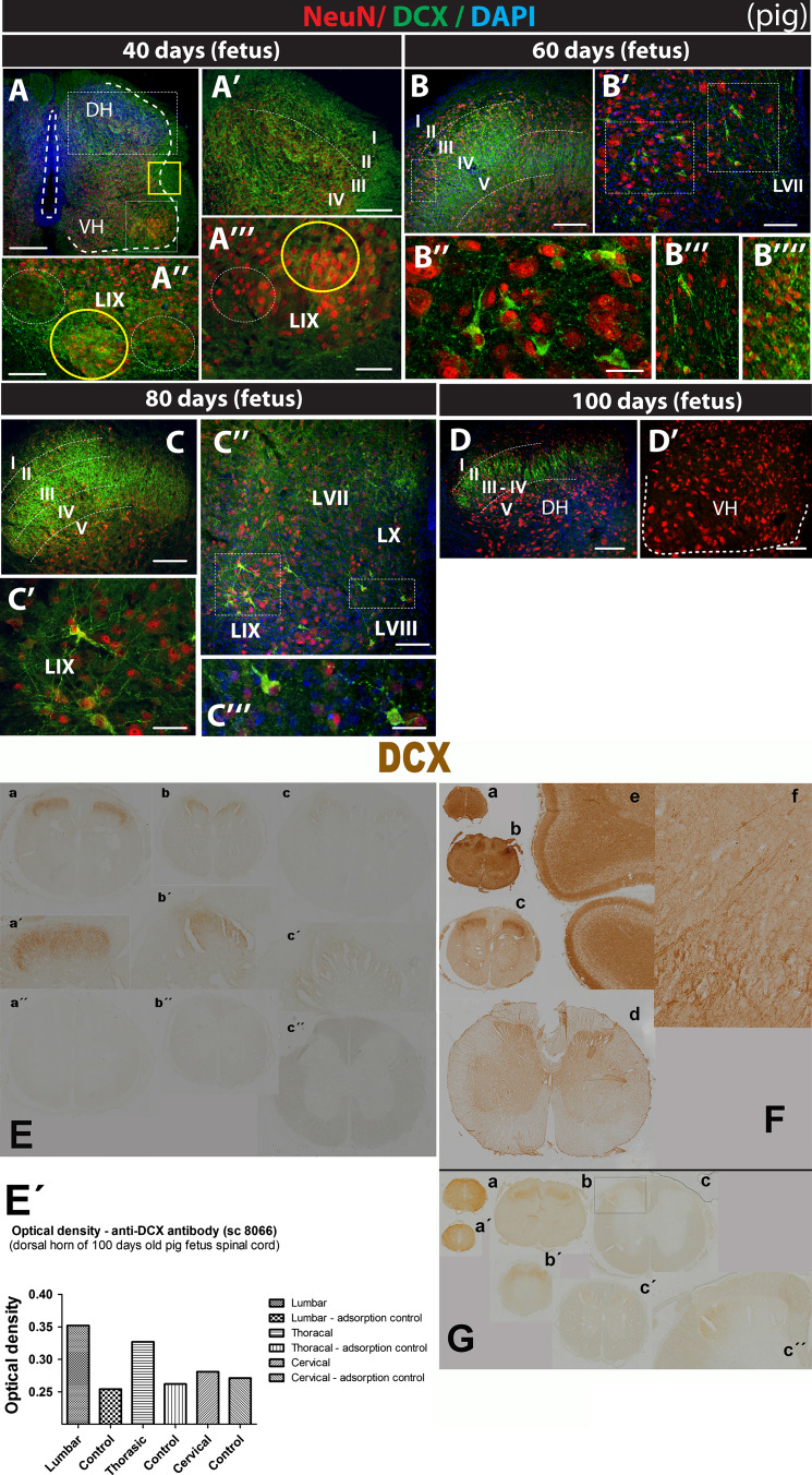
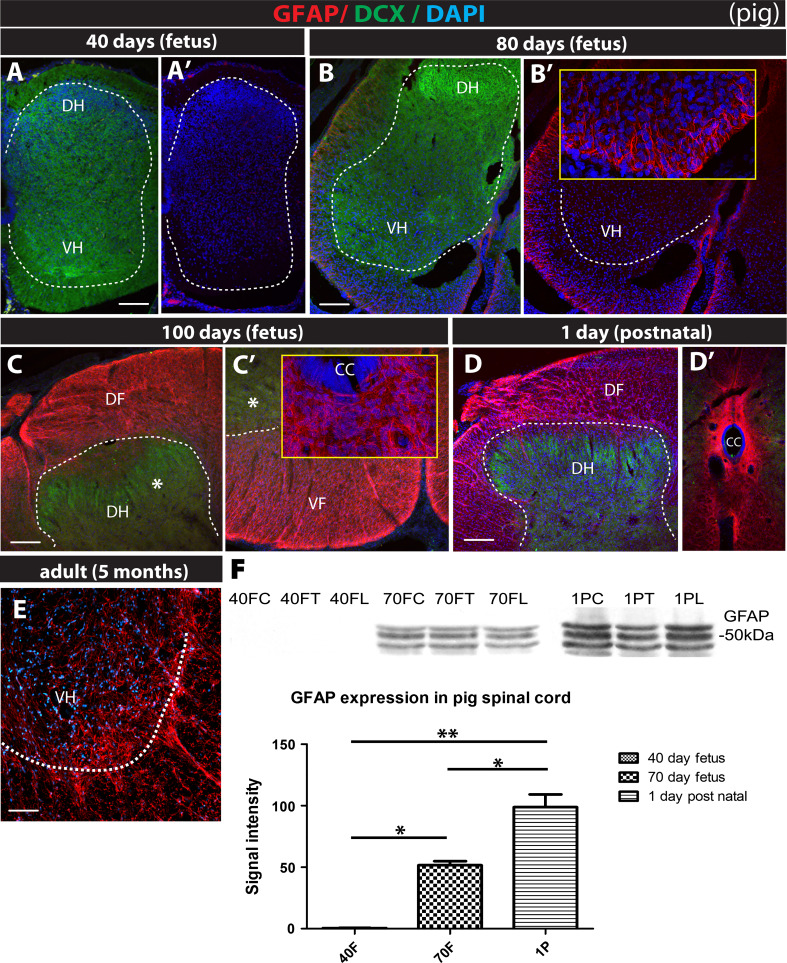
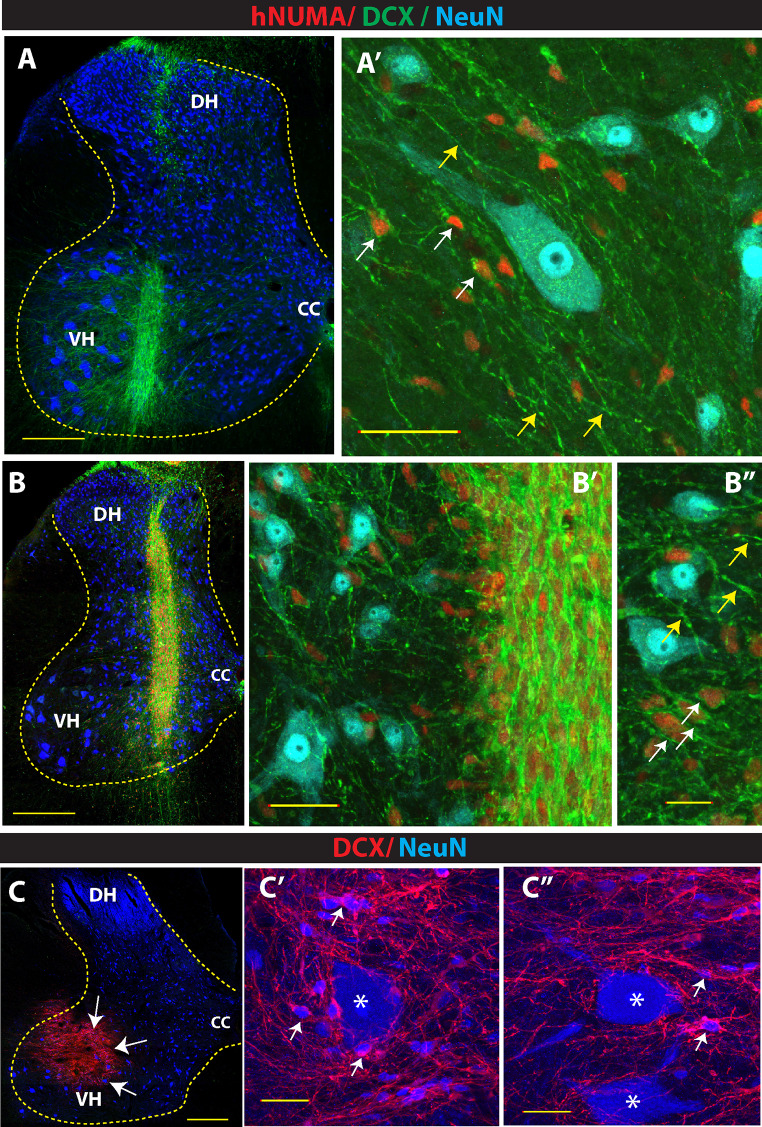
Expression of doublecortin (DCX), a 43-53 kDa microtubule binding protein, is frequently used as (i) an early neuronal marker to identify the stage of neuronal maturation of in vivo grafted neuronal precursors (NSCs), and (ii) a neuronal fate marker transiently expressed by immature neurons during development. Reliable identification of the origin of DCX-immunoreactive cells (i.e., host vs. graft) requires detailed spatial and temporal mapping of endogenous DCX expression at graft-targeted brain or spinal cord regions. Accordingly, in the present study, we analyzed (i) the time course of DCX expression in pre- and postnatal rat and porcine spinal cord, and (ii) the DCX expression in spinally grafted porcine-induced pluripotent stem cells (iPS)-derived NSCs and human embryonic stem cell (ES)-derived NSCs. In addition, complementary temporospatial GFAP expression study in porcine spinal cord was also performed. In 21-day-old rat fetuses, an intense DCX immunoreactivity distributed between the dorsal horn (DH) and ventral horn was seen and was still present in the DH neurons on postnatal day 20. In animals older than 8 weeks, no DCX immunoreactivity was seen at any spinal cord laminae. In contrast to rat, in porcine spinal cord (gestational period 113-114 days), DCX was only expressed during the pre-natal period (up to 100 days) but was no longer present in newborn piglets or in adult animals. Immunohistochemical analysis was confirmed with a comparable expression profile by western blot analysis. Contrary, the expression of porcine GFAP started within 70-80 days of the pre-natal period. Spinally grafted porcine iPS-NSCs and human ES-NSCs showed clear DCX expression at 3-4 weeks postgrafting. These data indicate that in spinal grafting studies which employ postnatal or adult porcine models, the expression of DCX can be used as a reliable marker of grafted neurons. In contrast, if grafted neurons are to be analyzed during the first 4 postnatal weeks in the rat spinal cord, additional markers or grafted cell-specific labeling techniques need to be employed to reliably identify grafted early postmitotic neurons and to differentiate the DCX expression from the neurons of the host.
Figures


Similar articles
-
Survival and differentiation of human embryonic stem cell-derived neural precursors grafted spinally in spinal ischemia-injured rats or in naive immunosuppressed minipigs: a qualitative and quantitative study.Cell Transplant. 2012;21(12):2603-19. doi: 10.3727/096368912X653200. Epub 2012 Aug 10. Cell Transplant. 2012. PMID: 22889456
-
Expression of doublecortin, a neuronal migration protein, in unipolar brush cells of the vestibulocerebellum and dorsal cochlear nucleus of the adult rat.Neuroscience. 2012 Jan 27;202:169-83. doi: 10.1016/j.neuroscience.2011.12.013. Epub 2011 Dec 17. Neuroscience. 2012. PMID: 22198017 Free PMC article.
-
Analysis of dosing regimen and reproducibility of intraspinal grafting of human spinal stem cells in immunosuppressed minipigs.Cell Transplant. 2010;19(9):1103-22. doi: 10.3727/096368910X503406. Epub 2010 Apr 21. Cell Transplant. 2010. PMID: 20412634
-
Preparation of Neural Stem Cells and Progenitors: Neuronal Production and Grafting Applications.Methods Mol Biol. 2021;2311:73-108. doi: 10.1007/978-1-0716-1437-2_7. Methods Mol Biol. 2021. PMID: 34033079 Free PMC article.
-
Progress in cerebral transplantation of expanded neuronal stem cells.J Neurosurg. 2004 Apr;100(4):659-71. doi: 10.3171/jns.2004.100.4.0659. J Neurosurg. 2004. PMID: 15070121 Review.
Cited by
-
The progress of induced pluripotent stem cells derived from pigs: a mini review of recent advances.Front Cell Dev Biol. 2024 Jun 24;12:1371240. doi: 10.3389/fcell.2024.1371240. eCollection 2024. Front Cell Dev Biol. 2024. PMID: 38979033 Free PMC article. Review.
-
Vasculature cells control neuroglial co-localization and synaptic connection in a central nervous system tissue mimic system.Hum Cell. 2023 Nov;36(6):1938-1947. doi: 10.1007/s13577-023-00955-x. Epub 2023 Jul 20. Hum Cell. 2023. PMID: 37470936
-
Physiological, pathological, and engineered cell identity reprogramming in the central nervous system.Wiley Interdiscip Rev Dev Biol. 2016 Jul;5(4):499-517. doi: 10.1002/wdev.234. Epub 2016 Jun 3. Wiley Interdiscip Rev Dev Biol. 2016. PMID: 27258392 Free PMC article. Review.
-
Chromatin establishes an immature version of neuronal protocadherin selection during the naive-to-primed conversion of pluripotent stem cells.Nat Genet. 2019 Dec;51(12):1691-1701. doi: 10.1038/s41588-019-0526-4. Epub 2019 Nov 18. Nat Genet. 2019. PMID: 31740836 Free PMC article.
-
In vivo reprogramming of NG2 glia enables adult neurogenesis and functional recovery following spinal cord injury.Cell Stem Cell. 2021 May 6;28(5):923-937.e4. doi: 10.1016/j.stem.2021.02.009. Epub 2021 Mar 5. Cell Stem Cell. 2021. PMID: 33675690 Free PMC article.
References
-
- Cao QL, Onifer SM, Whittemore SR (2008) Labeling stem cells in vitro for identification of their differentiated phenotypes after grafting into the CNS. Methods Mol Biol 438:361–374 - PubMed
-
- Couillard-Despres S, Winner B, Schaubeck S, Aigner R, Vroemen M, Weidner N, Bogdahn U, Winkler J, Kuhn HG, Aigner L (2005) Doublecortin expression levels in adult brain reflect neurogenesis. Eur J Neurosci 21(1):1–14 - PubMed
-
- Couillard-Despres S, Finkl R, Winner B, Ploetz S, Wiedermann D, Aigner R, Bogdahn U, Winkler J, Hoehn M, Aigner L (2008) In vivo optical imaging of neurogenesis: watching new neurons in the intact brain. Mol Imaging 7(1):28–34 - PubMed
Publication types
MeSH terms
Substances
LinkOut - more resources
Full Text Sources
Other Literature Sources
Molecular Biology Databases
Miscellaneous