

Dynamic properties of water in breast pathology depend on the histological compounds: distinguishing tissue malignancy by water diffusion coefficients
- PMID: 25487139
- PMCID: PMC4295355
- DOI: 10.1186/1756-0500-7-887
Dynamic properties of water in breast pathology depend on the histological compounds: distinguishing tissue malignancy by water diffusion coefficients
Abstract
Background: The parameters that characterize the intricate water diffusion in tumors may also reveal their distinct pathology. Specifically, characterization of breast cancer could be aided by diffusion magnetic resonance.The present in vitro study aimed to discover connections between the NMR biexponential diffusion parameters [fast diffusion phase (D(FDP)), slow diffusion phase (D(SDP)), and spin population of fast diffusion phase (P1)] and the histological constituents of nonmalignant (control) and malignant human breast tissue. It also investigates whether the diffusion coefficients indicate tissue status.
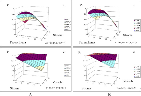
Methods: Post-surgical specimens of control (mastopathy and peritumoral tissues) and malignant human breast tissue were placed in an NMR spectrometer and diffusion sequences were applied. The resulting decay curves were analyzed by a biexponential model, and slow and fast diffusion parameters as well as percentage signal were identified. The same samples were also histologically examined and their percentage composition of several tissue constituents were measured: parenchyma (P), stroma (St), adipose tissue (AT), vessels (V) , pericellular edema (PCE), and perivascular edema (PVE). Correlations between the biexponential model parameters and tissue types were evaluated for different specimens. The effects of tissue composition on the biexponential model parameters, and the effects of histological and model parameters on cancer probability, were determined by non-linear regression.
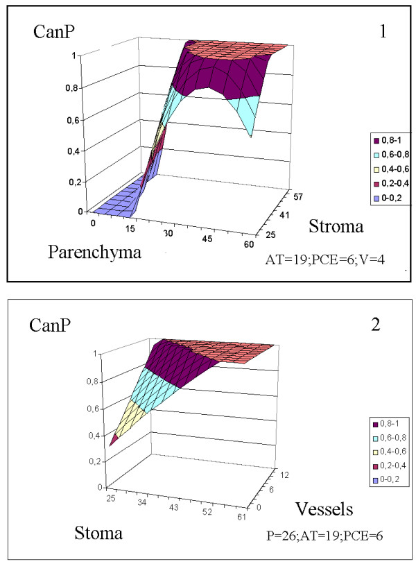
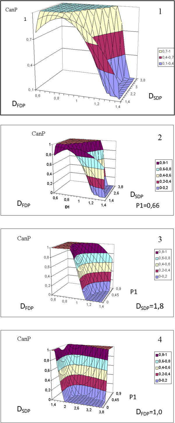
Results: Meaningful relationships were found among the in vitro data. The dynamic parameters of water in breast tissue are stipulated by the histological constituents of the tissues (P, St, AT, PCE, and V). High coefficients of determination (R2) were obtained in the non-linear regression analysis: D(FDP) (R2 = 0.92), D(SDP) (R2 = 0.81), and P1(R2 = 0.93).In the cancer probability analysis, the informative value (R2) of the obtained equations of cancer probability in distinguishing tissue malignancy depended on the parameters input to the model. In order of increasing value, these equations were: cancer probability (P, St, AT, PCE, V) (R2 = 0.66), cancer probability (D(FDP), D(SDP))(R2 = 0.69), cancer probability (D(FDP), D(SDP), P1) (R2 = 0.85).
Conclusion: Histological tissue components are related to the diffusion biexponential model parameters. From these parameters, the relative probability of cancer in a given specimen can be determined with some certainty.
Figures
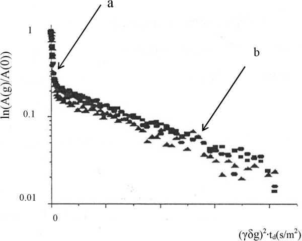
 ) <t
d3
(▲). a- fast diffusion phase, b- slow diffusion phase.
) <t
d3
(▲). a- fast diffusion phase, b- slow diffusion phase.


References
-
- Meiboom S. Nuclear magnetic resonance study of the proton transfer in water. J Chem Physiol. 1961;34:375–388. doi: 10.1063/1.1700960. - DOI
-
- Stejskal EO, Tanner JE. Spin diffusion measurements:spin echoes in the presence of a time-dependent field gradient. J Chem Phys. 1965;42:288–292. doi: 10.1063/1.1695690. - DOI
-
- Callaghan PT. Pulsed field gradient nuclear magnetic resonance as probe of liquid state molecular organization. Aust J Phys. 1984;37:359–387. doi: 10.1071/PH840539. - DOI
MeSH terms
Substances
LinkOut - more resources
Full Text Sources
Other Literature Sources
Medical
