. 2015 Jan 1;517(7532):94-98.
doi: 10.1038/nature14019. Epub 2014 Dec 10.

A PP1-PP2A phosphatase relay controls mitotic progression

Affiliations

A PP1-PP2A phosphatase relay controls mitotic progression

Agnes Grallert et al. Nature. .

Abstract

The widespread reorganization of cellular architecture in mitosis is achieved through extensive protein phosphorylation, driven by the coordinated activation of a mitotic kinase network and repression of counteracting phosphatases. Phosphatase activity must subsequently be restored to promote mitotic exit. Although Cdc14 phosphatase drives this reversal in budding yeast, protein phosphatase 1 (PP1) and protein phosphatase 2A (PP2A) activities have each been independently linked to mitotic exit control in other eukaryotes. Here we describe a mitotic phosphatase relay in which PP1 reactivation is required for the reactivation of both PP2A-B55 and PP2A-B56 to coordinate mitotic progression and exit in fission yeast. The staged recruitment of PP1 (the Dis2 isoform) to the regulatory subunits of the PP2A-B55 and PP2A-B56 (B55 also known as Pab1; B56 also known as Par1) holoenzymes sequentially activates each phosphatase. The pathway is blocked in early mitosis because the Cdk1-cyclin B kinase (Cdk1 also known as Cdc2) inhibits PP1 activity, but declining cyclin B levels later in mitosis permit PP1 to auto-reactivate. PP1 first reactivates PP2A-B55; this enables PP2A-B55 in turn to promote the reactivation of PP2A-B56 by dephosphorylating a PP1-docking site in PP2A-B56, thereby promoting the recruitment of PP1. PP1 recruitment to human, mitotic PP2A-B56 holoenzymes and the sequences of these conserved PP1-docking motifs suggest that PP1 regulates PP2A-B55 and PP2A-B56 activities in a variety of signalling contexts throughout eukaryotes.


Figures
Extended Data Figure 1
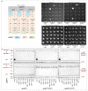
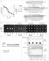
Extended Data Figure 1. PP1 phosphorylation by Cdk1-Cyclin B on T316 represses activity and reduces abundance in a manner that is mimicked by mutation of the target site to an acidic residue in PP1Dis2.T316D
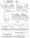
a) Clustal W alignments of the carboxyl terminal residues of the indicated PP1 isoforms. The Cdk1-Cyclin B phosphorylation site is highlighted by red shading. Red letters within this region of shading highlight the clear deviation from this consensus in the S. pombe PP1Sds21 isoform. PP1Sds21 is unable to bind to either B55Pab1 or B56Par1. The molecular basis for this inability of PP1Sds21 to bind the regulatory subunits of PP2A remains to be established. The initial threonine in the S. pombe PP1Dis2 shaded consensus sequence region is T316. b,c) Phosphatase assays of the indicated samples with activity detection by either EnzChek (b) or histone H3 serine 10 dephosphorylation (c – a second established PP1 activity assay). For each assay the graphs show arbitrary units. Bars=standard deviation. Identical results were obtained with each assay. The similarity between the recombinant Rabbit PP1γ and potato acid phosphatase (PAP) activities indicate that both reactions measure PP1 activity. The inhibition of the activity of PP1Dis2 precipitates by 1 μM, but not 10nM Okadaic acid (OA) and suggest that it is the PP1Dis2 activity rather than any coprecipitating PP2A activity that is being monitored in these assays. This view is consolidated by the inhibition of activity in both assays by the PP1 specific inhibitor NIPP1 (2, 5, 10 and 15 nM; panel c and Figure 1a). 5nM is routinely used to inhibit mammalian PP1,. Manufacturers instructions were followed for EnzChek assays. For Histone H3 serine 10 assays, histone H3 that had been phosphorylated by AuroraArk1 kinase was used as a substrate. The dephosphorylation reaction was conducted in 20mM Hepes, 100mM NaCl, 1mM EDTA 0.1% NP40 at 30 °C for 1 hour. The molarity of NaCl indicates the salt concentration of the buffer used for the immunoprecipitation, not the molarity within the phosphatase reaction. All phosphatase reactions for each substrate were conducted under identical conditions. A 1.2M NaCl, buffer was used to isolate the PP1Dis2 for the experiments in Figures 3c,d and Extended Data Figure 6d, giii, hii-iv. The level of PP1 activity detected in either assay shown in panel b and panel c was the same irrespective of the NaCl concentration in the buffer used to isolate the PP1Dis2. Phos stop is a universal phosphatase inhibitor. The key conclusion from all these assays is that the PP1Dis2.T316D protein has a similar level of activity as the Cdk1-Cyclin B phosphorylated enzyme and so behaves as a “genetically inhibited” phosphatase in the subsequent experiments described in the manuscript. d) Validation of the T316Phos antibody. PP1Dis2 immunoprecipitates from the indicated strains were treated as indicated and probed with either PP1Dis2 or T316Phos polyclonal antibodies. e-g) Cultures synchronised with respect to cell cycle progression by size selection through elutrient centrifugation probed with the PP1Dis2 antibodies at the high dilution that revealed the fluctuations in PP1Dis2 levels in Figure 1c. Graphs show the septation index alongside the PP1Dis2 levels in each sample normalised to the level of the α-tubulin loading control and a sample from an asynchronous culture that was run on each gel. e) Samples from this Rpn12mts3.1 culture was split into three immediately after elution and one third was maintained at the permissive temperature of 25°C for the remainder of the experiment, another third was immediately shifted to 36°C to inactivate this essential component of the proteasome 26S subunit, while the inactivating temperature shift of the remaining third was done 200 minutes later, after cells had completed one round of division at 25 °C. Note that the mitotic decline in PP1Dis2 levels at the permissive temperature as cells progress from mitotic commitment to the metaphase anaphase transition is not seen at 36 °C when the proteasome function is inactivated, despite the fact that the chromosome condensation index indicates that over 60% of cells have arrested cell cycle progression with a metaphase spindle. f,g) PP1Dis2 levels fail to oscillate when the Cdk1-Cyclin B phosphorylation site at position T316 is mutated. A switch to the phosphomimetic glutamic acid results in persistently low protein levels (f) while levels are persistently high upon mutation to alanine to block phosphorylation (g). Because these levels remain constant through the cell cycle these normalised levels have been superimposed upon the data from a wild type culture in Figure 1c to show each level relative to the oscillating wild type protein levels. The septation profile in Figure 1c is the profile for that wild type culture. The septation profile in Figure 1c does not contain any data from either of the cultures shown here in Extended Data Figures 1f, g. The septation profiles for each mutant culture are shown here in panels f and g.
Extended Data Figure 2
Extended Data Figure 2. Association of PP1Dis2 with the PP2A regulatory subunits B56Par1 and B55Pab1
a-d) Immunoprecipitation reactions in which PP1Dis2 polyclonal antibodies precipitated and detected PP1Dis2, GFP antibodies precipitated and detected either PP1Dis2 or PP1Sds functional fusion proteins or 12CA5 monoclonal antibodies precipitated HA tagged B55Pab1 and B56Par1 regulatory subunits of the PP2A phosphatase, as indicated. These blots establish that the association between PP1Dis2 and both B56Par1 and B55Pab1 was independent of PP1Sds function. No association was detected between PP1Sds and any PP2A regulatory subunit (including B56Par2 (data not shown)). In b and c the red asterisk indicates a non-specific band that is detected by the anti-GFP antibodies. d) No association was detected between B56Par2 and PP1Dis2, even when cell numbers in the precipitates were increased 10 fold (numbers shown in red under the panel are X × 108 cells per ml) to enhance the sensitivity of detection and the B56par1+ gene encoding the B56Par1 subunit was deleted in an attempt to remove any competition from this primary B56 isoform. e) Expression of B56par2.PkC and B56par2. L482VPkC genes from the pINT41PkC integration vector was derepressed by removal of thiamine. B56Par2 proteins (upper) or PP1Dis2 (lower) were precipitated with antibodies against the Pk epitope (upper) or PP1Dis2 peptide (lower) respectively and these immunoprecipitation reactions were run alongside aliquots of the equivalent whole cell extracts (WCE), before western blotting detected PP1Dis2 and B56Par2 fusion proteins. In both assays, the PP1 docking site mutant in which the leucine of B56Par2 was replaced with the valine of B56Par1 (B56Par2.L482V) associated with PP1 whereas the wild type B56Par2 protein did not.
Extended Data Figure 3
Extended Data Figure 3. Yeast two hybrid assays reveal docking site dependent association between B56Par1 and PP1Dis2
Yeast two hybrid assays in which PP1dis2+ and PP1sds21+ sequences were fused to the activating domain of Gal4 and the indicated version of the core homology domains of the PP2A regulatory subunits B56par1+ (encoding amino acids 112-424) and B56par2+ (encoding amino acids 213-525) were fused to the Gal4-DNA binding domain according to the procedures of the matchmaker gold yeast two hybrid system. b) Blotting the cell extracts with 12CA5 and 9E10 monoclonal antibodies that recognized HA and myc epitopes within the cassettes harbouring the activation (HA) and DNA binding domains (myc) indicated that equivalent protein expression levels were achieved for each version of the protein. Probing for the DNA replication factor Ctf4 was used as a loading control. Thus, the failure of the PP1 docking site mutants to interact with PP1dis2+ suggests that the change in the PP1 docking site abolished the affinity between the two molecules.
Extended Data Figure 4
Extended Data Figure 4. Validation of the SKEVLF and GLLR motifs of the PP1 docking consensus site of B56Par1 in the polarity protein Wsh3/Tea4 and the mitotic regulator Cut12
The morphology protein Wsh3/Tea4 is required to recruit PP1Dis2 to cell tips. Wsh3/Tea4 recruited PP1 promotes the de-phosphorylation of the DYRK family kinase Pom1. De-phosphorylation of Pom1 promotes its association with the cell cortex. Subsequent auto-phosphorylation diminishes this new-found affinity for the cortex until the kinase looses its affinity for the cortex. Because Wsh3/Tea4 is only found at cell tips, Pom1 only associates with the cortex at the tips. We exploited this relationship to test the ability of the SKEVLF sequence in the PP1 docking consensus site of B56Par1 to function as a PP1Dis2 docking site in vivo. a) The indicated wsh3 alleles were cloned into the pINTL41PkN vector and integrated at the leu1 locus before introduction into wsh3.Δ background in which the endogenous wsh3+/tea4+ gene had been deleted. b) Expression of the transgenes was repressed by the addition of thiamine to culture medium (uninduced) before filtration into thiamine free medium induced expression of each allele 24 hours later (induced). c) PP1Dis2.GFP was not enriched at cell tips in the absence of wsh3 induction. Induction of the wild type or wsh3.I232SR234ET236L allele in which the SKEVLF motif of the B56Par1 PP1Dis2 docking site was substituted for the native IFRVTF motif of Wsh3/Tea4 led to the recruitment of PP1Dis2.GFP to cell tips whereas expression of the PP1 docking site mutant wsh3. F237A failed to do so. d) Pom1 recruitment to cell tips upon expression of both the wild type and SKEVLF alleles, but not the PP1 docking site alleles indicated that the PP1Dis2 recruited to Wsh3/Tea4 by the SKEVLF allele was functionally indistinguishable from that recruited by the wild type molecule. e-g) Commitment to mitosis is promoted by the activation of Cdk1-CyclinB. Cdk1-CyclinB activity is regulated by the level of phosphorylation on threonine 14 and tyrosine 15 within its ATP binding site. Wee1 kinase phosphorylates these residues while Cdc25 removes the phosphate to trigger mitotic commitment. The lack of Cdc25 phosphatase activity achieved by incubating cells harbouring the temperature sensitive cdc25.22 mutation at temperatures above 34 °C kills cells because they are unable to remove the inhibitory phosphate from Cdk1. The cut12.s11 and cut12.s14 mutations enable cdc25.22 cells to divide at 34°C ,. cut12.s11 is a G71V mutation at the start of a highly conserved bipartite PP1 docking site GILKTPGTLQIKKTVNF. cut12.s14 is a F87L mutation at the end of the same docking motif. (e) Alignment of the conserved PP1 docking site of Cut12 homologues from the indicated fungi and the mutations generated in each of the indicated cut12 alleles. Mutation of the PP1 docking site of S. pombe Cut12 to abolish the function of either the GILK or the KxVxF motifs of Cut12 reduces PP1Dis2 association in immunoprecipitation assays (f) and suppresses the lethal mitotic commitment defect of cdc25.22 (g). Simultaneous mutation of both motifs abolishes PP1 association in immunoprecipitation assays and confers a higher level of suppression of cdc25.22 (g) that can enable cells to survive complete removal of cdc25+ coding sequences. We asked whether the GLLR sequence found upstream of the B56Par1 KxVxF motif (Figure 2a) would substitute for the GILK sequence of Cut12 in both the co-immunoprecipitation and cdc25.22 suppression assays. In both cases it could. The association between PP1Dis2 and Cut12 in the cut12.I72LK74R mutant that converts the GILK motif to a GLLR was indistinguishable from wild type. Also, the G71V mutation of cut12.s11 suppressed cdc25.22 whereas cut12.I72LK74R did not (g). Thus, the function bestowed on Cut12 by the GILK sequence is fully maintained upon substitution with GLLR indicating that GLLR is a functional variant of GILK. f) bars = standard deviation.
Extended Data Figure 5
Extended Data Figure 5. in vitro association of purified PP1Dis2 with PP2A-B55Pab1 and PP2A-B56Par1 complexes in a docking site dependent manner
The tandem affinity purification steps employed in Extended Data Figure 10e were followed by immuno-affinity with antibodies directed either against the HA epitope on B55Pab1.HA or B56Par1.HA fusion proteins or the unique sequence SQNWHMTPPRKNK in the C terminus of PP1Dis2 using anti-HA Affinity Matrix followed by elution with HA peptide, or Dynabeads A pre-loaded with PP1Dis2 antibodies followed by elution with the SQNWHMTPPRKNK peptide respectively. b) Coomassie stained 4 – 12 % SDS NUPAGE gradient gels. In each case the first two sample lanes show the purified wild type and PDSN PP2A holoenzymes and the fifth lane shows the purified PP1Dis2 enzyme. This quantity of PP1Dis2 was mixed with the quantity of each PP2A complex shown in lanes 1 and 2 before re-isolated of PP1Dis2 via affinity for beads bearing PP1Dis2 antibodies. This PP1Dis2 (and any associated partner molecules) was then eluted from these beads with the SQNWHMTPPRKNK peptide and the eluted proteins run in lanes 3 and 4 of each gel. For both the B55Pab1 (left) and B56Par1 (right) PP2A holoenzymes the wild type but not the PDSN complex bound to PP1Dis2. The numbered arrows indicate the lanes from which bands were excised for protein identification by mass spectrometry in panel c. c) Protein identification by mass spectrometry of the indicated bands from the lanes on the SDS PAGE gels highlighted by the numbered arrows in panel b. emPAI scores show that the purified PP1Dis2 preparations used for the in vitro reconstitution reaction and run in lanes 5 of the two gels in panel b also contained the conserved and well characterised PP1 partner Sds22,. emPAI scores for the bands found in the reconstituted PP2A holoenzyme/PP1 complexes show that the predominant molecule identified in each complexes corresponds with a known subunit of the PP2A complex, PP1Dis2 or Sds22. d) Western blotting to detect the indicated components in the complexes used for panels b and c. The epitope tagged regulatory B55Pab1 and B56Par1 molecules were detected with antibodies against the HA tag, while the catalytic CPpa1 and CPpa2 and scaffolding APaa1 subunits of PP2A were detected with commercial antibodies. e) Western blots of gel filtration chromatography fractions of either the isolated PP2A holo-enzyme complexes (i, ii) or the eluted PP2A holoenzyme/PP1 quaternary complexes (iii, iv) corresponding to lanes 1 and 3 of the two SDS NUPAGE gels of panel b. Note the very low level of PP2A-B55Pab1/PP1Dis2 and PP2A-B56Par1/PP1Dis2 quaternary complexes in the isolated PP2A holoenzyme preparations in panels i and ii. Migration of the different complexes at the appropriate size suggests that the procedures used isolated correctly folded proteins. f) Phosphatase assays of the isolated enzymes used in panels b-e show that these molecules are active phosphatases and so the procedures used isolated functional, correctly folded complexes rather than denatured, inappropriately folded, proteins. For each assay n = 5. bars = standard deviation. g) B56-Phos blots of the TAP and HA purified holoenzyme complex preparations used in panel c alongside a similarly purified PP2A-B56Par1.S378A holoenzyme complex.
Extended Data Figure 6
Extended Data Figure 6. Mitotic enhancement of PP2A-B56Par1 activity relies upon recruitment of active PP1Dis2 and is required for accurate chromosome segregation
a) DAPI staining of the Cdk1cdc2.33 strains in which mitotic progression has been synchronised by transient arrest at the restrictive temperature of 36°C. While two evenly sized chromatin masses are generated by the anaphase in wild type cells, chromosome segregation is uneven in the mutant lines and lagging chromosomes are frequently observed. The frequency of phenotypes is presented in Figure 2e. b) The PP2A-B56Par1 phosphatase assay of Ishiguro et al. in which recombinant GST-Rec8391-561 was phosphorylated by a fusion between glutathione and S. pombe casein kinase I (Hhp2) that had also been produced in E. coli. PP2A-B56Par1.HA was isolated from 2*108 cells with 12CA5 antibody under non-denaturing conditions. Phosphatase activity was calculated from the reduction of 32P incorporation in the GST-Rec8391-561 substrate per unit B56Par1.HA. The level obtained in this assay (and every assay presented in this study) was normalized to the reduction in substrate phosphorylation displayed by a B56Par1 precipitate from 2 × 10⁸ cells of an asynchronous B56par1.HA culture run on the same gel (the second lane in panel b)). OA = Okadaic acid. c) PP1Dis2 activity assays conducted in parallel with the same samples used in panel b. These assays established that both the recombinant Rabbit PP1γ and the PP1Dis2 samples that were added to the PP2A-B56Par1 assay in panel a both contained PP1 phosphatase activity. We conclude that we have successfully re-established the phosphatase assays described by Ishiguro et al and that the addition of PP1 to this assay did not alter the phosphorylation status of the phosphorylated GST-Rec8 substrate indicating that PP1Dis2 displays no activity towards phosphorylated GST-Rec8 used in this PP2A-B56Par1 enzyme assay. d) This panel presents blots of protein levels (left) PP1Dis2 activity assays (centre) and PP2A-B56Par1 phosphatase assays (right) of the PP1Dis2 samples used in the add back experiments in panels giii and hii-iv. The blot of PP1Dis2 levels on the left shows that similar levels of the different PP1Dis2 proteins were added in each case, while the central panel shows that these samples possessed PP1Dis2 activity. The panel on the right shows that none of the PP1Dis2 samples exhibited any activity in the PP2A-B56Par1 phosphatase assay. In other words, there was no PP2A-B56Par1 in these pull downs despite the fact that PP1Dis2 is able to bind to PP2A-B56Par1. This absence of PP2A-56Par1 from these samples is either due to the fact that only a minor fraction of the PP1Dis2 complex formed a complex with PP2A-B56Par1, or, as we anticipate, that the high salt (1.2M NaCl) conditions we used in the immunopreciptation reactions that isolated these PP1Dis2 molecules for the “add back” experiments (panels g,h and Figure 3c,d) had disassociated any PP2A-B56Par1 molecules that partnered these PP1Dis2 molecules in vivo. e-k) PP2A-B56Par1 assays as for panel b. A quantitative plot of the data in panel gi is shown in main Figure 3b while the data from giii and hi-hiv are represented in the plots in Figure 3c and d while data from i-k are shown in Figure 3g-i. For the experiments in panels g ii iii and h we exploit the redundancy between PP1Dis2 and PP1Sds21 to use PP1Sds21 to provide essential PP1 function and support the viability of PP1dis2 cells. The PP2A-B56Par1 complexes isolated from these cells have therefore never been exposed to PP1Dis2 regulation and so will be fully phosphorylated on PP1Dis2 target sites. The addition of PP1Dis2 in vitro enabled us to assesses the impact of dephosphorylation of these sites. To ensure that there could be no cell cycle dependency to any outcome in these assays we assessed the impact of PP1Dis2 addition on PP2A-B56Par1 samples from all stages of a Cdk1cdc2.33 synchronised mitosis. The data from these experiments that is presented in Figure 3 and this figure panels d, g and h clearly demonstrate that the addition of active PP1Dis2 to naïve PP2A-B56Par1 complexes that have never been exposed to PP1Dis2 in vivo was able to reactivate the PP2A activity as long as the PP1Dis2 docking site within B56Par1 was intact. In contrast PP1Dis2 addition failed to reactivate these naïve PP2A-B56Par1 complexes when the PP1Dis2 docking site in B56Par1 had been mutated to block PP1Dis2 recruitment, or the Cdk1-CyclinB inhibition site at T316 of PP1Dis2 had been mutated to aspartic acid to mimic the phosphorylated state.
Extended Data Figure 7
Extended Data Figure 7. Residue 378 within the PP1 docking site of B56Par1 was dephosphorylated by PP2A-B55Pab1 that had been activated by PP1Dis2 recruitment to its own PP1 docking site
a) Validation of the B56-Phos antibody that was raised against and affinity purified with a peptide in which residues corresponding to the serines at 377 and 378 were each phosphorylated. Immunoprecipitates in which the 12CA5 monoclonal antibody was used to recognise the HA epitope on a B56Par1.HA fusion protein or commercial rabbit antibodies raised against human B56ε were used to precipitate B56Par1 and probed as indicated. b) Validation of the PP2A-B55Pab1 activity assay. This assay exploits the fact that PP2A-B55Pab1 removes B56-Phos reactivity from B56Par1.HA. The phosphorylated B56Par1.HA substrate was precipitated from cells in which mitotic progression had been arrested by incubation of Eg5cut7.24 cells at the restrictive temperature for 3 hours. Eg5cut7.24 is a temperature sensitive mutation in the kinesin 5 motor protein that is required for inter-digitation of the two halves of the bipolar spindle. To ensure that these B56Par1 precipitates were free of any B56Par1 partners, the total protein content of intact cells was precipitated by TCA treatment before cells were homogenised and re-suspended in, 2% SDS. After a centrifugation clearing step, the addition of 1% Triton ×100 to the resultant supernatant sequestered the SDS into micelles thereby generating conditions for immuno-precipitation (IP) of phosphorylated B56Par1.HA substrate. Lane 2 in the right hand panel shows that that this procedure disassociated the B56Par1 subunit from the other subunits of the tri-partite PP2A-B56 complex whereas non-denaturing IP conditions (lane 1) do not. The CPpa1 and CPpa2 catalytic subunits are detected with a commercial antibody. The B56Par1 samples generated in this way were incubated with the indicated components. “eluted” indicates reactions in which the B55Pab1 immunoprecipitate was incubated with a peptide corresponding to the PP1Dis2 docking site on B56Par1 (CWPKVNSSKEVLF) to disassociate the PP1Dis2 enzyme from the PP2A-B55Pab1 complex before a re-purification isolated the PP1Dis2 complex once more. It was this re-isolated PP1Dis2 that was used for the assay in lane 9 while the residual PP2A-B55Pab1 was used in the assay in lane 6. The impact of this disassociation can be seen by comparing lanes 8 and 9. The PP1Dis2 immunoprecipitate exhibited a low level of B56-Phos phosphatase activity when the elution step was not performed. Displacement of partner molecules through peptide incubation removed the activity that reduced B56-Phos reactivity in the un-eluted PP1Dis2 precipitates. The inhibitor profiles, all support the conclusion that this assay specifically detected PP2A-B55Pab1 activity. Phos-Stop is a commercial pan-phosphatase inhibitor. c-f) PP2A-B55Pab1 activity assays of cultures in which mitotic progression had been synchronised by the Cdk1cdc2.33 arrest/release protocol. The quantification of these assays is presented in Figure 4f-i.
Extended Data Figure 8
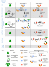
Extended Data Figure 8. Mitotic control of phosphatase activities
A schematic representation of the control and activity levels of the three phosphatase activities. This view is derived from bulk activity assays. We believe that localised reactions at particular times at discrete locations will differ, however the basic relationships between the three phosphatases outlined here will apply. Left) As previously established in a number of studies,- PP1Dis2 activity is repressed by Cdk1-Cyclin B phosphorylation upon mitotic commitment through phosphorylation on T316 at a rate that exceeds the rate at which auto-dephosphorylation re-activates inhibited PP1. PP1Dis2T316 phosphorylation also targets PP1Dis2 for destruction to further reduce PP1Dis2 activity although the majority of the protein persists. It is currently unclear whether this reflects a highly localised destruction of a particular pool of PP1Dis2 in its entirety or a rate of destruction across the population that is simply too slow to out compete with synthesis. Cyclin B destruction curtails the inhibitory phosphorylation on T316 enabling PP1Dis2 to auto-reactivate and persist in an active state for the remainder of mitosis. Middle) PP2A-B55Pab1 activity is inhibited upon mitotic commitment (Extended Data Figure 9a). The mechanism by which this inhibition is achieved is currently unclear. The inactivated PP2A-B55Pab1 recruits T316 phosphorylated (and therefore catalytically compromised) PP1Dis2 upon mitotic commitment. Cyclin B destruction upon the metaphase anaphase transition promotes PP1Dis2 reactivation (lefthand panel). The reactivation of the PP2A-B55Pab1 associated PP1Dis2 enables this PP1Dis2 to reactivate PP2A-B55Pab1. PP1Dis2 then disassociates from B55Pab1. The molecular basis for the staged association between PP1Dis2 and B55Pab1 upon mitotic commitment and dissociation at the metaphase anaphase transition remains to be established. PP2A-B55Pab1 persists in an active state for the remainder of mitosis. Right) PP2A-B56Par1 activity is inhibited upon mitotic commitment (Extended Data Figure 9a). The mechanism by which this inhibition is achieved is currently unclear. PoloPlo1 activation upon mitotic commitment enables it to phosphorylate serine 378 within the PP1 docking site on B56Par1 to block PP1Dis2 recruitment to this B’ regulatory subunit of the PP2A-B56 holoenzyme. This phosphorylation persists to block PP1Dis2 recruitment until telophase when the balance between PoloPlo1 kinase and PP2A-B55Pab1 phosphatase activities tips in favour of PP2A-B55Pab1 to remove phosphate from serine 378, thereby promoting the recruitment of active, T316 dephosphorylated PP1Dis2. PP1Dis2 recruitment enables this phosphatase to reactivate the PP2A-B56Par1 holoenzyme.
Extended Data Figure 9
Extended Data Figure 9. PP1Dis2, PP2A-B55Pab1 and PP2A-B56Par1 activities decline as cells arrest cell cycle progression in mitosis and association between human PP1 and PP2A B56 isoforms
a) The temperature of Eg5cut7.24 B55Pab1.HA, Eg5cut7.24 B55Par1.HA and Eg5cut7.24 cultures that had been grown overnight to early-log phase in supplemented EMM2 medium at 25°C was increased to 36°C at t=0 to inactivate this kinesin 5 and so arrest cell cycle progression in mitosis. Samples were taken for the phosphatase assays and to monitor the degree of mitotic arrest by scoring the frequency of cells with condensed chromosomes. Protein phosphatase assays were conducted as described for Extended Data Figures 1b, 6b and 7b. Each activity declined as the frequency of cells in which the inability to form a bipolar spindle has triggered a mitotic arrest due to activation of the spindle assembly checkpoint. b) Two hybrid assays with the indicated human PP1 isoforms with wild type and mutant isoforms of the conserved core domain (sequences encoding amino acids 84-400) of human B56δ. c) The mutations introduced in each case. d) Blotting the cell extracts from the two hybrid clones shown in panel e with 12CA5 and 9E10 monoclonal antibodies that recognized HA and myc epitopes within the cassettes harbouring the Gal4 activation and DNA binding domains respectively indicated that equivalent protein expression levels were achieved for each version of the protein. Thus, the failure of the PP1 docking site mutants indicates that the change in the PP1 docking site abolished the affinity between the two molecules.
Extended Data Figure 10
Extended Data Figure 10. PP1 docking site mutations do not alter the stoichiometry of PP2A subunits in immunoprecipitation assays or of holoenzymes isolated by TAP tag purification
a-c) Immunoprecipitation reactions from cell extracts of the indicated strains in which the 12CA5 antibodies were used to precipitate epitope tagged versions of the B55Pab1 or B56Par1 PP2A regulatory subunits or poly-clonal anti-GFP antibodies were used to precipitate a GFP tagged version of the APaa1 PP2A scaffolding subunit. Although mutation of either the PP1 docking site motif sequences or the B56par1.S378D mutation abolished PP1 precipitation with each PP2A holoenzyme, they had no impact upon the stoichiometry of PP2A subunits precipitating with either the regulatory or the scaffolding subunits. We conclude that the PP1 docking site mutations do not alter the integrity of the PP2A-B55 or PP2A-B56 holoenzyme complexes. The ability to detect S377/S378 phosphorylation in APaa1 precipitates consolidates the data in Extended Data Figure 5g to demonstrate that this site between the two elements of the PP1 docking site of B56Par1 can be phosphorylated in the PP2A-B56Par1 holoenzyme complexes. Catalytic CPpa1 and CPpa2 and the scaffolding APaa1 subunits of PP2A were detected with commercial antibodies. d) The tandem affinity purification scheme used to isolate PP2A phosphatases from yeast cultures via the TAP tag fused to the scaffolding APaa1 subunit. e) Coomassie stained 4 – 12 % SDS NUPAGE gradient gels of Paa1.TAP purifications of the indicated strains. The disappearance of bands upon gene deletion combines with Mass spectrometric analysis of isolated bands after a further round of purification (Extended Data Figure 5b,c) to confirm that this procedure isolates PP2A enzymes. The persistence of the wild type pattern in B55pab1.PDSN, B56par1.PDSN and B56par1.S378A strains indicates that these mutations do not alter the composition of either PP2A holoenzyme. i.e. they are not altering the structural bonds that maintain the integrity of the PP2A holo-enzyme complexes as predicted from existing current crystal structures of PP2A holoenzyme complexes-.
Figure 1
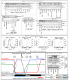
Figure 1. PP1Dis2 threonine 316 phosphorylation and stability
a) EnzChek phosphatase assays (Extended data Figure 1b) of PP1Dis2 isolated from the indicated strains by antibody precipitation. “+ Cdk1-Cyclin B” indicates the addition of sepharose beads to which covalently linked p13Suc1 protein had recruited Cdk1-Cyclin B from S. pombe cell extracts. NIPP1 is a highly specific PP1 inhibitor. Purvalanol is a Cdk1 inhibitor. b, c) PP1Dis2 and T316 phosphorylation levels (Rpn12mts3.1 is a temperature sensitive proteasome mutation). For b,c) n=5; Bars=standard deviation c) Size selected cells transit the cell division cycle. The septation profile is for the wild type culture. Dis2.T316D and Dis2.T316A levels from the experiments in Extended Data Figures 1f,g are superimposed on this wild type dataset.
Figure 2
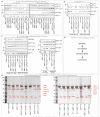
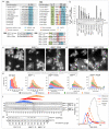
Figure 2. PP1Dis2 recruitment to PP2A-B55Pab1 and PP2A-B56Par1 regulates chromosome segregation
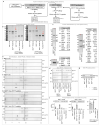
a) Clustal W alignments of PP1 docking motifs in B55 and B56 molecules. b,f,g) Immunoprecipitation reactions probed with PP1Dis2 20 and T316Phos polyclonal and 12CA5 and 366 monoclonal (HA and Pk epitopes respectively) antibodies. For f, mitotic progression was scored by tubulin staining with “Telophase” defined by post-anaphase arrays of microtubules. For b) n=6, bars=standard deviation. c,d) DAPI staining. e) Tubulin immunofluorescence staining for Cdk1cdc2.33 “arrest release” experiments scored mitotic progression.
Figure 3
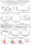
Figure 3. Phosphorlyation status of serine 378 determines competence to recruit active PP1Dis2 to PP2A-B56Par1 to promote PP2A-B56Par1 activity
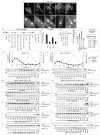
Assays of PP2A-B56Par1 activity following B56Par1.HA immunoprecipitation (with 12CA5 antibodies) of Cdk1cdc2.33 arrest release synchronised cultures followed for one (b, g-i) or two (a) cycles. Activity changes (loss of GST-Rec8391-561 32P radioactivity) at each point were normalised to the activity of B56Par1.HA from an asynchronous culture processed in parallel on the same gel (penultimate lane). (c,d) Aliquots of PP1Dis2 protein immunoprecipitates (isolated under high salt (1.2M NaCl) extraction conditions to disassociate partners) from Cdk1cdc2.33 arrested (interphase) cultures were added to PP2A-B56Par1 assays from Cdk1cdc2.33 dis2.Δ cultures (Extended Data Figure 6). (e,f) Co-immunoprecipitation assays as for Figure 2b. (j) Phenotype analysis of Cdk1cdc2.33 synchronised mitoses as for Figure 2e.
Figure 4
Figure 4. The mitotic phosphatase relay: PP2A-B55Pab1 de-phosphorylation of B56Par1 promotes PP1Dis2 recruitment to activate PP2A-B56Par1 phosphatase
a, e-i) Cdk1cdc2.33 synchronised mitoses. a-c, e) B56-Phos antibodies (Extended Data Figure 7a) detected phosphorylation on B56Par1 following immunoprecipitation from the indicated strains and the indicated treatments. PP1Dis2 levels were monitored where indicated. PoloPlo1.KDHA = HA tagged catalytically inactive. d) B56Par1 precipitates probed for PP1Dis2. f-i) PP2A-B55Pab1 phosphatase assays (Extended Data Figure 7c-f). j) Bulk activities are illustrated: local activities at any given time will depend on the specific balance of each activity at a particular site. 1) Cdk1-CyclinB activation represses all three phosphatase activities (Extended Data Figure 9a). Direct phosphorylation of PP1Dis2 TPRR motif inhibits catalytic activity,- and promotes destruction. PP1Dis2 binds B55Pab1 but cannot reactivate PP2A-B55Pab1 while TPRR phosphorylation persists. PoloPlo1 phosphorylation of B56Par1 prevents PP1Dis2 recruitment to PP2A-B56Par1. 2) Declining Cdk1-Cyclin B activity facilitates PP1 auto-reactivation. 3) Re-activated PP1Dis2 promotes PP2A-B55Pab1 reactivation. 4) PP2A-B55Pab1 de-phosphorylates the PP1 docking site in B56Par1 less efficiently than PoloPlo1 phosphorylates it to keep PP2A-B56Par1 activity low. 5) Declining PoloPlo1 activity enables PP2A-B55Pab1 to dephosphorylate B56Par1 and promote reactivating recruitment of PP1Dis2 (Extended Data Figure 8). k) Flag tagged B56γ and B56δ isoforms were stably expressed in HeLa-FRT cell lines, immunoprecipitated from mitotic cells isolated by shake-off from nocodazoletreated cells, and the immunoprecipitates probed with an antibody that recognises all forms of PP1.

Comment in

References

    1. Wu JQ, Guo JY, Tang W, Yang CS, Freel CD, Chen C, Nairn AC, Kornbluth S. PP1-mediated dephosphorylation of phosphoproteins at mitotic exit is controlled by inhibitor-1 and PP1 phosphorylation. Nature cell biology. 2009;11:644–651. doi:10.1038/ncb1871. - PMC - PubMed
    1. Ohkura H, Kinoshita N, Miyatani S, Toda T, Yanagida M. The Fission Yeast dis2+ Gene Required For Chromosome Disjoining Encodes One Of 2 Putative Type-1 Protein Phosphatases. Cell. 1989;57:997–1007. - PubMed
    1. Mochida S, Hunt T. Calcineurin is required to release Xenopus egg extracts from meiotic M phase. Nature. 2007;449:336–340. doi:10.1038/nature06121. - PubMed
    1. Mochida S, Ikeo S, Gannon J, Hunt T. Regulated activity of PP2A-B55 delta is crucial for controlling entry into and exit from mitosis in Xenopus egg extracts. EMBO J. 2009;28:2777–2785. - PMC - PubMed
    1. Schmitz MH, Held M, Janssens V, Hutchins JR, Hudecz O, Ivanova E, Goris J, Trinkle-Mulcahy L, Lamond AI, Poser I, Hyman AA, Mechtler K, Peters JM, Gerlich DW. Live-cell imaging RNAi screen identifies PP2A-B55alpha and importin-beta1 as key mitotic exit regulators in human cells. Nature cell biology. 2010;12:886–893. doi:10.1038/ncb2092. - PMC - PubMed

Publication types

MeSH terms