. 2014 Dec 9:11:63.
doi: 10.1186/s12989-014-0063-3.

Titanium dioxide nanoparticles promote arrhythmias via a direct interaction with rat cardiac tissue

Affiliations

Titanium dioxide nanoparticles promote arrhythmias via a direct interaction with rat cardiac tissue

Monia Savi et al. Part Fibre Toxicol. .

Abstract

Background: In light of recent developments in nanotechnologies, interest is growing to better comprehend the interaction of nanoparticles with body tissues, in particular within the cardiovascular system. Attention has recently focused on the link between environmental pollution and cardiovascular diseases. Nanoparticles <50 nm in size are known to pass the alveolar-pulmonary barrier, enter into bloodstream and induce inflammation, but the direct pathogenic mechanisms still need to be evaluated. We thus focused our attention on titanium dioxide (TiO₂) nanoparticles, the most diffuse nanomaterial in polluted environments and one generally considered inert for the human body.

Methods: We conducted functional studies on isolated adult rat cardiomyocytes exposed acutely in vitro to TiO₂ and on healthy rats administered a single dose of 2 mg/Kg TiO₂ NPs via the trachea. Transmission electron microscopy was used to verify the actual presence of TiO₂ nanoparticles within cardiac tissue, toxicological assays were used to assess lipid peroxidation and DNA tissue damage, and an in silico method was used to model the effect on action potential.

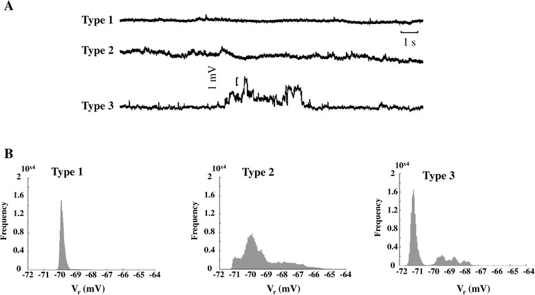
Results: Ventricular myocytes exposed in vitro to TiO₂ had significantly reduced action potential duration, impairment of sarcomere shortening and decreased stability of resting membrane potential. In vivo, a single intra-tracheal administration of saline solution containing TiO₂ nanoparticles increased cardiac conduction velocity and tissue excitability, resulting in an enhanced propensity for inducible arrhythmias. Computational modeling of ventricular action potential indicated that a membrane leakage could account for the nanoparticle-induced effects measured on real cardiomyocytes.

Conclusions: Acute exposure to TiO₂ nanoparticles acutely alters cardiac excitability and increases the likelihood of arrhythmic events.


Figures

Figure 1
Figure 1
Atomic Force Microscopy analysis of titanium dioxide nanoparticles (TiO 2 -NPs) deposited on poly-ornithine-treated mica. A. Image of deposed TiO2-NPs. B. Height profile along the white line shown in A. C. Height distribution of TiO2-NPs. D. Images of TiO2-NP aggregates (scale bars =100 nm).
Figure 2
Figure 2
Representative traces of sarcomere shortening recorded in CTRL (black) and NP C (red) cardiomyocytes field-stimulated at 0.5 (A), 1 (B) and 2 (C) Hz. Graphs of resting sarcomere length (D), sarcomere fractional shortening (FS) (E), maximal rate of shortening (−dl/dtmax) (F), and maximal rate of re-lengthening (+dl/dtmax) (G). H. Pie charts of the percentages of cardiomyocytes exhibiting spontaneous contractions (SCs, stippled areas) in CTRL (white) and NPC (red) cells after 60 s of conditional training at 0.5 Hz. I. Graph of number of SCs/cardiomyocyte in the 60 s measurement period. *, p <0.01 vs. CTRL.
Figure 3
Figure 3
Variability in resting membrane potential in cardiomyocytes exposed to TiO 2 NPs. A. Traces representative of the three types of Vr behavior found over a 60 s recording period subsequent to conditioning training at 5 Hz for 40 beats. B. Frequency distribution of Vr for the three ΔVr types.
Figure 4
Figure 4
TiO 2 NPs-induced changes in cellular electrophysiology. A. Representative action potential (AP) waveforms recorded from control (CTRL, black line) and TiO2-NP (NPC, red line) cardiomyocytes at the physiological driving rate of rat heart (5 Hz). B–E. Graphs of action potential duration (APD) measured at 20 mV (APD20) and 60 mV (APD60), beat-to-beat variability of APD60 (CVAPD60), AP upstroke (UPS) and membrane capacitance (Cm). In all graphs, CTRL is given by white columns, and NPC by the red columns (n = 37 NPC and n = 49 CTRL). *, p < 0.05 vs. CTRL. F. APs simulated with the Pandit model, without (black trace, CTRL) and with (red trace, NPC) a simulated 1.5 nS constant potassium leakage.
Figure 5
Figure 5
Instillation of TiO 2 -NPs and in vivo recordings of cardiac electrical performance. A. Time-scale (hours) of the experimental protocol. B. Representative EGs recorded from an 8×8 epicardial electrode array. Each waveform of the grid represents the time-course of extracellular potential at the corresponding position. The scheme on the right hand side explains the EG parameters, as measured from their root mean square (RMS)-derived signals. Magenta thin trace represents the first time derivative, whose minimum value is taken as a marker of the end of the QT interval C. Representative activation time maps (isochrones, ms) from Vehicle (left) and NPR (right), showing differences in longitudinal (red arrows) and transverse (blue arrows) propagation.
Figure 6
Figure 6
Susceptibility to arrhythmias in Vehicle and NP R rats. A. Ventricular ectopic couplet (top) and ventricular fibrillation (bottom) recorded during evaluation of the effective refractory period (ERP). Scale bar =500 ms. B. Evaluation of ERP in Vehicle and NPR. **, p < 0.005. C. Percentage of inducible arrhythmc events.
Figure 7
Figure 7
Presence of titanium dioxide (TiO 2 ) nanoparticles (NPs) in the rat ventricular myocardium after tracheal instillation: TEM analysis. A. Right Ventricle. Electron-dense NPs in two longitudinally oriented cardiomyocytes and in the wall of a vascular structure. B. Left Ventricle. NPs accumulating at the edge of longitudinally oriented cardiomyocytes, as well as in the sarcolemma. NPs are also present in the interstitial space, in endothelial cells and within the capillary lumen (L). C. Left ventricle. The lumen of a capillary neighboring a cardiomyocyte containing TiO2 NPs, which also appear to be connected to and engulfed by endothelial cells. GJ marks a gap junction location. Blue rectangles include areas shown at higher magnification in the lower panels (A1, B1 and C1). Scale Bars: A and B =5 μm; A1 and B1 = 2 μm; C =1 μm; C1 = 200 nm. Bottom. Ultrathin sections of lung samples from NP-exposed treated rats. D. The bronchial epithelium is apparent by the presence of ciliated cells (*). Electron-dense NPs are best seen in cytoplasm at high magnification (D1). Clusters of NPs were found within the lung parenchyma (E) and in macrophages (F). N, nucleus. G,H. The typical shape of titanium NPs is apparent at higher magnification. Scale Bars: D =5 μm; D1 = 2 μm; E =2 μm; F =1 μm; G =200 nm; H =100 nm.
Figure 8
Figure 8
TiO 2 NPs-induced toxicological effects. A. DNA damage detected in single isolated cardiomyocytes by Comet assay (pH >13) in CTRL (white columns) and NPC (red columns) after 1 h and 5 h of exposure. DNA damage is expressed as tail intensity (TI%; *p < 0.05). B. Percent increase of ROS in single isolated cardiomyocytes, NPC (red column) after 1 h. C. Evaluation of TBARS in trachea, lungs and heart tissue after tracheal instillation of saline solution (Vehicle) or saline solution containing TiO2-NPs (2 mg/Kg, NPR). *, p < 0.05 vs. Vehicle.

References

    1. Shi H, Magaye R, Castranova V, Zhao J. Titanium dioxide nanoparticles: a review of current toxicological data. Part Fibre Toxicol. 2013;10:15. doi: 10.1186/1743-8977-10-15. - DOI - PMC - PubMed
    1. Zhen S, Qian Q, Jia G, Zhang J, Chen C, Wei Y. A panel study for cardiopulmonary effects produced by occupational exposure to inhalable titanium dioxide. J Occup Environ Med. 2012;54:1389–1394. doi: 10.1097/JOM.0b013e3182611a49. - DOI - PubMed
    1. Participants IRSIW. The relevance of the rat lung response to particle overload for human risk assessment: a workshop consensus report. ILSI risk science institute workshop participants. Inhal Toxicol. 2000;12:1–17. - PubMed
    1. Inoue K, Takano H, Ohnuki M, Yanagisawa R, Sakurai M, Shimada A, Mizushima K, Yoshikawa T. Size effects of nanomaterials on lung inflammation and coagulatory disturbance. Int J Immunopathol Pharmacol. 2008;21:197–206. - PubMed
    1. Yazdi AS, Guarda G, Riteau N, Drexler SK, Tardivel A, Couillin I, Tschopp J. Nanoparticles activate the NLR pyrin domain containing 3 (Nlrp3) inflammasome and cause pulmonary inflammation through release of IL-1alpha and IL-1beta. Proc Natl Acad Sci U S A. 2010;107:19449–19454. doi: 10.1073/pnas.1008155107. - DOI - PMC - PubMed

Publication types

MeSH terms