Review
. 2015 May;360(2):363-78.
doi: 10.1007/s00441-014-2061-z. Epub 2014 Dec 9.

Molecular architecture and function of the hemidesmosome

Affiliations
Review

Molecular architecture and function of the hemidesmosome

Gernot Walko et al. Cell Tissue Res. 2015 May.

Abstract

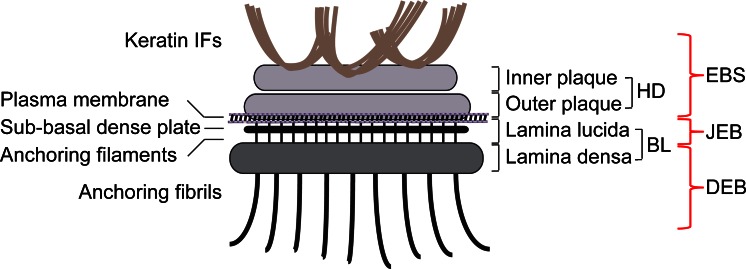
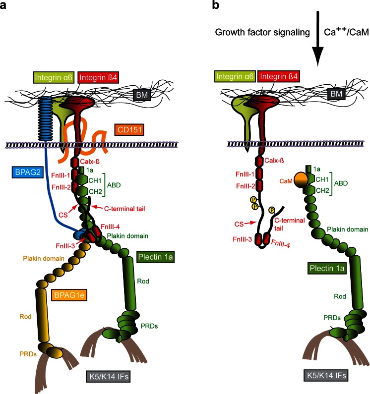
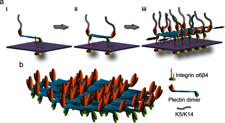
Hemidesmosomes are multiprotein complexes that facilitate the stable adhesion of basal epithelial cells to the underlying basement membrane. The mechanical stability of hemidesmosomes relies on multiple interactions of a few protein components that form a membrane-embedded tightly-ordered complex. The core of this complex is provided by integrin α6β4 and P1a, an isoform of the cytoskeletal linker protein plectin that is specifically associated with hemidesmosomes. Integrin α6β4 binds to the extracellular matrix protein laminin-332, whereas P1a forms a bridge to the cytoplasmic keratin intermediate filament network. Other important components are BPAG1e, the epithelial isoform of bullous pemphigoid antigen 1, BPAG2, a collagen-type transmembrane protein and CD151. Inherited or acquired diseases in which essential components of the hemidesmosome are missing or structurally altered result in tissue fragility and blistering. Modulation of hemidesmosome function is of crucial importance for a variety of biological processes, such as terminal differentiation of basal keratinocytes and keratinocyte migration during wound healing and carcinoma invasion. Here, we review the molecular characteristics of the proteins that make up the hemidesmosome core structure and summarize the current knowledge about how their assembly and turnover are regulated by transcriptional and post-translational mechanisms.


Figures

Fig. 1
Fig. 1
Schematic representation of a hemidesmosome and location of blisters within the skin in different types of EB. Keratin IFs are attached to the inner plaque of the hemidesmosome (HD). The outer plaque is parallel to the inner plaque, is slightly larger and lies on the plasma membrane of basal keratinocytes. Adjacent to the membrane are an electron-clear zone, the lamina lucida and an electron-dense zone, the lamina densa, which together make up the basal lamina (BL). Within the lamina lucida and directly below the plasma membrane, there is a thin electron dense line corresponding to the sub-basal dense plate. Fine anchoring filaments originate at HDs, traverse the lamina lucida and insert into the lamina densa. From the lamina densa collagenous anchoring fibrils extend into the dermal tissue. Outer brackets on the right indicate blister formation at different levels within the skin as is characteristic for the three major types of EB: EBS, within the epidermis; JEB, at the dermal-epidermal junction within the lamina lucida; DEB, in the upper layers of the dermis
Fig. 2
Fig. 2
Molecular organization of HDs and a model of the HD disassembly mechanism. a Schematically drawn are the six key components of type I HDs (integrin subunits α6 and β4, CD151, BPAG2, BPAG1e and plectin 1a), the extracellular basement membrane (BM) containing the ligand laminin-332 and the intracellular binding partner, intermediate filaments (IFs) of the keratin (K5/K14) type. The various protein domains involved in binding of integrin β4 to plectin 1a and in the linkage of the HD complex to keratin IFs are indicated. b Mechanism of growth factor-induced dissociation of the integrin β4–plectin 1a complex. For simplicity, only integrin α6β4, plectin 1a and K5/K14 IFs are shown. Growth factor-induced phosphorylation of serine residues in the C-terminal integrin β4 tail and CS domains results in the dissociation of integrin β4 from plectin 1a’s plakin and ABD domains. Subsequent interaction of plectin 1a’s ABD with the Ca++-bound form of calmodulin (CaM) prevents re-association of the integrin β4-plectin 1a complex. 1a, isoform-specific N-terminal domain preceding the ABD of plectin 1a; Calx-β, Na+–Ca2+ exchanger motif; CH1,2, calponin homology domains 1 and 2; CS, connecting segment; FnIII-1,-2,-3,-4, fibronectin type III domains 1–4; PRDs, plectin repeat domains
Fig. 3
Fig. 3
Hypothetical model of HD stabilization through plectin self-association. a Three consecutive stages (iiii) of inner plaque formation are depicted; for simplicity, only integrin α6β4 and plectin 1a molecules are shown. In a first step (i) a parallel plectin 1a dimer binds, via its N-terminal β4-binding domains (green), to the cytoplasmic tail (red) of plasma membrane (violet)-embedded integrin β4; plectin’s C-terminal domain (orange) makes the connection to keratin filaments. In a second step (ii), two plectin 1a dimers form a tetramer by lateral association of their central rod domains in an anti-parallel fashion, creating integrin β4 and K5/K14 binding sites at both ends of the tetramer. Further self-association of plectin rod segments leads to oligomeric sheet-like structures (iii). Note that self-association of plectin 1a molecules could lead to the clustering of integrin α6β4 (as depicted), or vice versa, clustered integrins may facilitate the focal recruitment of plectin molecules promoting their alignment and lateral association; also, targeting of plectin 1a molecules to integrin β4-could occur in their keratin IF-bound (as indicated) or unbound state. Opposing black arrows, plaque-stabilizing force component provided by laterally associated rod segments. b Structural model of an inner plaque assembled by the staggered lateral association of plectin dimers. Note the focal density of potential K5/K14 and integrin β4 binding sites on opposite sides of the plate structure. This model is based on the ultrastructural analysis of paracrystalline structures formed from recombinant full-length versions of plectin’s rod domain and the lateral association potential of intact plectin molecules isolated from cells (Walko et al. 2011)

References

    1. Ackerl R, Walko G, Fuchs P, Fischer I, Schmuth M, Wiche G. Conditional targeting of plectin in prenatal and adult mouse stratified epithelia causes keratinocyte fragility and lesional epidermal barrier defects. J Cell Sci. 2007;120:2435–2443. - PubMed
    1. Adhikary G, Chew YC, Reece EA, Eckert RL. PKC-delta and -eta, MEKK-1, MEK-6, MEK-3, and p38-delta are essential mediators of the response of normal human epidermal keratinocytes to differentiating agents. J Invest Dermatol. 2010;130:2017–2030. - PMC - PubMed
    1. Alt A, Ohba M, Li L, Gartsbein M, Belanger A, Denning MF, Kuroki T, Yuspa SH, Tennenbaum T. Protein kinase Cdelta-mediated phosphorylation of alpha6beta4 is associated with reduced integrin localization to the hemidesmosome and decreased keratinocyte attachment. Cancer Res. 2001;61:4591–4598. - PubMed
    1. Ambler CA, Watt FM. Adult epidermal Notch activity induces dermal accumulation of T cells and neural crest derivatives through upregulation of jagged 1. Development. 2010;137:3569–3579. - PMC - PubMed
    1. Andrä K, Lassmann H, Bittner R, Shorny S, Fässler R, Propst F, Wiche G. Targeted inactivation of plectin reveals essential function in maintaining the integrity of skin, muscle, and heart cytoarchitecture. Genes Dev. 1997;11:3143–3156. - PMC - PubMed

Publication types

MeSH terms

Substances

LinkOut - more resources