

4-D flow magnetic resonance imaging: blood flow quantification compared to 2-D phase-contrast magnetic resonance imaging and Doppler echocardiography
- PMID: 25487721
- PMCID: PMC4450116
- DOI: 10.1007/s00247-014-3246-z
4-D flow magnetic resonance imaging: blood flow quantification compared to 2-D phase-contrast magnetic resonance imaging and Doppler echocardiography
Abstract
Background: Doppler echocardiography (echo) is the reference standard for blood flow velocity analysis, and two-dimensional (2-D) phase-contrast magnetic resonance imaging (MRI) is considered the reference standard for quantitative blood flow assessment. However, both clinical standard-of-care techniques are limited by 2-D acquisitions and single-direction velocity encoding and may make them inadequate to assess the complex three-dimensional hemodynamics seen in congenital heart disease. Four-dimensional flow MRI (4-D flow) enables qualitative and quantitative analysis of complex blood flow in the heart and great arteries.
Objectives: The objectives of this study are to compare 4-D flow with 2-D phase-contrast MRI for quantification of aortic and pulmonary flow and to evaluate the advantage of 4-D flow-based volumetric flow analysis compared to 2-D phase-contrast MRI and echo for peak velocity assessment in children and young adults.
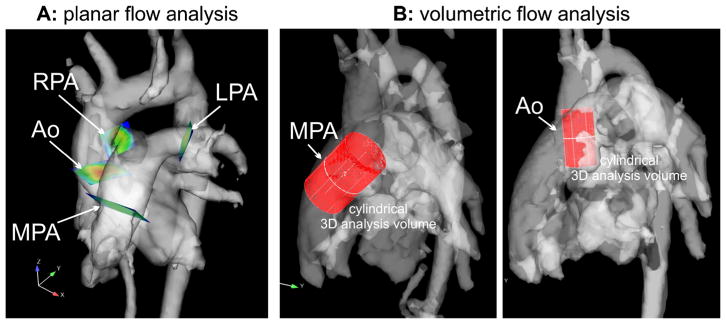
Materials and methods: Two-dimensional phase-contrast MRI of the aortic root, main pulmonary artery (MPA), and right and left pulmonary arteries (RPA, LPA) and 4-D flow with volumetric coverage of the aorta and pulmonary arteries were performed in 50 patients (mean age: 13.1 ± 6.4 years). Four-dimensional flow analyses included calculation of net flow and regurgitant fraction with 4-D flow analysis planes similarly positioned to 2-D planes. In addition, 4-D flow volumetric assessment of aortic root/ascending aorta and MPA peak velocities was performed and compared to 2-D phase-contrast MRI and echo.
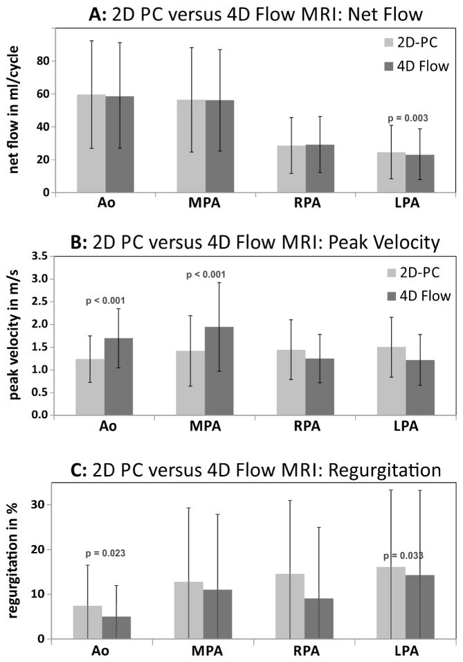
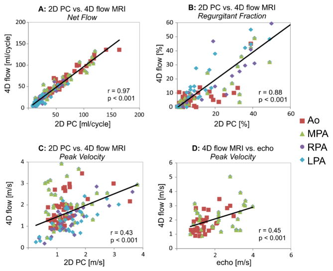
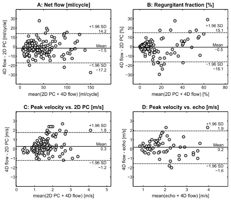
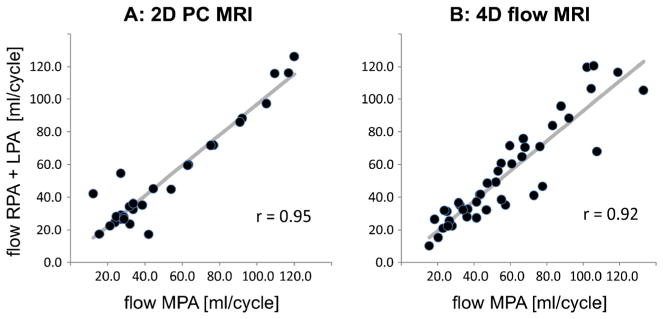
Results: Excellent correlation and agreement were found between 2-D phase-contrast MRI and 4-D flow for net flow (r = 0.97, P < 0.001) and excellent correlation with good agreement was found for regurgitant fraction (r = 0.88, P < 0.001) in all vessels. Two-dimensional phase-contrast MRI significantly underestimated aortic (P = 0.032) and MPA (P < 0.001) peak velocities compared to echo, while volumetric 4-D flow analysis resulted in higher (aortic: P = 0.001) or similar (MPA: P = 0.98) peak velocities relative to echo.
Conclusion: Excellent flow parameter agreement between 2-D phase-contrast MRI and 4-D flow and the improved volumetric 4-D flow velocity analysis relative to echo suggests that 4-D flow has the potential to become a clinical alternative to 2-D phase-contrast MRI.
Conflict of interest statement
Figures
Similar articles
-
Analysis of an automated background correction method for cardiovascular MR phase contrast imaging in children and young adults.Pediatr Radiol. 2014 Mar;44(3):265-73. doi: 10.1007/s00247-013-2830-y. Epub 2013 Dec 5. Pediatr Radiol. 2014. PMID: 24306733
-
Efficient method for volumetric assessment of peak blood flow velocity using 4D flow MRI.J Magn Reson Imaging. 2016 Dec;44(6):1673-1682. doi: 10.1002/jmri.25305. Epub 2016 May 18. J Magn Reson Imaging. 2016. PMID: 27192153 Free PMC article.
-
Peak velocity measurements in tortuous arteries with phase contrast magnetic resonance imaging: the effect of multidirectional velocity encoding.Invest Radiol. 2014 Apr;49(4):189-94. doi: 10.1097/RLI.0000000000000013. Invest Radiol. 2014. PMID: 24300842
-
Quantification of peak blood flow velocity at the cardiac valve and great thoracic vessels by four-dimensional flow and two-dimensional phase-contrast MRI compared with echocardiography: a systematic review and meta-analysis.Clin Radiol. 2021 Nov;76(11):863.e1-863.e10. doi: 10.1016/j.crad.2021.07.011. Epub 2021 Aug 14. Clin Radiol. 2021. PMID: 34404516
-
Imaging of the thoracic aorta with time-resolved three-dimensional phase-contrast MRI: a review.Semin Thorac Cardiovasc Surg. 2008 Winter;20(4):358-64. doi: 10.1053/j.semtcvs.2008.11.013. Semin Thorac Cardiovasc Surg. 2008. PMID: 19251177 Review.
Cited by
-
Right and left ventricular function and flow quantification in pediatric patients with repaired tetralogy of Fallot using four-dimensional flow magnetic resonance imaging.BMC Med Imaging. 2021 Oct 31;21(1):161. doi: 10.1186/s12880-021-00693-2. BMC Med Imaging. 2021. PMID: 34719378 Free PMC article.
-
In vitro evaluation of cerebrospinal fluid velocity measurement in type I Chiari malformation: repeatability, reproducibility, and agreement using 2D phase contrast and 4D flow MRI.Fluids Barriers CNS. 2021 Mar 18;18(1):12. doi: 10.1186/s12987-021-00246-3. Fluids Barriers CNS. 2021. PMID: 33736664 Free PMC article.
-
Age-Related Changes of Normal Cerebral and Cardiac Blood Flow in Children and Adults Aged 7 Months to 61 Years.J Am Heart Assoc. 2016 Jan 4;5(1):e002657. doi: 10.1161/JAHA.115.002657. J Am Heart Assoc. 2016. PMID: 26727967 Free PMC article.
-
Four-dimensional flow cardiac magnetic resonance assessment of left ventricular diastolic function.Front Cardiovasc Med. 2022 Jul 22;9:866131. doi: 10.3389/fcvm.2022.866131. eCollection 2022. Front Cardiovasc Med. 2022. PMID: 35935619 Free PMC article. Review.
-
Clinical assessment of aortic valve stenosis: Comparison between 4D flow MRI and transthoracic echocardiography.J Magn Reson Imaging. 2020 Feb;51(2):472-480. doi: 10.1002/jmri.26847. Epub 2019 Jun 30. J Magn Reson Imaging. 2020. PMID: 31257647 Free PMC article.
References
-
- Atkinson DJ, Edelman RR. Cineangiography of the heart in a single breath hold with a segmented turboFLASH sequence. Radiology. 1991;178:357–360. - PubMed
-
- Beerbaum P, Korperich H, Barth P, et al. Noninvasive quantification of left-to-right shunt in pediatric patients: phase-contrast cine magnetic resonance imaging compared with invasive oximetry. Circulation. 2001;103:2476–2482. - PubMed
-
- Chai P, Mohiaddin R. How we perform cardiovascular magnetic resonance flow assessment using phase-contrast velocity mapping. J Cardiovasc Magn Reson. 2005;7:705–716. - PubMed
-
- Didier D. Assessment of valve disease: qualitative and quantitative. Magn Reson Imaging Clin N Am. 2003;11:115–134. - PubMed
-
- Gatehouse PD, Keegan J, Crowe LA, et al. Applications of phase-contrast flow and velocity imaging in cardiovascular MRI. Eur Radiol. 2005;15:2172–2184. - PubMed
Publication types
MeSH terms
Substances
Grants and funding
LinkOut - more resources
Full Text Sources
Other Literature Sources
Miscellaneous
