Review
. 2015 Feb;38(2):124-30.
doi: 10.1002/clc.22346. Epub 2014 Dec 8.

Direct myocardial ischemia imaging: a new cardiovascular nuclear imaging paradigm

Affiliations
Review

Direct myocardial ischemia imaging: a new cardiovascular nuclear imaging paradigm

Diwakar Jain et al. Clin Cardiol. 2015 Feb.

Abstract

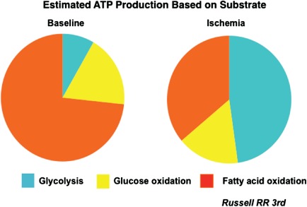
Myocardial perfusion imaging (MPI), using radiotracers, has been in routine clinical use for over 40 years. This modality is used for the detection of coronary artery disease (CAD), risk stratification, optimizing therapy, and follow-up of patients with CAD. Molecular cardiovascular imaging using targeted radiotracers provides a unique opportunity for imaging biochemical and metabolic processes, and cell membrane transporter and receptor functions at a cellular and molecular level in experimental animal models as well as in humans. Cardiac imaging using radiolabeled free fatty acid analogues and glucose analogues enable us to image myocardial ischemia directly as an alternative to stress-rest MPI. Direct ischemia imaging techniques can avoid and overcome some of the limitations of standard stress-rest MPI. This article describes recent studies using (18) F-fluorodeoxyglucose ((18) FDG) for myocardial ischemia imaging.


Figures

Figure 1
Figure 1
A diagrammatic representation of the relative proportion of myocardial substrate utilization. Under resting conditions, fatty acid metabolism contributes to nearly 70% to 75% energy generation, whereas the remaining comes from glucose metabolism (approximately 8% from glycolysis and 18% from oxidative metabolism). Myocardial substrate utilization changes dramatically with the onset of myocardial ischemia, with glycolysis contributing to nearly 50% of the energy production and a significant reduction in fatty acid uptake and metabolism. Courtesy of Dr. Raymond R. Russell, III, Yale University School of Medicine. Abbreviations: ATP, adenosine triphosphate.
Figure 2
Figure 2
Representative exercise (Ex) and rest (R) technetium‐99 m‐sestamibi and exercise 18 F‐fluorodeoxyglucose (18FDG) images of a patient with angina in the short axis, and vertical and horizontal long axes. This patient had no history of myocardial infarction. A large area of partially reversible perfusion abnormality involving the inferior and lateral walls is seen on the perfusion images. Intense 18FDG uptake is seen in the areas corresponding to the perfusion abnormalities. On coronary angiography, 100% occlusion of the right coronary artery and a 50% narrowing of the left anterior descending coronary artery were observed. Reproduced with permission from Jain and He.34
Figure 3
Figure 3
Representative exercise (Ex) and rest (R) technetium‐99 m‐sestamibi and exercise 18 F‐fluorodeoxyglucose (18FDG) images of a patient with angina in the short axis, and vertical and horizontal long axes. This patient had no prior myocardial infarction. No perfusion abnormality is observed on the stress and rest perfusion images. An intense global 18FDG uptake is observed in all 3 vascular territories (solid arrowheads). On coronary angiography, 3‐vessel disease was observed (70% narrowing of the left anterior descending, 60% narrowing of the left circumflex, and 60% narrowing of the right coronary arteries). Reproduced with permission from He et al.30
Figure 4
Figure 4
This 49‐year‐old male with exertional angina underwent exercise (Ex) and rest 99mTc‐sestamibi (Perf) and 18 F‐fluorodeoxyglucose (FDG) imaging 24 hours apart. Representative short axis, and vertical (Verti) and horizontal (Horiz) long (Lg) axes slices of the heart are shown. Reversible perfusion abnormality in the posterior septum and inferior walls is observed on this study. Intense FDG uptake on the exercise images (yellow arrows) is seen in the corresponding segments. No FDG uptake is seen in the heart on rest images. This patient was found to have 85% narrowing of the right coronary artery. Reproduced with permission from Dou et al.40

References

    1. Iskandrian AE, Hage FG, Shaw LJ, et al. Serial myocardial perfusion imaging: defining a significant change and targeting management decisions. JACC Cardiovasc Imaging. 2014;7:79–96. - PubMed
    1. Zaret BL, Strauss HW, Martin ND, et al. Noninvasive evaluation of regional myocardial perfusion with radioactive potassium: study of patients at rest, exercise, and during anginal pectoris. N Engl J Med. 1973;288:809–812. - PubMed
    1. Cremer P, Hachamovitch R, Tamarappoo B. Clinical decision making with myocardial perfusion imaging in patients with known or suspected coronary artery disease. Semin Nucl Med. 2014;44:320–329. - PMC - PubMed
    1. Jain D, Zaret BL. Nuclear imaging in cardiovascular medicine In: Rosendorf C, ed. Essential Cardiology 3rd ed New York, NY: Springer; 2013:195–220.
    1. Jain D, Wackers FJ, Mattera J, et al. Biokinetics of technetium‐99 m‐tetrofosmin: myocardial perfusion imaging agent: implications for a one‐day imaging protocol. J Nucl Med. 1993;34:1254–1259. - PubMed

MeSH terms

Substances

LinkOut - more resources