

Segmentation of tongue muscles from super-resolution magnetic resonance images
- PMID: 25487963
- PMCID: PMC4294977
- DOI: 10.1016/j.media.2014.11.006
Segmentation of tongue muscles from super-resolution magnetic resonance images
Abstract
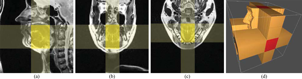
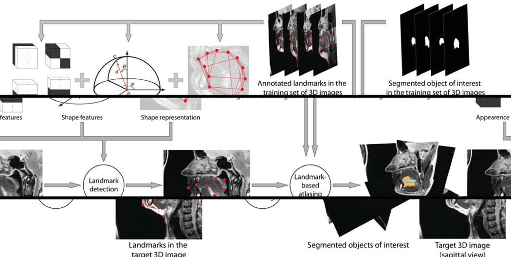
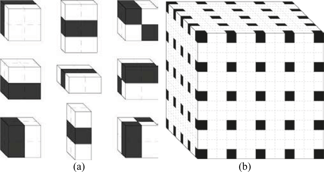
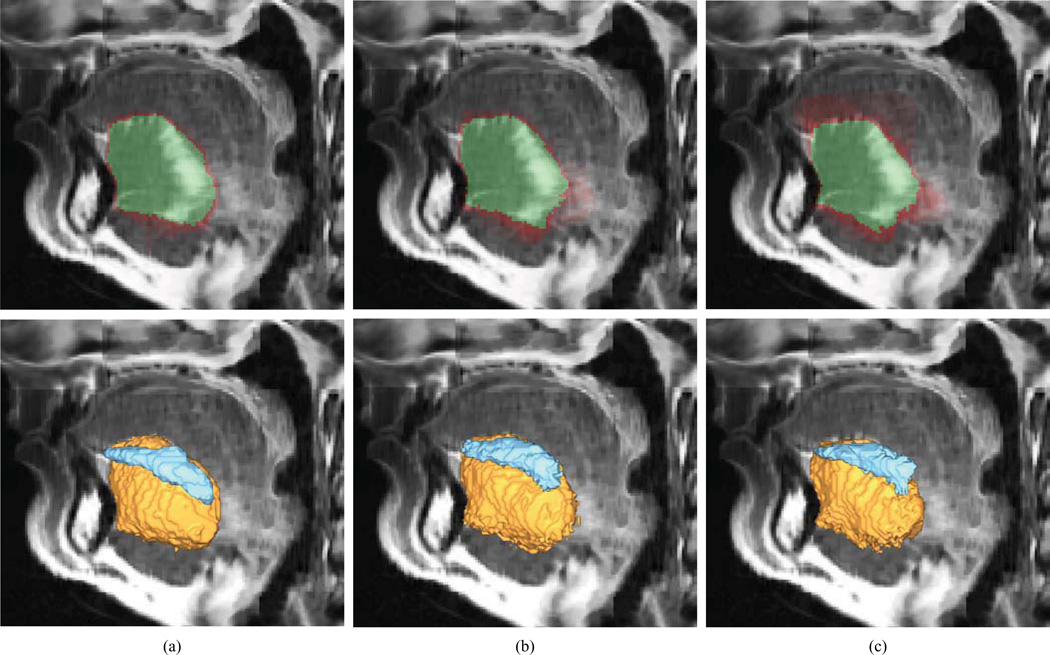
Imaging and quantification of tongue anatomy is helpful in surgical planning, post-operative rehabilitation of tongue cancer patients, and studying of how humans adapt and learn new strategies for breathing, swallowing and speaking to compensate for changes in function caused by disease, medical interventions or aging. In vivo acquisition of high-resolution three-dimensional (3D) magnetic resonance (MR) images with clearly visible tongue muscles is currently not feasible because of breathing and involuntary swallowing motions that occur over lengthy imaging times. However, recent advances in image reconstruction now allow the generation of super-resolution 3D MR images from sets of orthogonal images, acquired at a high in-plane resolution and combined using super-resolution techniques. This paper presents, to the best of our knowledge, the first attempt towards automatic tongue muscle segmentation from MR images. We devised a database of ten super-resolution 3D MR images, in which the genioglossus and inferior longitudinalis tongue muscles were manually segmented and annotated with landmarks. We demonstrate the feasibility of segmenting the muscles of interest automatically by applying the landmark-based game-theoretic framework (GTF), where a landmark detector based on Haar-like features and an optimal assignment-based shape representation were integrated. The obtained segmentation results were validated against an independent manual segmentation performed by a second observer, as well as against B-splines and demons atlasing approaches. The segmentation performance resulted in mean Dice coefficients of 85.3%, 81.8%, 78.8% and 75.8% for the second observer, GTF, B-splines atlasing and demons atlasing, respectively. The obtained level of segmentation accuracy indicates that computerized tongue muscle segmentation may be used in surgical planning and treatment outcome analysis of tongue cancer patients, and in studies of normal subjects and subjects with speech and swallowing problems.
Keywords: Atlasing; Game theory; Human tongue; Magnetic resonance imaging; Segmentation.
Copyright © 2014 Elsevier B.V. All rights reserved.
Figures

References
-
- Akgul YS, Kambhamettu C, Stone M. Extraction and tracking of the tongue surface from ultrasound image sequences; Proc, IEEE Comput. Soc. Conf. on Comput. Vis. and Pattern Recogn; 1998. pp. 298–303.
-
- Bozma H, Duncan J. A game-theoretic approach to integration of modules. IEEE Trans. Pattern Anal. Mach. Intell. 1994;16(11):1074–1086.
-
- Bresch E, Kim Y-C, Nayak K, Byrd D, Narayanan S. Seeing speech: capturing vocal tract shaping using real-time magnetic resonance imaging. IEEE Signal Process. Mag. 2008;25:123–132.
-
- Chong VFH, Zhou J-Y, Khoo JBK, Huang J, Lim T-K. Tongue carcinoma: tumor volume measurement. Int. J. Radiat. Oncol. Biol. Phys. 2004;59:59–66. - PubMed
Publication types
MeSH terms
Grants and funding
LinkOut - more resources
Full Text Sources
Other Literature Sources
Medical
