. 2014 Dec 9:4:7382.
doi: 10.1038/srep07382.

Ultrasensitive non-enzymatic glucose sensor based on three-dimensional network of ZnO-CuO hierarchical nanocomposites by electrospinning

Affiliations

Ultrasensitive non-enzymatic glucose sensor based on three-dimensional network of ZnO-CuO hierarchical nanocomposites by electrospinning

Chunyang Zhou et al. Sci Rep. .

Abstract

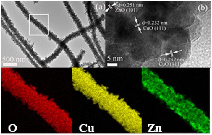
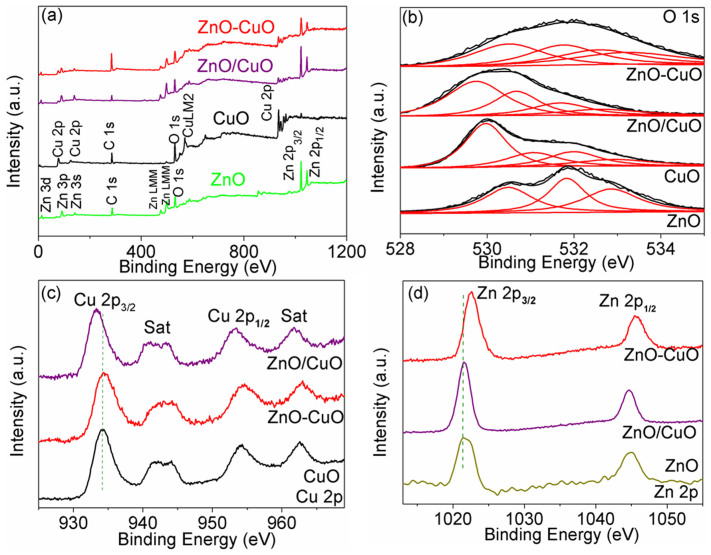
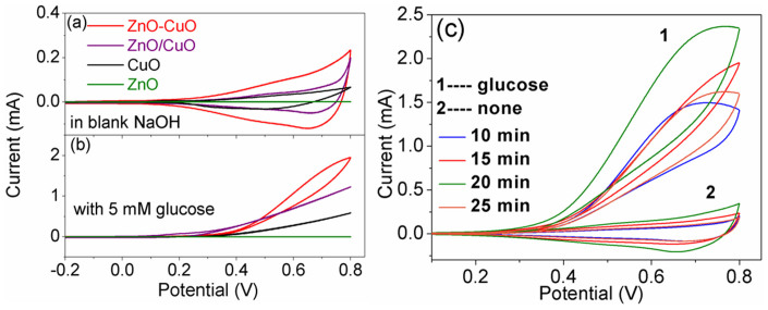
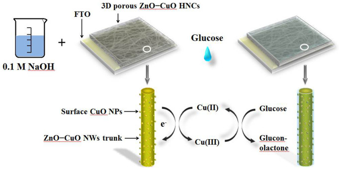
Three-dimensional (3D) porous ZnO-CuO hierarchical nanocomposites (HNCs) nonenzymatic glucose electrodes with different thicknesses were fabricated by coelectrospinning and compared with 3D mixed ZnO/CuO nanowires (NWs) and pure CuO NWs electrodes. The structural characterization revealed that the ZnO-CuO HNCs were composed of the ZnO and CuO mixed NWs trunk (~200 nm), whose outer surface was attached with small CuO nanoparticles (NPs). Moreover, a good synergetic effect between CuO and ZnO was confirmed. The nonenzymatic biosensing properties of as prepared 3D porous electrodes based on fluorine doped tin oxide (FTO) were studied and the results indicated that the sensing properties of 3D porous ZnO-CuO HNCs electrodes were significantly improved and depended strongly on the thickness of the HNCs. At an applied potential of + 0.7 V, the optimum ZnO-CuO HNCs electrode presented a high sensitivity of 3066.4 μAmM(-1)cm(-2), the linear range up to 1.6 mM, and low practical detection limit of 0.21 μM. It also showed outstanding long term stability, good reproducibility, excellent selectivity and accurate measurement in real serum sample. The formation of special hierarchical heterojunction and the well-constructed 3D structure were the main reasons for the enhanced nonenzymatic biosensing behavior.


Figures

Figure 1
Figure 1. The XRD of 3D ZnO–CuO HNCs, mixed ZnO/CuO NWs, pure CuO and ZnO NWs samples.
The XRD peaks of CuO (JCPDS no.48-1548) are marked with “#” and ZnO (JCPDS no.36-1451) are marked with “*”.
Figure 2
Figure 2
(a) and (b) typical SEM image of 3D porous ZnO–CuO HNCs electrode (20 min) on the surface of FTO. (c) and (d) the low- and high-magnification cross section SEM images of (b). (e) The SEM image of 3D pure CuO NWs electrode. The inset of (e) is its high-magnification SEM image. (f) The SEM image of 3D mixed ZnO/CuO NWs and its inset is high-magnification SEM image.
Figure 3
Figure 3
(a) TEM image and (b) HR-TEM image of ZnO–CuO HNCs. (c), (d), and (e), EDX elemental mapping images of O, Cu, and, Zn in the ZnO–CuO HNCs sample taken from the box of (a).
Figure 4
Figure 4
(a) Survey, (b) O 1s, (c) Cu 2p, and (d) Zn 2p high resolution XPS spectrum of the corresponding samples.
Figure 5
Figure 5
The CV curves of 3D porous ZnO–CuO HNCs as well as mixed ZnO/CuO NWs, pure CuO and ZnO NWs electrodes both fabricated for 20 min in (a) blank NaOH solution and (b) NaOH solution with 5 mM glucose; (c) CV curves of 3D porous ZnO–CuO HNCs electrodes (10, 15, 20, 25 min) in 0.1 M NaOH in the absence (trace 2) and presence of 5 mM glucose (trace 1), respectively. The scan rate is 100 mV/s.
Figure 6
Figure 6. Reaction mechanism of 3D porous ZnO–CuO HNCs electrodes.
(the image of the flask are painting according to the model in our experiment by Lin Xu, one of the auther of this article).
Figure 7
Figure 7. CV curves of the 3D porous ZnO–CuO HNCs at various scan rates (50, 100, 150, 200, and 250 mV/s), inset is the plots of peak current vs. scan rate.
Figure 8
Figure 8
(a) Amperometric response of 3D porous ZnO–CuO HNCs (10, 15, 20, and 25 min) electrodes as well as 3D mixed ZnO/CuO, 3D pure CuO and ZnO NWs electrodes at an applied potential of 0.7 V upon successive additions of different concentration of glucose in a step of 10, 50, and 200 μM, respectively for each current step, inset is the current response of 3D porous ZnO–CuO HNCs (20 min) to 0.47 and 1 μM glucose). (b) The corresponding calibration curve of current vs. concentration of glucose. The error bars denote the standard deviation of triplicate determination of each concentration of glucose.
Figure 9
Figure 9
The amperometric response of the 3D porous ZnO–CuO HNCs electrode (20 min) (a) with successive additions of different interfering species (UA, AA, DA, NADH, Mg2+, and Ca2+) and (b) with sequential addition of 10 μΜ various interfering sugars (maltose, mannose, lactose, and galactose) after initial addition of 100 μΜ glucose. The inset of (a) is its current response towards to 100 µM glucose in human serum.

References

    1. Zhai D. Y. et al. Highly sensitive glucose sensor based on Pt nanoparticle/polyaniline hydrogel heterostructures. ACS Nano 4, 3540–3546 (2013). - PubMed
    1. Kros A. et al. Poly (3, 4–ethylenedioxythiophene)–Based Glucose Biosensors. Adv. Mater. 13, 1555–1557 (2001).
    1. Shafer–Peltier K. E. et al. Toward a glucose biosensor based on surface-enhanced raman scattering. J. Am. Chem. Soc. 125, 588–593 (2003). - PubMed
    1. Niu X. H. et al. Electrochemical sensing interfaces with tunable porosity for nonenzymatic glucose detection: A Cu foam case. Biosens. Bioelectron. 51, 22–28 (2014). - PubMed
    1. Niu X. H. et al. Highly sensitive and selective nonenzymatic detection of glucose using three-dimensional porous nickel nanostructures. Anal. Chem. 85, 3561–3569 (2013). - PubMed

Publication types