. 2014 Dec 9;18(6):676.
doi: 10.1186/s13054-014-0676-9.

Assessment of brain midline shift using sonography in neurosurgical ICU patients

Affiliations

Assessment of brain midline shift using sonography in neurosurgical ICU patients

Julie Motuel et al. Crit Care. .

Abstract

Introduction: Brain midline shift (MLS) is a life-threatening condition that requires urgent diagnosis and treatment. We aimed to validate bedside assessment of MLS with Transcranial Sonography (TCS) in neurosurgical ICU patients by comparing it to CT.

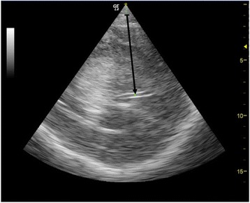
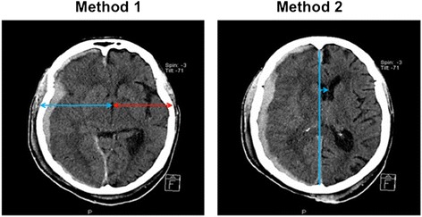
Methods: In this prospective single centre study, patients who underwent a head CT were included and a concomitant TCS performed. TCS MLS was determined by measuring the difference between the distance from skull to the third ventricle on both sides, using a 2 to 4 MHz probe through the temporal window. CT MLS was measured as the difference between the ideal midline and the septum pellucidum. A significant MLS was defined on head CT as > 0.5 cm.

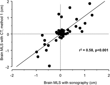
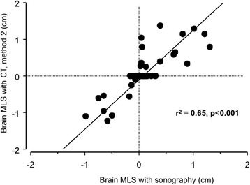
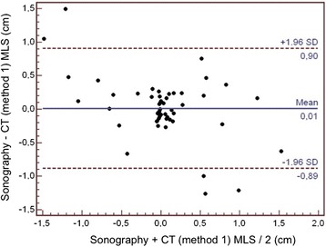
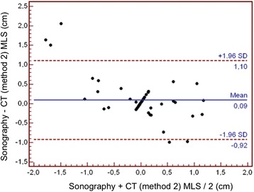
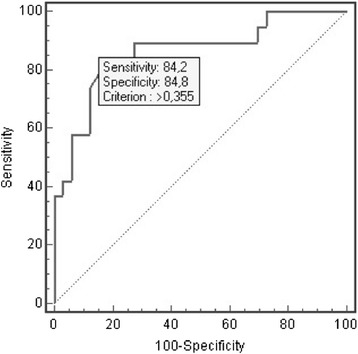
Results: A total of 52 neurosurgical ICU patients were included. The MLS (mean ± SD) was 0.32 ± 0.36 cm using TCS and 0.47 ± 0.67 cm using CT. The Pearson's correlation coefficient (r(2)) between TCS and CT scan was 0.65 (P < 0.001). The bias was 0.09 cm and the limits of agreements were 1.10 and -0.92 cm. The area under the ROC curve for detecting a significant MLS with TCS was 0.86 (95% CI = 0.74 to 0.94), and, using 0.35 cm as a cut-off, the sensitivity was 84.2%, the specificity 84.8% and the positive likelihood ratio was 5.56.

Conclusions: This study suggests that TCS could detect MLS with reasonable accuracy in neurosurgical ICU patients and that it could serve as a bedside tool to facilitate early diagnosis and treatment for patients with a significant intracranial mass effect.


Figures

Figure 1
Figure 1
Example of visualisation of the third ventricle with brain sonography, the arrow pointing to the centre of the third ventricle.
Figure 2
Figure 2
Example of MLS measure with CT scan: on the left, method 1 (in the same plane as sonography through the third ventricle), and on the right, method 2 (measuring the distance between the ideal median line and the septum pellucidum). CT, computed tomography; MLS, midline shift.
Figure 3
Figure 3
Correlation between sonography and CT method 1 for MLS assessment. CT, computed tomography; MLS, midline shift.
Figure 4
Figure 4
Correlation between sonography and CT method 2 for MLS assessment. CT, computed tomography; MLS, midline shift.
Figure 5
Figure 5
Bland and Altman plot for the agreement between sonography and CT method 1 for MLS assessment. CT, computed tomography; MLS, midline shift.
Figure 6
Figure 6
Bland and Altman plot for the agreement between sonography and CT method 2 for MLS assessment. CT, computed tomography; MLS, midline shift.
Figure 7
Figure 7
Receiver operating characteristic curve for the detection of a CT MLS >0.5 cm (method 2) with TCS. CT, computed tomography; MLS, midline shift; TCS, transcranial sonography.

References

    1. Becker DP, Miller JD, Ward JD, Greenberg RP, Young HF, Sakalas R. The outcome from severe head injury with early diagnosis and intensive management. J Neurosurg. 1977;47:491–502. doi: 10.3171/jns.1977.47.4.0491. - DOI - PubMed
    1. Vollmer G, Torner JC, Jane JA, Sadovnic B, Charlebois D, Eisenberg HM, Foulkes MA, Marmarou A, Marshall LF. Age and outcome following traumatic coma: why do older patients fare worse? J Neurosurg. 1991;75:S37–S49.
    1. Quattrocchi KB, Prasad P, Willits NH, Wagner FC., Jr Quantification of midline shift as a predictor of poor outcome following head injury. Surg Neurology. 1991;35:183–188. doi: 10.1016/0090-3019(91)90069-L. - DOI - PubMed
    1. Young B, Rapp RP, Norton JA, Haack D, Tibbs PA, Bean JR. Early prediction of outcome in head-injured patients. J Neurosurg. 1981;54:300–303. doi: 10.3171/jns.1981.54.3.0300. - DOI - PubMed
    1. Ross DA, Olsen WL, Ross AM, Andrews BT, Pitts LH. Brain shift, level of consciousness, and restoration of consciousness in patients with acute intracranial hematoma. J Neurosurg. 1989;71:498–502. doi: 10.3171/jns.1989.71.4.0498. - DOI - PubMed

LinkOut - more resources