

Surviving mossy cells enlarge and receive more excitatory synaptic input in a mouse model of temporal lobe epilepsy
- PMID: 25488607
- PMCID: PMC4412767
- DOI: 10.1002/hipo.22396
Surviving mossy cells enlarge and receive more excitatory synaptic input in a mouse model of temporal lobe epilepsy
Abstract
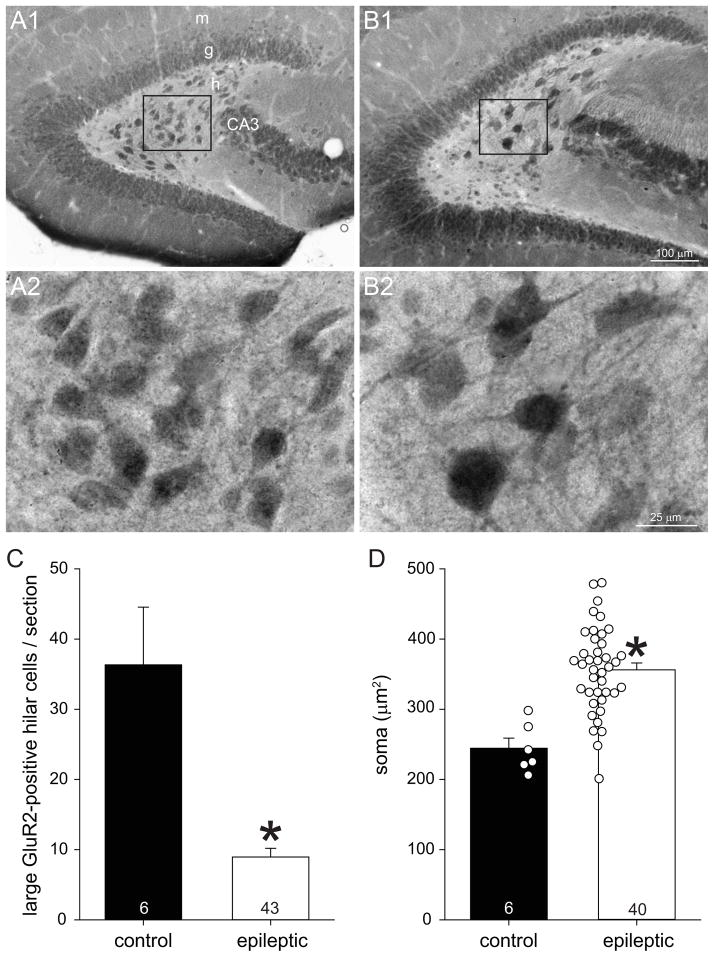
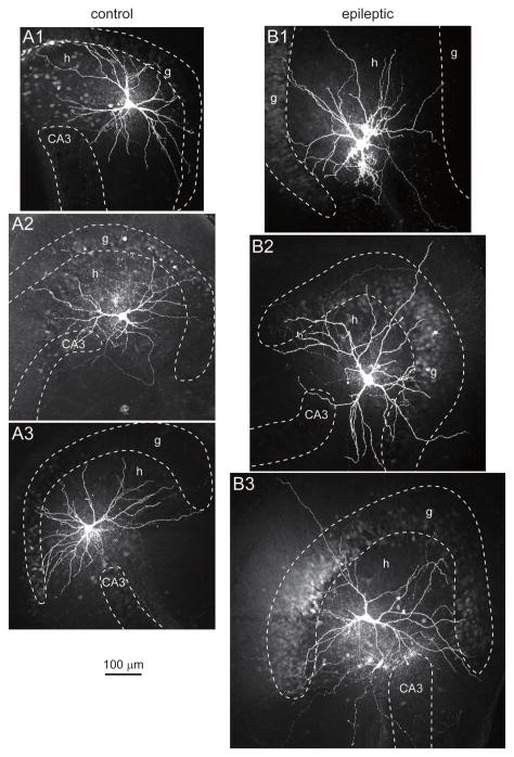
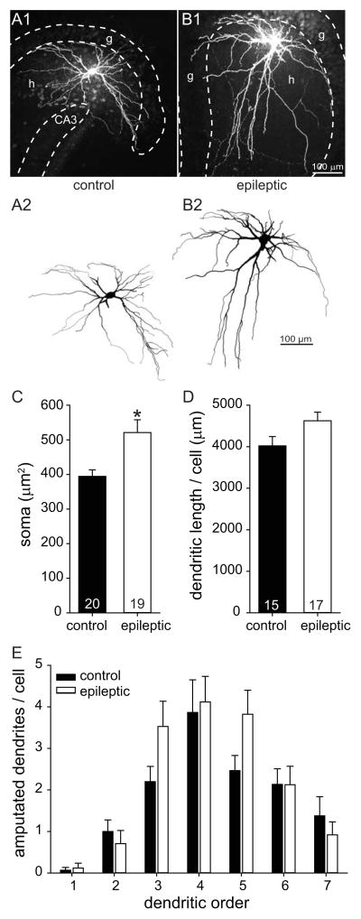
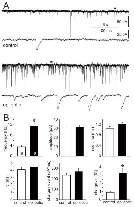
Numerous hypotheses of temporal lobe epileptogenesis have been proposed, and several involve hippocampal mossy cells. Building on previous hypotheses we sought to test the possibility that after epileptogenic injuries surviving mossy cells develop into super-connected seizure-generating hub cells. If so, they might require more cellular machinery and consequently have larger somata, elongate their dendrites to receive more synaptic input, and display higher frequencies of miniature excitatory synaptic currents (mEPSCs). To test these possibilities pilocarpine-treated mice were evaluated using GluR2-immunocytochemistry, whole-cell recording, and biocytin-labeling. Epileptic pilocarpine-treated mice displayed substantial loss of GluR2-positive hilar neurons. Somata of surviving neurons were 1.4-times larger than in controls. Biocytin-labeled mossy cells also were larger in epileptic mice, but dendritic length per cell was not significantly different. The average frequency of mEPSCs of mossy cells recorded in the presence of tetrodotoxin and bicuculline was 3.2-times higher in epileptic pilocarpine-treated mice as compared to controls. Other parameters of mEPSCs were similar in both groups. Average input resistance of mossy cells in epileptic mice was reduced to 63% of controls, which is consistent with larger somata and would tend to make surviving mossy cells less excitable. Other intrinsic physiological characteristics examined were similar in both groups. Increased excitatory synaptic input is consistent with the hypothesis that surviving mossy cells develop into aberrantly super-connected seizure-generating hub cells, and soma hypertrophy is indirectly consistent with the possibility of axon sprouting. However, no obvious evidence of hyperexcitable intrinsic physiology was found. Furthermore, similar hypertrophy and hyper-connectivity has been reported for other neuron types in the dentate gyrus, suggesting mossy cells are not unique in this regard. Thus, findings of the present study reveal epilepsy-related changes in mossy cell anatomy and synaptic input but do not strongly support the hypothesis that mossy cells develop into seizure-generating hub cells.
Keywords: GluR2; dendrites; dentate gyrus; hypertrophy; miniature EPSC.
© 2014 Wiley Periodicals, Inc.
Figures

References
-
- Amaral DG. A Golgi study of cell types in the hilar region of the hippocampus in the rat. J Comp Neurol. 1978;182:851–914. - PubMed
-
- Babb TL, Brown WJ, Pretorious J, Davenport C, Lieb JP, Crandall PH. Temporal lobe volumetric cell densities in temporal lobe epilepsy. Epilepsia. 1984;25:729–740. - PubMed
-
- Backman SA, Stambolic V, Suzuki A, Haight J, Elia A, Pretorius J, Tsao M-S, Shannon P, Bolon B, Ivy GO, Mak TW. Deletion of Pten in mouse brain causes seizures ataxia and defects in soma size resembling Lhermitte-Duclos disease. Nat Genet. 2001;29:396–403. - PubMed
-
- Blümcke I, Suter B, Behle K, Kuhn R, Schramm J, Elger CE, Wiestler OD. Loss of hilar mossy cells in Ammon’s horn sclerosis. Epilepsia. 2000;41(s6):S174–S180. - PubMed
-
- Blümcke I, Zuschratter W, Schewe J-C, Suter B, Lie AA, Riederer BM, Meyer B, Schramm J, Elger CE, Wiestler OD. Cellular pathology of hilar neurons in Ammon’s horn sclerosis. J Comp Neurol. 1999;414:437–453. - PubMed
Publication types
MeSH terms
Substances
Grants and funding
LinkOut - more resources
Full Text Sources
Other Literature Sources
