. 2015 Jan 23;290(4):2379-94.
doi: 10.1074/jbc.M114.607341. Epub 2014 Dec 8.

The C-terminal 50 amino acid residues of dengue NS3 protein are important for NS3-NS5 interaction and viral replication

Affiliations

The C-terminal 50 amino acid residues of dengue NS3 protein are important for NS3-NS5 interaction and viral replication

Moon Y F Tay et al. J Biol Chem. .

Abstract

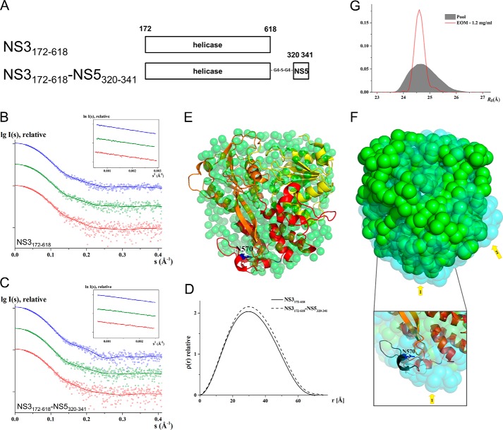
Dengue virus multifunctional proteins NS3 protease/helicase and NS5 methyltransferase/RNA-dependent RNA polymerase form part of the viral replication complex and are involved in viral RNA genome synthesis, methylation of the 5'-cap of viral genome, and polyprotein processing among other activities. Previous studies have shown that NS5 residue Lys-330 is required for interaction between NS3 and NS5. Here, we show by competitive NS3-NS5 interaction ELISA that the NS3 peptide spanning residues 566-585 disrupts NS3-NS5 interaction but not the null-peptide bearing the N570A mutation. Small angle x-ray scattering study on NS3(172-618) helicase and covalently linked NS3(172-618)-NS5(320-341) reveals a rigid and compact formation of the latter, indicating that peptide NS5(320-341) engages in specific and discrete interaction with NS3. Significantly, NS3:Asn-570 to alanine mutation introduced into an infectious DENV2 cDNA clone did not yield detectable virus by plaque assay even though intracellular double-stranded RNA was detected by immunofluorescence. Detection of increased negative-strand RNA synthesis by real time RT-PCR for the NS3:N570A mutant suggests that NS3-NS5 interaction plays an important role in the balanced synthesis of positive- and negative-strand RNA for robust viral replication. Dengue virus infection has become a global concern, and the lack of safe vaccines or antiviral treatments urgently needs to be addressed. NS3 and NS5 are highly conserved among the four serotypes, and the protein sequence around the pinpointed amino acids from the NS3 and NS5 regions are also conserved. The identification of the functionally essential interaction between the two proteins by biochemical and reverse genetics methods paves the way for rational drug design efforts to inhibit viral RNA synthesis.

Keywords: Dengue Virus; Flavivirus; Nonstructural Protein Interaction; Plus-stranded RNA Virus; Protein-Protein Interaction; Replication Complex; Viral Helicase; Viral Polymerase; Viral Protein; Viral Replication.


Figures

FIGURE 1.
FIGURE 1.
Competitive NS3-NS5 interaction ELISA with NS3-truncated proteins. A, schematic representation and diagram of the recombinant NS3 and NS5 proteins that were expressed in E. coli and used in B for competitive NS3-NS5 interaction ELISA. C, during incubation of NS2B18NS3 protein with coated NS5 RdRP, 2-fold serial dilution of either NS3 or GST protein (starting from 20 μm) was added in triplicate competition experiments. GST protein was included as negative control. Data are shown as the mean ± S.D. of triplicates from two independent experiments.
FIGURE 2.
FIGURE 2.
In vitro functional characterization of NS3 residue Asn-570. A, array of overlapping 15-mer peptides that spanned subdomain III were tested in the competitive ELISA as in Fig. 1B. 20 μm helicase was included as negative control and is shown as the mean ± S.D. of duplicates from one experiment. B, 2-fold serial dilution of 20-mer NS3(566–585) or NS3(566–585)(N570A) peptide (starting from 480 μm) was tested in the competitive ELISA as shown in Fig. 1B. NS3(86–100), which has a similar net charge as NS3(566–585) and NS3(571–585), was included as negative control. Data are shown as the mean ± S.D. of triplicates from two independent experiments. C, viral inhibition assay was performed with NS3 (panel i) or NS5 (panel ii) peptide that spanned the NS3-NS5 interaction site. For NS3 peptides, they formed a nonconvalent complex with penetratin peptide, which also enables NS3 peptide to be transported into the cells (57). For NS5 peptides, they were synthesized as penetratin fusion peptide as penetratin has been shown to have cell-penetrating property (73), and this enables the NS5 peptide to be transported into the cells. 6 h post-infection, the cells were treated with the peptides. Infected cells were harvested at 24 h post-infection for cellular viral RNA quantification by real time RT-PCR analysis. Fold-change was normalized to 24-h control (penetratin alone) and was plotted, and data are shown as the mean ± S.D. of duplicate from one independent experiment. The x axis labels are as follows: penetratin (p), penetratin and NS3(566–585) complex (NS3566–585+p), and penetratin and NS3(86–100) complex (NS386–100+p) for panel i and penetratin (p), penetratin fused to NS5(320–341) (pNS5320–341), and penetratin fused to scrambled NS5(320–341) (pNS5s320–341) for panel ii. D, sequence alignment of NS3 residues 566–585 of DENV2 with other DENV serotypes and representative members of the Flavivirus genus (74). NS3 residue Asn-570 that is critical for NS3-NS5 interaction is highlighted in gray and bold. The alignment was performed using ClustalW (74). The virus sequences and their GenBankTM accession numbers are as follows: DENV2 (AF038403), DENV1 (U88535), DENV3 (M93130), DENV4 (AF326573), yellow fever virus (YFV; X15062), Japanese encephalitis virus (JEV; M55506), Murray Valley encephalitis virus (MVEV; AF161266), and West Nile virus (WNV; M12294). The numbering of residues is based on DENV2 protein sequence. E, ATPase assay of NS2B18NS3 WT and N570A proteins was carried out with 2.5 nm protein, in the presence of the indicated concentrations of ATP. The amount of inorganic phosphate released during reaction was measured with the malachite green reagent, and the initial rates were computed (moles of phosphate released/s). The data points were fitted using Michaelis-Menten Equation 1 (see under “Experimental Procedures”).
FIGURE 3.
FIGURE 3.
SAXS of NS3(172–618)-NS5(320–341) indicates NS3-NS5 interaction. A, schematic representation of recombinant NS3 and NS3-NS5 fusion proteins that were expressed in E. coli and used in SAXS study. SAXS scattering pattern (○) and its corresponding experimental fit (—) of NS3(172–618) (B) and NS3(172–618)-NS5(320–341) (C) at protein concentrations of 1.2 mg/ml (red), 2.1 mg/ml (olive), and 4 mg/ml (blue). The curves are displayed in logarithmic unit for clarity. The insets in B and C show the respective Guinier plot. D, distance distribution functions of NS3(172–618) (—) and NS3(172–618)-NS5(320–341) (—). E, superposition of the determined solution shape of NS3(172–618) with the crystallographic structure of NS3(172–618) (Protein Data Bank code 2JLS (12)). Subdomains I, II, and III are colored in yellow, orange, and red, respectively. F, superposition of the NS3(172–618) and (green) and NS3(172–618)-NS5(320–341) (cyan) solution shapes. The Rg of the NS3(172–618) and NS3(172–618)-NS5(320–341) is 25.2 and 25.4 Å, respectively. The arrows indicate the two protrusions, one at the bottom of the NS3(172–618)-NS5(320–341) solution shape, which leads to a more elongated shape in this protein, reflected by the increased Dmax value (see Fig. 3D). This protrusion is in proximity to the Asn-570 (blue sticks) and NS3 peptide region, NS3(566–585) (black schematic) (inset in F). The second protrusion of the NS3(172–618)-NS5(320–341) may be caused by a conformational alteration due to NS3(172–618) and NS5(320–341) interactions. G, EOM Rg distribution of the selected ensemble (red line) contains a narrow peak at 24.6 Å, which is slightly smaller than the center Rg of the random pool (gray filled area), 24.8 Å, suggesting NS3(172–618)-NS5(320–341) is rigid and compact in solution, and peptide NS5(320–341) is bound to NS3(172–618).
FIGURE 4.
FIGURE 4.
Construction of DENV2 cDNA infectious clone. A, schematic representation of full-length DENV2 cDNA clone generation. The DENV genome (represented approximately to scale) contains a single open reading frame that encodes three structural proteins (c, premembrane protein, and E) and seven NS proteins (NS1, NS2A, NS2B, NS3, NS4A, NS4B, and NS5). Three fragments (fragment boundaries indicated by nucleotide numbers) covering the entire viral genome were made and digested using unique restriction sites to facilitate the assembly of full-length cDNA clone (refer to “Experimental Procedures” for details). The complete DENV2 clone has a T7 promoter at 5′ end for in vitro transcription and SacI site at 3′ end for linearization of plasmid. B and C, BHK-21 cells were electroporated with 10 μg of genomic length RNA, and samples were harvested daily for 3 days. B, infected cells were analyzed for the presence of E protein by IFA with mouse anti-E 4G2 antibody on 48 and 72 h post-transfection, and percentage infection was determined. C, culture supernatants were used to determine viral titer by plaque assay on BHK-21 cells. The size of plaque on BHK-21 cells that was derived from the supernatant of either BHK-21 (left inset) or C6/36 (right inset) cells, which was transfected with DENV2 WT RNA, is shown.
FIGURE 5.
FIGURE 5.
Real time RT-PCR and plaque assay of supernatant, and Western blot and IFA against NS3 proteins of cells transfected with DENV2 WT, NS5:K330A, or NS3:N570A RNAs. A–C, BHK-21 cells were electroporated with 10 μg of WT and mutant (NS3:N570A and NS5:K330A as control) genomic length RNA, and samples were harvested daily for 5 days. Culture supernatants were used to determine the absolute number of extracellular viral RNA copy by real time RT-PCR (A) and to check for plaque production on BHK-21 cells (B). Data are shown as the mean ± S.D. of duplicates from two independent experiments. Plotted absolute number of viral RNA copy in log scale per ml of supernatant used for real time RT-PCR is shown. The detection limit of real time RT-PCR assay is indicated as a gray line in the graph. C and D, Western blot (C) and IFA against NS3 protein (D) were performed with the use of human anti-NS3 3F8 antibody (33), and one representative experiment out of two independent studies was shown.
FIGURE 6.
FIGURE 6.
Real time RT-PCR and IFA against dsRNA of cells transfected with DENV2 WT, NS5:K330A, or NS3:N570A RNAs. A–C, same experiment setup was performed as mentioned in Fig. 5. A, RNA was extracted from infected cells, and the absolute number of intracellular viral RNA copy was determined by real time RT-PCR. B, infected cells were analyzed for the presence of dsRNA by IFA with mouse anti-dsRNA antibody on 48 and 72 h post-transfection, and percentage infection was determined. Data from one independent experiment are shown, and it is a separate experiment from Figs. 4B and 5D. C, extracted RNA was also used to determine positive- and negative-strand synthesis by real time RT-PCR, and absolute number of intracellular viral RNA copy was plotted (one independent experiment). Data are shown as the mean ± S.D. of duplicate from one independent experiment. Plotted absolute number of viral RNA copy in log scale per μg of RNA used for real time RT-PCR. The gray line in A and C indicates the amount of residual negative strand from transfected RNA.
FIGURE 7.
FIGURE 7.
Schematic representation of NS3-NS5-RNA interaction. A, surface electrostatic potential presentation of NS5(273–900) (RdRP) and NS3(172–618) (helicase). The protein backbones of NS5(320–341) and NS3(566–585) are shown in ribbon presentation. The side chains of NS5 Lys-330 and NS3 Asn-570 are displayed. B, simplified schematic model of NS3-NS5 interaction complex with RNA. Dashed line (green) is used to denote that the uncertain path that exiting template RNA from NS3 takes to enter NS5 for complementary daughter strand synthesis. Dark blue line denotes the unwound parental strand; green line denotes the parental strand that serves as template; gray line denotes the newly synthesized daughter strand.

Similar articles

Cited by

References

    1. Bhatt S., Gething P. W., Brady O. J., Messina J. P., Farlow A. W., Moyes C. L., Drake J. M., Brownstein J. S., Hoen A. G., Sankoh O., Myers M. F., George D. B., Jaenisch T., Wint G. R., Simmons C. P., Scott T. W., Farrar J. J., Hay S. I. (2013) The global distribution and burden of dengue. Nature 496, 504–507 - PMC - PubMed
    1. Lindenbach B. D., Thiel H., Rice C. M. (2007) in Fields Virology (Knipe D. M., Howley P. M., eds) 5th Ed., pp. 1101–1152, Wolters Kluwer/Lippincott Williams & Wilkins, Philadelphia
    1. Bartholomeusz A. I., Wright P. J. (1993) Synthesis of dengue virus RNA in vitro: initiation and the involvement of proteins NS3 and NS5. Arch. Virol. 128, 111–121 - PubMed
    1. Falgout B., Pethel M., Zhang Y. M., Lai C. J. (1991) Both nonstructural proteins NS2B and NS3 are required for the proteolytic processing of dengue virus nonstructural proteins. J. Virol. 65, 2467–2475 - PMC - PubMed
    1. Preugschat F., Yao C. W., Strauss J. H. (1990) In vitro processing of dengue virus type 2 nonstructural proteins NS2A, NS2B, and NS3. J. Virol. 64, 4364–4374 - PMC - PubMed

Publication types

MeSH terms

Associated data