. 2015 Feb 6;290(6):3693-707.
doi: 10.1074/jbc.M114.603431. Epub 2014 Dec 8.

Matrix metalloproteinase-14 both sheds cell surface neuronal glial antigen 2 (NG2) proteoglycan on macrophages and governs the response to peripheral nerve injury

Affiliations

Matrix metalloproteinase-14 both sheds cell surface neuronal glial antigen 2 (NG2) proteoglycan on macrophages and governs the response to peripheral nerve injury

Tasuku Nishihara et al. J Biol Chem. .

Abstract

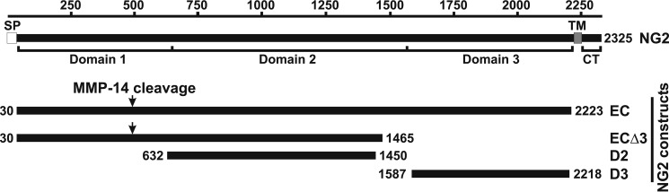
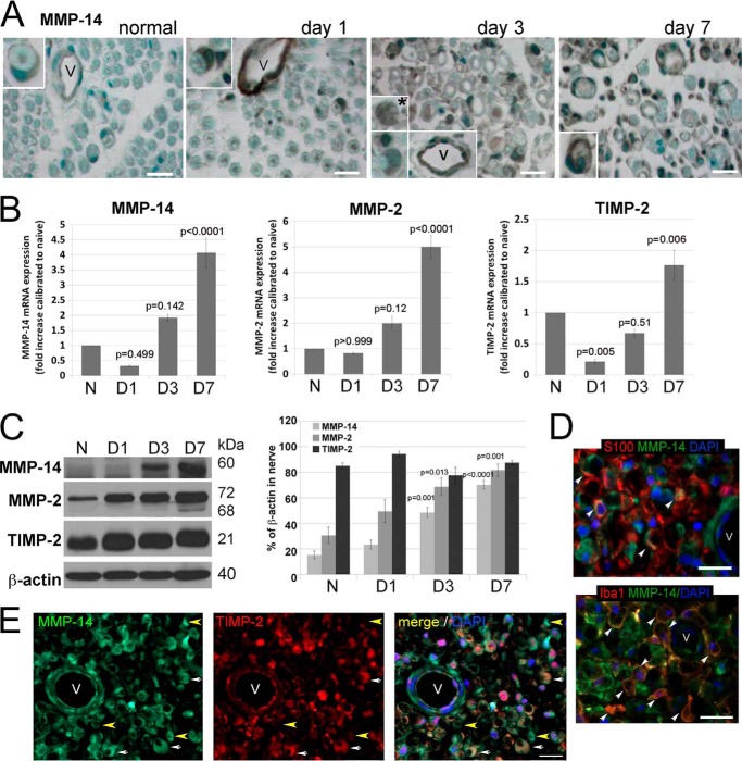
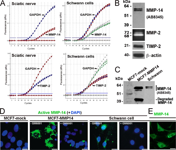
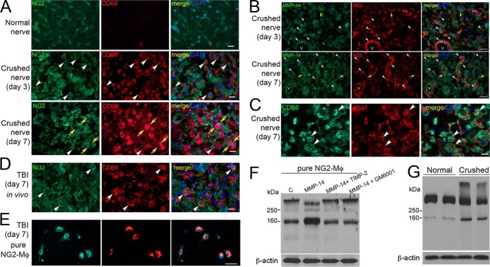
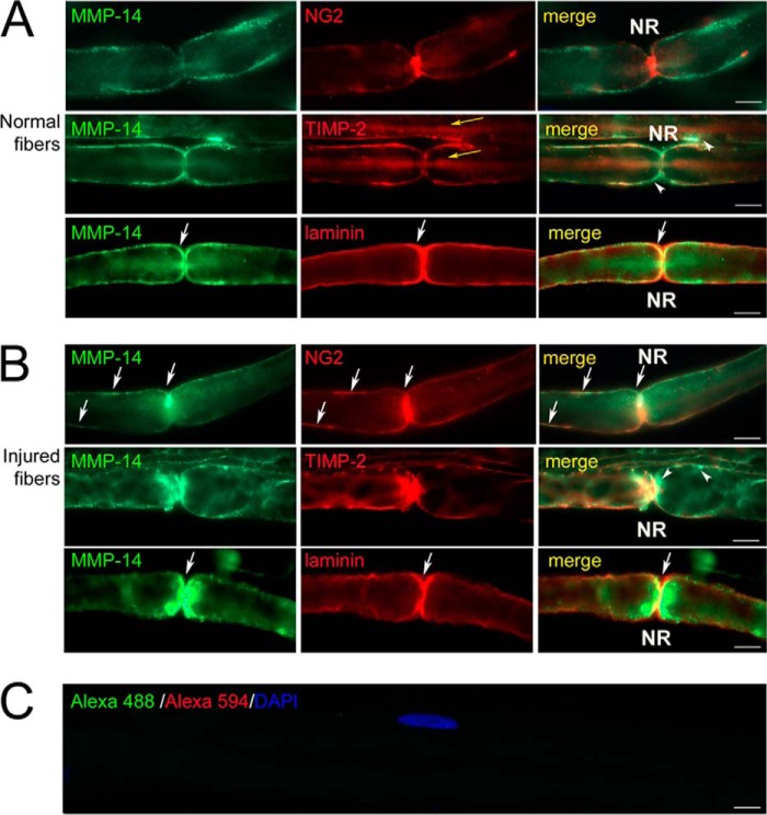
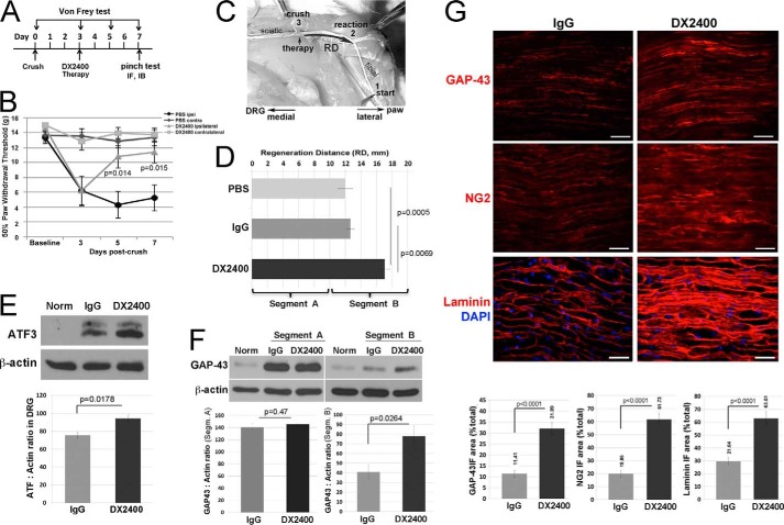
Neuronal glial antigen 2 (NG2) is an integral membrane chondroitin sulfate proteoglycan expressed by vascular pericytes, macrophages (NG2-Mφ), and progenitor glia of the nervous system. Herein, we revealed that NG2 shedding and axonal growth, either independently or jointly, depended on the pericellular remodeling events executed by membrane-type 1 matrix metalloproteinase (MT1-MMP/MMP-14). Using purified NG2 ectodomain constructs, individual MMPs, and primary NG2-Mφ cultures, we demonstrated for the first time that MMP-14 performed as an efficient and unconventional NG2 sheddase and that NG2-Mφ infiltrated into the damaged peripheral nervous system. We then characterized the spatiotemporal relationships among MMP-14, MMP-2, and tissue inhibitor of metalloproteinases-2 in sciatic nerve. Tissue inhibitor of metalloproteinases-2-free MMP-14 was observed in the primary Schwann cell cultures using the inhibitory hydroxamate warhead-based MP-3653 fluorescent reporter. In teased nerve fibers, MMP-14 translocated postinjury toward the nodes of Ranvier and its substrates, laminin and NG2. Inhibition of MMP-14 activity using the selective, function-blocking DX2400 human monoclonal antibody increased the levels of regeneration-associated factors, including laminin, growth-associated protein 43, and cAMP-dependent transcription factor 3, thereby promoting sensory axon regeneration after nerve crush. Concomitantly, DX2400 therapy attenuated mechanical hypersensitivity associated with nerve crush in rats. Together, our findings describe a new model in which MMP-14 proteolysis regulates the extracellular milieu and presents a novel therapeutic target in the damaged peripheral nervous system and neuropathic pain.

Keywords: Extracellular Matrix; MT1-MMP; Macrophage; Matrix Metalloproteinase (MMP); NG2; Nerve; Pain; Proteoglycan; Regeneration; Schwann Cells.


Figures

FIGURE 1.
FIGURE 1.
Schematic representation of the domain structure of NG2 and the purified NG2 ectodomain fragments. The residue numbering is shown at the top. SP, TM, and CT, the signal peptide, the transmembrane domain, and the cytoplasmic tail, respectively. EC, the full-length NG2 ectodomain; ECΔ3, the NG2 ectodomain lacking D3; D2, central domain 2 of NG2; D3, juxtamembrane domain 3 of NG2. The numbers at the beginning and end of each construct refer to the residue numbering. The arrows indicate the putative MMP-14 cleavage site in the D1 domain.
FIGURE 2.
FIGURE 2.
In vitro cleavage of the purified NG2 ectodomain fragments by MMP-14. A, cleavage of rat NG2 by MMP-2, MMP-9, and MMP-14. The proteases were co-incubated for 1 h at 37 °C with purified EC construct at the indicated enzyme-substrate molar ratio. The resulting cleavage products are shown by asterisks. B, the purified EC and ECΔ3 constructs were co-incubated for 1 h at 37 °C with MMP-14 at a 1:10 enzyme-substrate molar ratio. The digests were separated by SDS-gel electrophoresis followed by Coomassie staining. The ∼51–52-kDa NG2 cleavage fragment is shown by an asterisk. Where indicated, GM6001 was added to the reactions. C, confirmation of the specificity of the NG2 antibodies. The D2 antibody recognized the intact D2 domain. The D3 and 05-710 antibodies recognized the intact D3 domain. The anti-D1 1088 antibody did not recognize the intact D2 and D3 domains. D, MMP-14 cleaves the N-terminal portion of NG2. The purified EC and ECΔ3 constructs were each co-incubated with MMP-14 at a 1:10 enzyme-substrate molar ratio. The digests were analyzed by immunoblotting using the indicated NG2 antibodies. The N-terminal ∼51–52-kDa cleavage fragment is shown by an asterisk. The vertical arrow points to a nonspecific band.
FIGURE 3.
FIGURE 3.
The MMP-14/MMP-2/TIMP-2 axis in the PNS. A, immunostaining for MMP-14 using 3G4 antibody (2′2-diaminobenzidine; brown) in rat sciatic nerves at day 0 (normal) and days 1, 3, and 7 after crush injury (the crush site). MMP-14 is observed in Schwann cells (crescent-shaped; insets) and vessel (V) endothelial cells in all nerves and in macrophage-like cells at day 3 postinjury (asterisk). Images are representative of n = 3–4/group. Scale bars are 25 μm. B, TaqMan qPCR of MMP-14, MMP-2, and TIMP-2 in rat sciatic nerves at day 0 (normal (N)) and days 1, 3, and 7 after crush injury. The mean relative mRNA of n = 4/group are normalized to GAPDH and compared with the normal nerve samples (p values by analysis of variance and Bonferroni post hoc test) is shown. C, immunoblotting for MMP-14 (60 kDa), MMP-2 (72 and 68 kDa, latent and active, respectively), and TIMP-2 (21 kDa) in rat sciatic nerve at day 0 (normal) and days 1, 3, and 7 after crush injury. The graph represents the mean optical density of n = 4/group as a percentage of β-actin (p values by analysis of variance and Bonferroni post hoc test). D, immunostaining for MMP-14 (3G4 antibody; green) in the injured nerve with Schwann cells (S100; top, red) or macrophages (Iba1; bottom, red) depicts co-localization of the signals in the injured nerve (arrowhead). E, immunostaining of MMP-14 (3G4 antibody; green) and TIMP-2 (red) in rat sciatic nerve at day 3 postcrush. Schwann cells co-express MMP-14 and TIMP-2 (crescent-shaped structures; white arrows). TIMP-2/MMP-14+ structures are observed (yellow arrowhead). D and E, all sections show DAPI-stained nuclei (blue) and vessels (V). Images are representative of n = 3/group. Scale bars are 25 μm. Error bars represent S.E.
FIGURE 4.
FIGURE 4.
The MMP-14/MMP-2/TIMP-2 axis in Schwann cells in vitro. A, TaqMan qPCR amplification plots for MMP-14, TIMP-2, and GAPDH (normalizer) in primary rat Schwann cell cultures (grown in DMEM containing 10% FBS for 24 h) and normal rat sciatic nerve. MMP-14 and TIMP-2 amplification closely follows GAPDH amplification (i.e. 0–5 cycle intervals between threshold cycle (Ct) values), suggesting high baseline expression of both the enzyme and its inhibitor. Data shown are from duplicate Schwann cell samples (same color curves) from two independent experiments or duplicate nerve samples (same color curves) pooled from n = 5/sample. B, immunoblotting for MMP-14 (AB8345 antibody) and TIMP-2 of Schwann whole cell lysate aliquots (5 μg/lane) and gelatin zymography of the Schwann cell medium aliquots (20 μl) for the activation status of MMP-2. β-Actin is used as a loading control. dRn, the fluorescence emission of the baseline. C, immunoblotting of MMP-14 (AB8345 antibody) in MCF7-mock, MCF7-MMP14, and Schwann cell whole cell lysate aliquots (equal amounts; 3.5 μg/lane each). D, imaging of the catalytically active cellular MMP-14. MCF7-mock, MCF7-MMP14, and Schwann cells were co-incubated for 3 h with the MP-3653 fluorescent reporter alone or jointly with the non-fluorescent hydroxamate inhibitor GM6001 (+GM6001). The resulting fluorescence of the cell-bound MP-3653 reporter recorded active MMP-14 (green). DAPI stains the nuclei (blue). Scale bars are 8 μm. E, Schwann cells immunostaining with the MMP-14 3G4 antibody are reactive with both active and inactive enzyme (green). Scale bars are 15 μm.
FIGURE 5.
FIGURE 5.
NG2-Mφ in the PNS and in cultures stimulated with MMP-14. A, immunostaining of NG2 (green) and CD68 (red) in rat sciatic nerve at day 0 (normal) and days 3 and 7 after crush injury. NG2+/CD68+ cells (white arrowheads) and NG2+/CD68 cells adjacent to NG2/CD68+ Mφ (yellow arrows) are observed. Scale bars are 25 μm. B, immunostaining of MMP-14 (3G4 antibody; green) and NG2 (AB5320 antibody; red) in sciatic nerve at days 3 and 7 postcrush. White arrowheads, NG2 co-localized with MMP-14 in fibroblast-like cells and microvascular (V) pericytes and/or endothelial cells especially at day 7 postcrush. Yellow arrows, MMP-14 and NG2 interface when expressed by adjacent cells. Scale bars are 30 μm. C, immunostaining of Iba1 (red) and CD68 (green) in rat sciatic nerve at day 7 postcrush. Macrophages recruited into the injury site are predominantly CD68+ phagocytes. Scale bars are 20 μm. D, immunostaining of NG2 (AB5320 antibody; green) and CD68 (red) in the TBI lesion epicenter shows NG2+/CD68+ cells (white arrowheads). Scale bar are 20 μm. A–D, images are representative of n = 4/group. E, immunostaining of NG2 (AB5320 antibody; green) and CD68 (red) in the fixed (4% PFA) NG2-Mφ cultures isolated from the TBI lesion epicenter from D. Images are representative of brain tissues from about n = 5/group. Scale bars are 25 μm. A–E, DAPI stains the nuclei (blue). F, immunoblotting of NG2 (05-710 antibody) in the cultured NG2-Mφ lysates from E. Where indicated, the cells were incubated with MMP-14 (1 μg/ml) alone or jointly with TIMP-2 (50 ng) or GM6001 (10 μm) for 30 min. Data are representative of two independent experiments with brain tissues from about n = 5/group. G, immunoblotting of NG2 (05-710 antibody) in sciatic nerve at days 0 (normal) and 7 postcrush. Duplicate representative samples of n = 4/group are shown. F and G, β-actin is used as a loading control.
FIGURE 6.
FIGURE 6.
MMP-14 translocation toward the node of Ranvier (NR). Immunostaining of MMP-14 (3G4 antibody; green) and NG2 (AB5320 antibody; red), TIMP-2 (red), or laminin (red) in teased out myelinated nerve fibers in rat sciatic nerves at days 0 (normal; A) and 3 after transection (B) is shown. Control (C) is stained using species-specific secondary antibodies conjugated to Alexa Fluor 488 (green) and Alexa Fluor 594 (red). White arrows indicate MMP-14 co-localized with its substrates, NG2 and laminin, postinjury. White arrowheads indicate TIMP-2/MMP-14+ reactivity. Yellow arrows indicate intraaxonal TIMP-2 staining. Images are representative of ∼40/group. Scale bars are 5 μm.
FIGURE 7.
FIGURE 7.
Function-blocking human MMP-14 antibody (DX2400) enhanced axon regeneration in the PNS. A, experimental schedule of the intraneural administration of DX2400 (1.1 mg/ml), control human IgG1 (1.1 mg/ml), or PBS alone (5 μl each) into rat sciatic nerve (crush site) once at day 3 postcrush. von Frey testing was done at days −5, −3, −1, 0, 3, 5, and 7 postcrush. Sensory axon regeneration was assessed by a nerve pinch test at day 7 postcrush followed by immunostaining (IF) or immunoblotting (IB) analyses at day 7 postcrush. B, MMP-14 inhibition reversed mechanical hypersensitivity as assessed by von Frey testing as described in A. The mean withdrawal threshold (gram-force; g) of n = 10/group (p values by Student's t test) is shown. C, an illustration of a nerve pinch test in rats is shown in the dorsal plane. The exposed sciatic nerve and its tibial branch are pinched with forceps in 1-mm-long consecutive segments starting from the distal end of the tibial nerve (1), proceeding in the proximal direction until a reflex response is observed. The distance between the reaction site (2) and the crush site (3) is defined as the regeneration distance (RD). D, MMP-14 inhibition increased the speed of sensory nerve regrowth. The mean regeneration distance (mm) of n = 5–8/group increased after treatment with DX2400 compared with control IgG1 or PBS (p values by analysis of variance and Bonferroni post hoc test). The 10-mm nerve segments at and immediately distal to the crush/injection site (0–10 mm; segment A) and consecutively distal (10–20 mm; segment B) were analyzed. E, MMP-14 inhibition increased intraganglionic ATF3. Immunoblotting of ATF3 in L4/L5 DRG at day 7 postcrush and after therapy described in A contralateral to nerve crush after IgG1 treatment (Norm) and ipsilateral to nerve crush after control IgG1 or DX2400 treatment. β-Actin is used as a loading control. The graph represents the mean ATF3 to actin ratio of n = 5/group (p value by Student's t test). F, immunoblotting of GAP-43 in the nerve samples contralateral to nerve crush after IgG1 treatment (Norm) and in the injured nerve segments (Segm.) A and B after control IgG1 and DX2400 treatment described in D. β-Actin is used as a loading control. The graph represents the mean GAP-43 to actin ratio of n = 4/group (p value by Student's t test). G, immunostaining of GAP-43, NG2 (AB5320 antibody), and laminin (red for each) in sciatic nerve after IgG1 or DX2400 injection at day 7 postcrush as described in A. DAPI stains nuclei (blue). Scale bars are 40 μm. The graphs represent the mean immunofluorescence (IF) area as a percentage of total area of n = 5/group (p value by Student's t test). Error bars represent S.E.

References

    1. Chen Z. L., Yu W. M., Strickland S. (2007) Peripheral regeneration. Annu. Rev. Neurosci. 30, 209–233 - PubMed
    1. Busch S. A., Silver J. (2007) The role of extracellular matrix in CNS regeneration. Curr. Opin. Neurobiol. 17, 120–127 - PubMed
    1. Fawcett J. W. (2006) Overcoming inhibition in the damaged spinal cord. J. Neurotrauma 23, 371–383 - PubMed
    1. Nishiyama A., Komitova M., Suzuki R., Zhu X. (2009) Polydendrocytes (NG2 cells): multifunctional cells with lineage plasticity. Nat. Rev. Neurosci. 10, 9–22 - PubMed
    1. Morgenstern D. A., Asher R. A., Naidu M., Carlstedt T., Levine J. M., Fawcett J. W. (2003) Expression and glycanation of the NG2 proteoglycan in developing, adult, and damaged peripheral nerve. Mol. Cell. Neurosci. 24, 787–802 - PubMed

Publication types

MeSH terms