. 2015 Jan 23;290(4):2007-23.
doi: 10.1074/jbc.M114.619007. Epub 2014 Dec 8.

The short stature homeobox 2 (Shox2)-bone morphogenetic protein (BMP) pathway regulates dorsal mesenchymal protrusion development and its temporary function as a pacemaker during cardiogenesis

Affiliations

The short stature homeobox 2 (Shox2)-bone morphogenetic protein (BMP) pathway regulates dorsal mesenchymal protrusion development and its temporary function as a pacemaker during cardiogenesis

Cheng Sun et al. J Biol Chem. .

Abstract

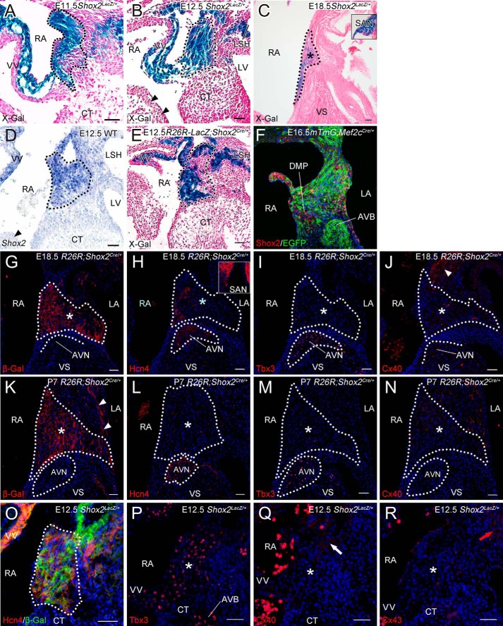
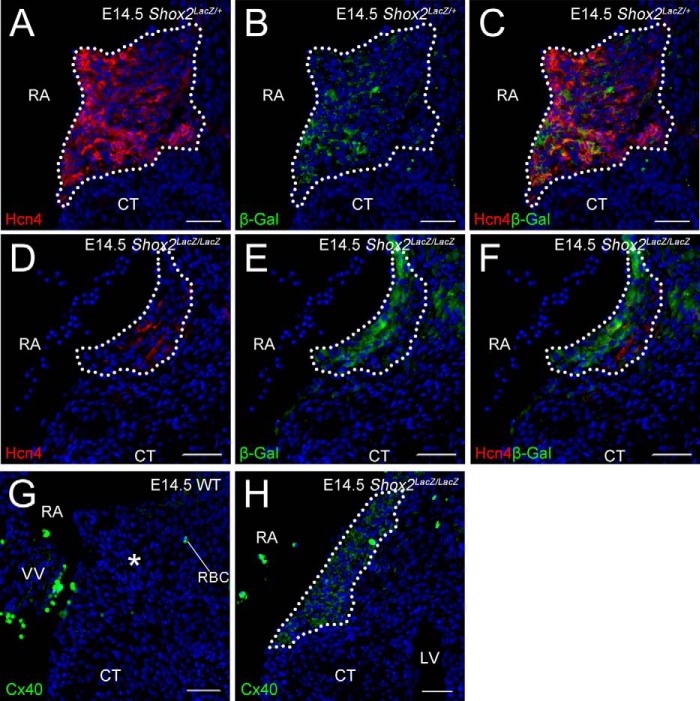
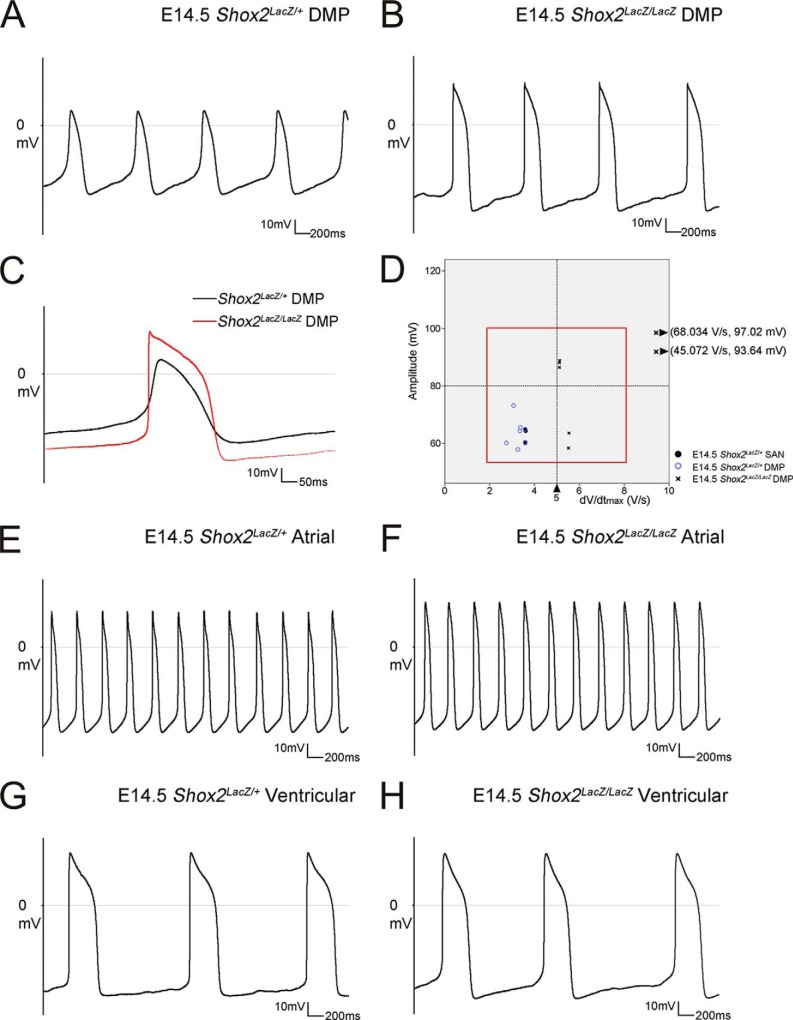
The atrioventricular (AV) junction plays a critical role in chamber septation and transmission of cardiac conduction pulses. It consists of structures that develop from embryonic dorsal mesenchymal protrusion (DMP) and the embryonic AV canal. Despite extensive studies on AV junction development, the genetic regulation of DMP development remains poorly understood. In this study we present evidence that Shox2 is expressed in the developing DMP. Intriguingly, this Shox2-expressing domain possesses a pacemaker-specific genetic profile including Hcn4 and Tbx3. This genetic profile leads to nodal-like electrophysiological properties, which is gradually silenced as the AV node becomes matured. Phenotypic analyses of Shox2(-/-) mice revealed a hypoplastic and defectively differentiated DMP, likely attributed to increased apoptosis, accompanied by dramatically reduced expression of Bmp4 and Hcn4, ectopic activation of Cx40, and an aberrant pattern of action potentials. Interestingly, conditional deletion of Bmp4 or inhibition of BMP signaling by overexpression of Noggin using a Shox2-Cre allele led to a similar DMP hypoplasia and down-regulation of Hcn4, whereas activation of a transgenic Bmp4 allele in Shox2(-/-) background attenuated DMP defects. Moreover, the lack of Hcn4 expression in the DMP of mice carrying Smad4 conditional deletion and direct binding of pSmad1/5/8 to the Hcn4 regulatory region further confirm the Shox2-BMP genetic cascade in the regulation of DMP development. Our results reveal that Shox2 regulates DMP fate and development by controlling BMP signaling through the Smad-dependent pathway to drive tissue growth and to induce Hcn4 expression and suggest a temporal pacemaking function for the DMP during early cardiogenesis.

Keywords: Bone Morphogenetic Protein (BMP); Dorsal Mesenchymal Protrusion; Gene Regulation; Heart Development; SMAD Transcription Factor; Shox2; Signal Transduction; Smad4-dependent.


Figures

FIGURE 1.
FIGURE 1.
Expression of Shox2 and pacemaker markers in the developing DMP. A–C, Shox2 expression in the developing AV junction was revealed by X-gal staining at E11.5 (A), E12.5 (B), and E18.5 when the expression became down-regulated as compared with the strong expression in the SAN (C and inset). D, in situ hybridization confirms Shox2 expression the AV junction. Arrowheads in B and D point to the absent Shox2 expression in the lateral AV canal tissue. E, X-gal staining shows localization of Shox2-expressing cells in the AV junction an E12.5 R26R-LacZ;Shox2Cre/+ mice. F, an E16.5 Mef2C-Cre;mTmG embryo exhibits Shox2 expression in the DMP, revealed by immunohistochemistry, which is part of second heart field-derived tissues labeled by EGFP. G–J, lineage tracing by Shox2-Cre allele shows that Shox2-expressing cells (asterisk) do not contribute to the forming AVN, as assessed by Hcn4 and Tbx3 expression. Although Cx40 expression is seen in the adjacent atrial septal tissue (white arrowhead), it is absent in both Shox2-expressing domain, and the AVN (J). The inset in H shows strong Hcn4 expression in the SAN at the same stage as positive control. K–N, immunohistochemistry shows the Hcn4+/Tbx3+/Cx40 AVN and the LacZ-positive domain (asterisk) that is Hcn4/Tbx3/Cx40+ in the AV junction of P7 R26R;Shox2Cre/+ mice. Note that LacZ-positive cells are also found near the orifice of the pulmonary vein (white arrowheads in K) at this stage. O–R, immunohistochemistry reveals that Hcn4 (dotted line in O) and Tbx3 (asterisk in P) but not Cx40 (asterisk in Q) and Cx43 (asterisk in R) are expressed within the Shox2-expressing DMP at E12.5. By contrast, Cx40 is expressed in the wall of coronary sinus (white arrow in Q) and Cx43 (red arrow in R) is expressed in the wall of LSH. The DMP domain is demarcated by dotted lines based on the expression domain of β-Gal and the AVN domain, also outlined by dotted lines, is defined by its expression of Hcn4 and Tbx3. CT, cushion tissue; LA, left atrium; LV, left ventricle; RA, right atrium; VS, ventricular septum; VV, venus valve; AVB, atrioventricular bundle; LSH, left sinus horn. Scale bar = 40 μm.
FIGURE 2.
FIGURE 2.
Multiple types of action potentials. A–E, action potential patterns of E14.5 SAN myocyte, nodal-like (A), E14.5 atrial myocyte, embryonic atrial-like (B), E14.5 ventricular myocyte, embryonic ventricular-like (C), E14.5 DMP myocyte, nodal-like (D), and E18.5 DMP myocyte showing a changed firing pattern other than that of nodal-like cell (E). F, a two-dimensional diagram showing the action potential amplitude and the maximum rate of rise of the AP (dV/dtmax) for each cell type studied. In E14.5 SAN and DMP cells, action potential amplitudes were smaller than 75, and dV/dtmaxs were smaller than 4. In E14.5 atrial and ventricular cells, action potential amplitudes and dV/dtmaxs were much larger. In E18.5 DMP cells, action potential amplitudes and dV/dtmaxs were considerably increased. The red-lined region will be revisited in Fig. 4D.
FIGURE 3.
FIGURE 3.
Hypoplasia and altered cellular processes in the developing DMP of Shox2 mutant. A–J, Shox2 null embryos display hypoplasia in the developing DMP, as shown by X-gal staining and three-dimensional reconstructions from X-gal-stained images at E12.5 (A–E) and E14.5 (F-J). DMP tissue volume reduction is shown by statistical results of volume comparison in the same region between Shox2LacZ/+ embryos and Shox2LacZ/LacZ embryos at E12.5 (E) and E14.5 (J). K–M, Ki67 immunofluorescence on representative sections of E12.5 wild-type (K) and Shox2 null (L) embryos shows no significant alteration of cell proliferation in the DMP between control and mutant animals, as determined by statistical analysis (M). NS, not significant. N–P, TUNEL assay on representative sections of E12.5 wild-type (N) and Shox2 null (O) embryos shows significantly increased cell apoptosis (black arrowheads) in the DMP region, as determined by statistical analysis (P). However, a comparable level of cell apoptosis (red arrowheads in N and O) in the adjacent cushion tissue of both control and Shox2 mutant was observed. Q and R, enhanced cell apoptosis was further confirmed by immunofluorescence of cleaved Caspase-3 (c-Caspase-3) (white arrowheads) in the DMP (asterisk) of E12.5 Shox2 null embryos (R) as compared with control (Q). Red arrowheads point to cleaved Caspase-3 positive cells in the adjacent cushion tissue. Dotted lines demarcate the DMP based on expression of β-Gal (A, B, F, and G). The DMP region in K, L, N, and O is defined based on β-gal expression at comparable stage. *, p < 0.05; ***, p < 0.0001. CT, cushion tissue; LV, left ventricle; RA, right atrium; VV, venus valve; LSH, left sinus horn. Scale bar = 40 μm.
FIGURE 4.
FIGURE 4.
Altered gene expression in the Shox2 null DMP at E11.5 and E12.5. A–C, Hcn4 and Shox2 expression in the DMP of an E11.5 Shox2LacZ/+ embryo, as detected by double immunofluorescence of Hcn4 and β-Gal reporter. D–F, Hcn4 expression and tissue morphology exhibit a wild-type like pattern in an E11.5 Shox2 mutant DMP and overlap with β-Gal reporter. G–I, overlapped expression of Hcn4 and Shox2 in the DMP of an E12.5 Shox2LacZ/+ embryo. J–L, Hcn4 expression (J) is almost diminished within the β-Gal-labeled hypoplastic DMP of an E12.5 Shox2LacZ/LacZ embryo. M–O, absence of Cx40 expression (N) in the Hcn4-positive DMP (M and O) of an E12.5 Shox2LacZ/+ embryo. P–R, ectopic Cx40 expression (Q and R) is seen in the Hcn4-negative DMP (P) of an E12.5 Shox2LacZ/LacZ embryo. Dotted lines demarcate the DMP, based on the expression domains of β-Gal (A–L), Hcn4 (M–O), and Cx40 (P–R). CT, cushion tissue; LV, left ventricle; RA, right atrium; VV, venus valve; LSH, left sinus horn. Scale bar = 40 μm.
FIGURE 5.
FIGURE 5.
Altered gene expression in the Shox2 mutant DMP at E14.5. A–F, immunohistochemistry reveals down-regulation of Hcn4 in the β-Gal labeled hypoplastic DMP (dashed line) of an E14.5 Shox2LacZ/LacZ embryo (D–F) as compared with controls (A–C). G and H, immunohistochemistry shows the absence of Cx40 expression in the DMP (asterisk) of an E14.5 wild-type embryo but ectopic Cx40 expression (dashed line) in the DMP of an E14.5 Shox2LacZ/LacZ embryo. CT, cushion tissue; LV, left ventricle; RA, right atrium; VV, venus valve; RBC, red blood cell. Scale = 40 μm.
FIGURE 6.
FIGURE 6.
Aberrant APs of Shox2-deficient DMP myocardium. A and B, typical APs of Shox2LacZ/+ (A) and Shox2LacZ/LacZ (B) DMP cells of E14.5 mice. C, overlying of a typical AP of Shox2LacZ/+ DMP cells with that of Shox2LacZ/LacZ DMP cells demonstrates the increased action potential amplitude and the rate of upstroke in Shox2 mutant DMP cells. D, a two-dimensional diagram showing the action potential amplitude and the maximum rate of rise of the AP (dV/dtmax) for each wild-type SAN, Shox2LacZ/+ DMP, and Shox2LacZ/LacZ DMP cell studied. Please note the red lined region is identical to that mentioned in Fig. 2F. E–H, by contrast, in both mice, the atrial and ventricular myocytes show the similar action potential amplitude and the maximum rate of rise of the AP (dV/dtmax).
FIGURE 7.
FIGURE 7.
Disrupted BMP signaling in the Shox2 mutant DMP. A and B, in situ hybridization assay reveals Bmp2 expression in the AV canal tissue (red arrowheads) but not in the DMP of both E11.5 Shox2LacZ/+ (A) and Shox2LacZ/LacZ (B) embryos. C and D, Bmp4 expression (arrowheads), as assayed by in situ hybridization, was observed in the DMP and the wall of the left sinus horn of an E11.5 Shox2LacZ/+ embryo (C) but was down-regulated in Shox2LacZ/LacZ embryo at the same stage (D). E–H, at E12.5, expressions of Bmp2 (E) and Bmp4 (G) is found in the developing DMP of wild-type embryos (E and G) but was dramatically reduced in Shox2 mutants (F and H). I and J, immunohistochemistry reveals a significantly reduced level of phosphorylated Smad1/5/8 in the DMP of Shox2 mutant (J) as compared with control (I). Asterisks indicate the DMP. CT, cushion tissue; LV, left ventricle; RA, right atrium; VV, venus valve; AVC, atrioventricular canal; LSH, left sinus horn. Scale = 40 μm.
FIGURE 8.
FIGURE 8.
Altered gene expression in the DMP of mice carrying disrupted BMP signaling. A–D, E12.5 Shox2-Cre;Bmp4F/F embryos exhibit an hypoplastic DMP (B) and compromised Hcn4 expression (D) as compared with control counterpart (A and C). E–H, immunohistochemistry reveals a significantly reduced level of pSmad1/5/8 in the DMP cells of an E12.5 Shox2-Cre;pMes-Nog mouse (F and H), as compared with control (asterisk in E). EGFP expression (F and H) by the pMes-Nog transgenic allele is used to demarcate the DMP domain. I–P, Shox2-Cre;pMes-Nog embryos show hypoplasia and ablation of Hcn4 expression (J and N) in the DMP labeled by EGFP at E12.5 (J–L) and E14.5 (N–P) compared with controls (I and M). Q–X, aberrant ectopic Cx40 expression (R–T) and activation of Caspase-3 (V–X) are seen in the DMP (demarcated by EGFP expression) of E12.5 Shox2-Cre;pMes-Nog embryos, as compared with controls (asterisk in Q and U). White arrowheads point to the apoptotic cells within the DMP, and red arrowheads point to the apoptotic cells in the cushion tissue. Dotted lines demarcate the DMP, defined by the expression of Hcn4 (A–D and M) and EGFP (F–H, J–L, N–P, R–T, V–X). CT, cushion tissue; LV, left ventricle; RA, right atrium; VV, venus valve; LSH, left sinus horn; RBC, red blood cell. Scale bar = 40 μm.
FIGURE 9.
FIGURE 9.
Smad4-mediated signaling is essential for Hcn4 expression in the DMP. A and B, an E12.5 Shox2-Cre;Smad4F/F embryo exhibits absent Hcn4 expression (white arrowhead) in the DMP (B) as compared with control (A). C and D, ChIP assay and schematic diagrams of genomic regions of the Hcn4 locus show the position of the conserved Smad binding site highlighted in the red rectangle and conserved binding sequence highlighted in background among several placental mammals (D) and binding of pSmad1/5/8 to conserved Smad binding site (C). E and F, immunohistochemistry reveals an elevated pSmad1/5/8 level in the forming AV node but a significantly reduced pSmad1/5/8 level in the DMP (asterisk) derived from Shox2-expressing lineage cells. Dotted lines demarcate the DMP, defined by the expression domains of Hcn4 (A and B) and β-Gal (E and F). CT, cushion tissue; RA, right atrium; VV, venus valve; VS, ventricular septum; LSH, left sinus horn. Scale = 40 μm.
FIGURE 10.
FIGURE 10.
Resumed Hcn4 expression in the DMP of pMes-Bmp4;Shox2Cre/LacZ mice. A–C, immunohistochemistry shows abundant pSmad1/5/8 in the DMP of an E13.5 control mouse (asterisk in A), the absence of pSmad1/5/8 in an E13.5 Shox2 null DMP (asterisk in B), but resumed pSmad1/5/8 staining in the DMP with rescued size in pMes-Bmp4;Shox2Cre/LacZ embryo at the same stage (asterisk in C). D–F, immunohistochemistry shows resumed Hcn4 expression in the DMP of an E13.5 pMes-Bmp4;Shox2Cre/LacZ embryo (F) as compared with wild-type control (D) and Shox2 mutant (asterisk in E) at the same stage. G–I, typical APs of Shox2LacZ/+ (G), Shox2LacZ/LacZ (H), and BMP4-treated Shox2LacZ/LacZ (I) DMP cells of E14.5 mice, demonstrating the decreased rate of upstroke in Shox2 mutant DMP cells. Dotted lines demarcate the DMP, defined by the expression domain of Hcn4. CT, cushion tissue; RA, right atrium; LSH, left sinus horn; RBC, red blood cell. Scale = 40 μm.

Comment in

Similar articles

Cited by

References

    1. Mangoni M. E., Nargeot J. (2008) Genesis and regulation of the heart automaticity. Physiol. Rev. 88, 919–982 - PubMed
    1. Wessels A., Markman M. W., Vermeulen J. L., Anderson R. H., Moorman A. F., Lamers W. H. (1996) The development of the atrioventricular junction in the human heart. Circ. Res. 78, 110–117 - PubMed
    1. Christoffels V. M., Smits G. J., Kispert A., Moorman A. F. (2010) Development of the pacemaker tissues of the heart. Circ. Res. 106, 240–254 - PubMed
    1. Munshi N. V. (2012) Gene regulatory networks in cardiac conduction system development. Circ. Res. 110, 1525–1537 - PMC - PubMed
    1. Briggs L. E., Phelps A. L., Brown E., Kakarla J., Anderson R. H., van den Hoff M. J., Wessels A. (2013) Expression of the BMP receptor Alk3 in the second heart field is essential for development of the dorsal mesenchymal protrusion and atrioventricular septation. Circ. Res. 112, 1420–1432 - PMC - PubMed

Publication types

MeSH terms

Substances