Review
. 2015 Jan;16(1):24-43.
doi: 10.15252/embr.201439363. Epub 2014 Dec 8.

Exosomes and other extracellular vesicles in host-pathogen interactions

Affiliations
Review

Exosomes and other extracellular vesicles in host-pathogen interactions

Jeffrey S Schorey et al. EMBO Rep. 2015 Jan.

Abstract

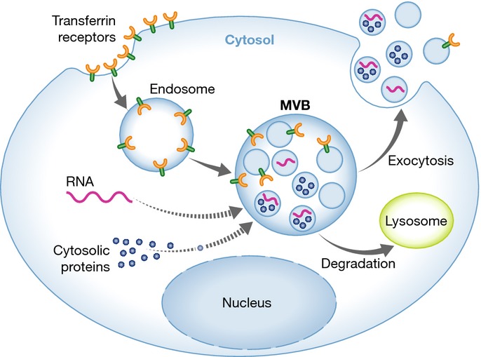
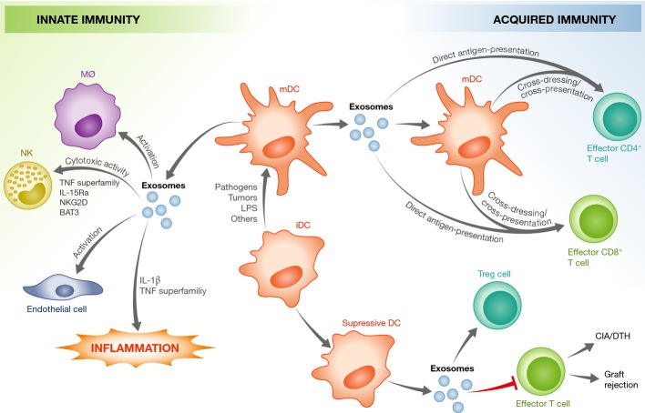
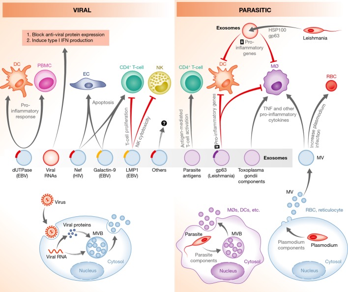
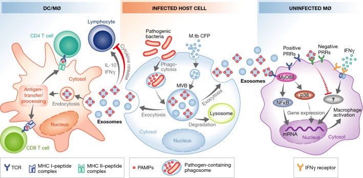
An effective immune response requires the engagement of host receptors by pathogen-derived molecules and the stimulation of an appropriate cellular response. Therefore, a crucial factor in our ability to control an infection is the accessibility of our immune cells to the foreign material. Exosomes-which are extracellular vesicles that function in intercellular communication-may play a key role in the dissemination of pathogen- as well as host-derived molecules during infection. In this review, we highlight the composition and function of exosomes and other extracellular vesicles produced during viral, parasitic, fungal and bacterial infections and describe how these vesicles could function to either promote or inhibit host immunity.

Keywords: exosomes; extracellular vesicles; immunity; pathogens.


Figures

Figure 1
Figure 1. Exosome biogenesis
Lipids, proteins and nucleic acids are transported to MVBs and onto or into the intraluminal vesicles, which upon fusion of the MVB with the plasma membrane are released as exosomes. Originally identified as a way to release transferrin receptor from maturing reticulocytes, other plasma membrane proteins have been shown to be targeted to MVBs through various mechanisms and released on exosomes. RNA and cytoplasmic proteins are also transported to MVBs, although the mechanisms mediating this transport are less understood (indicated by dashed line). See Glossary for definitions.
Figure 2
Figure 2. DC-derived exosomes modulate innate and acquired immune responses
Exosomes from mature DCs (mDCs) can provide antigen to T cells, stimulate innate immune responses in various immune and non-immune cells and promote a pro-inflammatory response. This is mediated by host factors present within the exosomes, such as those indicated in the figure. In contrast, exosomes from immature DCs (iDCs) seem to be immuno-suppressive, and induce apoptosis in effector T cells or promote the activation of regulatory T cells. CIA, collagen-induced arthritis; DTH, delayed-type hypersensitivity. See Glossary for definitions.
Figure 3
Figure 3. Modulation of host immunity by exosomes during a viral or parasitic infection
Virus- or parasite-infected cells, or the parasites themselves, release exosomes or microvesicles that can stimulate T-cell activation by providing antigens to APCs. In contrast, exosomes containing microbial molecules, such as HIV Nef or Leishmania GP63, can block T-cell activation or induce the apoptosis of immune effector cells. Extracellular vesicles released from virus- or parasite-infected cells can modulate both the innate and acquired immune response. In some cases, this is to the benefit of the pathogen, whereas in others, this is to the benefit of the host. Dashed lines indicate unknown mechanisms. See Glossary for definitions.
Figure 4
Figure 4. Bacteria-infected cells release exosomes that modify T-cell and macrophage function
Exosomes from bacteria-infected macrophages release exosomes containing antigens that induce cross-priming to activate antigen-specific CD4+ and CD8+ T cells. In contrast, some exosomes released from infected cells inhibit cytokine production by T cells. Exosomes from infected cells also contain PAMPs that stimulate macrophage production of pro-inflammatory mediators like TNF-α, or limit the macrophage response to IFN-γ stimulation. Dashed lines indicate unknown mechanisms. See Glossary for definitions.

Similar articles

Cited by

References

    1. Kumar H, Kawai T, Akira S. Pathogen recognition by the innate immune system. Int Rev Immunol. 2011;30:16–34. - PubMed
    1. Bliska JB, Wang X, Viboud GI, Brodsky IE. Modulation of innate immune responses by Yersinia type III secretion system translocators and effectors. Cell Microbiol. 2013;15:1622–1631. - PMC - PubMed
    1. Rana RR, Simpson P, Zhang M, Jennions M, Ukegbu C, Spear AM, Alguel Y, Matthews SJ, Atkins HS, Byrne B. Yersinia pestis TIR-domain protein forms dimers that interact with the human adaptor protein MyD88. Microb Pathog. 2011;51:89–95. - PubMed
    1. Schmolke M, Patel JR, de Castro E, Sánchez-Aparicio MT, Uccellini MB, Miller JC, Manicassamy B, Satoh T, Kawai T, Akira S, et al. RIG-I detects mRNA of intracellular Salmonella enterica serovar Typhimurium during bacterial infection. MBio. 2014;5:e01006–e01014. - PMC - PubMed
    1. Yadav M, Roach SK, Schorey JS. Increased mitogen-activated protein kinase activity and TNF-alpha production associated with Mycobacterium smegmatis- but not Mycobacterium avium-infected macrophages requires prolonged stimulation of the calmodulin/calmodulin kinase and cyclic AMP/protein kinase A pathways. J Immunol. 2004;172:5588–5597. - PubMed

Publication types