. 2014 Dec 15;211(13):2537-47.
doi: 10.1084/jem.20141759. Epub 2014 Dec 8.

Heterozygous splice mutation in PIK3R1 causes human immunodeficiency with lymphoproliferation due to dominant activation of PI3K

Affiliations

Heterozygous splice mutation in PIK3R1 causes human immunodeficiency with lymphoproliferation due to dominant activation of PI3K

Carrie L Lucas et al. J Exp Med. .

Abstract

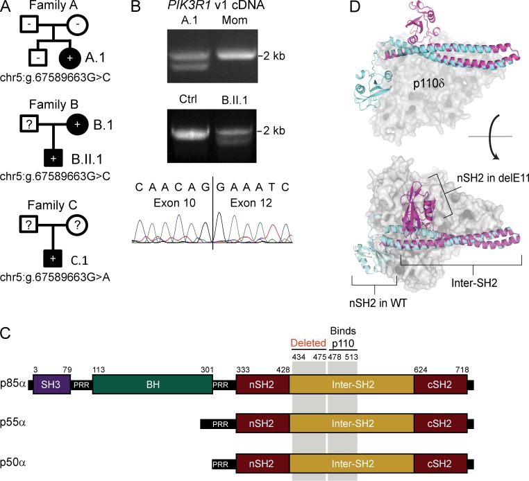
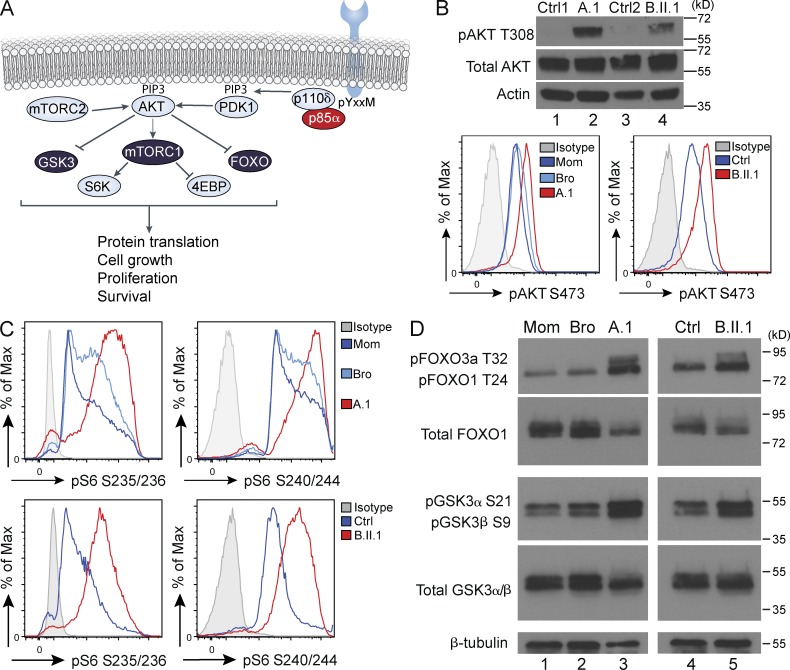
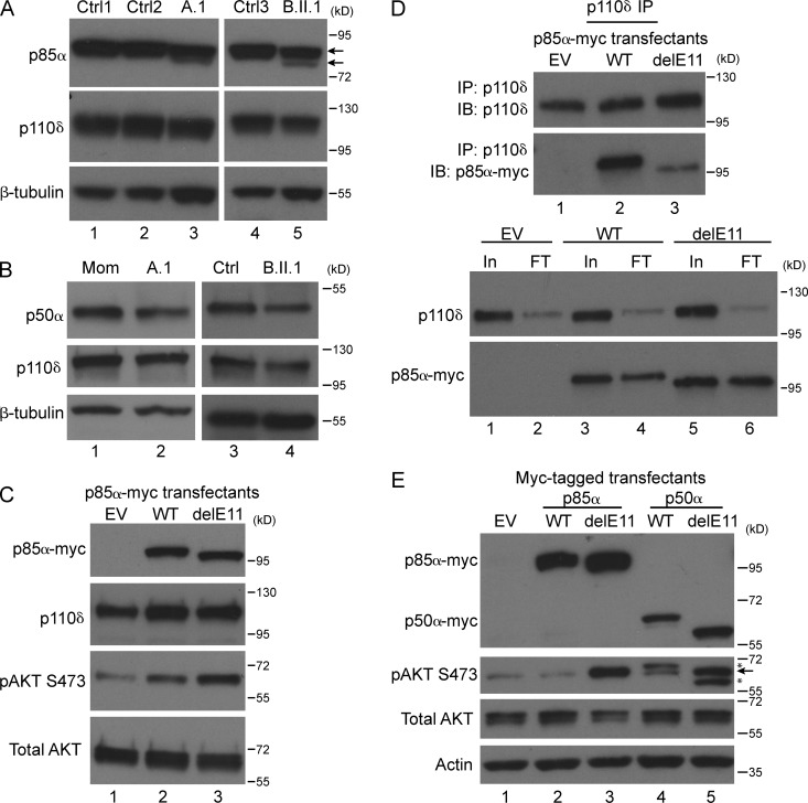
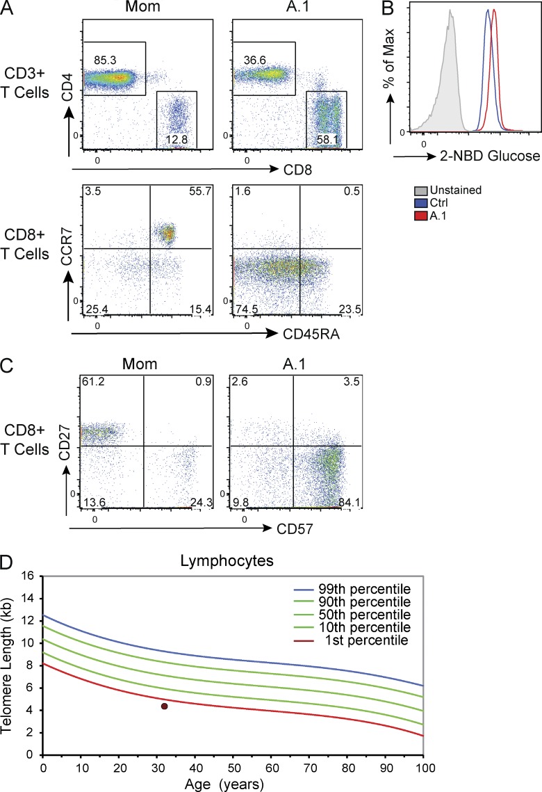
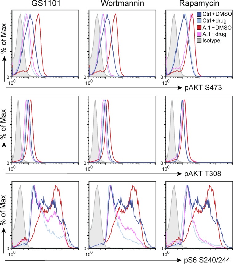
Class IA phosphatidylinositol 3-kinases (PI3K), which generate PIP3 as a signal for cell growth and proliferation, exist as an intracellular complex of a catalytic subunit bound to a regulatory subunit. We and others have previously reported that heterozygous mutations in PIK3CD encoding the p110δ catalytic PI3K subunit cause a unique disorder termed p110δ-activating mutations causing senescent T cells, lymphadenopathy, and immunodeficiency (PASLI) disease. We report four patients from three families with a similar disease who harbor a recently reported heterozygous splice site mutation in PIK3R1, which encodes the p85α, p55α, and p50α regulatory PI3K subunits. These patients suffer from recurrent sinopulmonary infections and lymphoproliferation, exhibit hyperactive PI3K signaling, and have prominent expansion and skewing of peripheral blood CD8(+) T cells toward terminally differentiated senescent effector cells with short telomeres. The PIK3R1 splice site mutation causes skipping of an exon, corresponding to loss of amino acid residues 434-475 in the inter-SH2 domain. The mutant p85α protein is expressed at low levels in patient cells and activates PI3K signaling when overexpressed in T cells from healthy subjects due to qualitative and quantitative binding changes in the p85α-p110δ complex and failure of the C-terminal region to properly inhibit p110δ catalytic activity.


Figures

Figure 1.
Figure 1.
Affected patients are heterozygous for a PIK3R1 splice site mutation that causes an in-frame deletion of exon 11. (A) Family pedigrees with clinically affected individuals in black and unaffected individuals in white. The mutation status is indicated within the symbol for each individual, where − indicates confirmed mutation-negative, + indicates confirmed mutation-positive, and ? indicates unknown mutation status. The specific nucleotide changes are indicated beneath each pedigree. (B) PIK3R1 variant 1 cDNA (encoding p85α) amplified between exons 2 and 15 and imaged in an agarose gel (top) or sequenced using the Sanger approach after excision of the lower band (bottom). Three independent experiments on the two indicated patients and controls were performed. (C) Protein schematic for p85α, p55α, and p50α isoforms of PIK3R1 indicating amino acid residue numbers (top), structural domains, region deleted by the patient splice mutation, and the p110-binding region. SH3: Src homology 3; PRR: proline-rich region; BH: breakpoint cluster region homology; nSH2 and cSH2: N-terminal and C-terminal Src homology 2. (D) Provisional structural model of the PI3K p85α protein showing an overlay of a WT (cyan) and delE11 (magenta) p85α protein fragment (nSH2 plus inter-SH2) in association with p110δ (gray). Top: view from side; Bottom: view from top.
Figure 2.
Figure 2.
PI3K signaling is constitutively active in patient immune cells. (A) Signaling diagram showing the major molecules downstream of the p110δ/p85α PI3K complex. PDK1: phosphoinositide-dependent kinase-1; mTORC: mechanistic target of rapamycin complex; GSK3: glycogen synthase kinase 3; S6K: ribosomal protein S6 kinase; 4EBP1: eukaryotic translation initiation factor 4E-binding protein 1; FOXO: forkhead box O. (B) Immunoblot for AKT phosphorylated at threonine 308, total AKT, and actin (top), and flow cytometric analysis of AKT phosphorylation at serine 473 (bottom) in T cell blasts from healthy relatives or control subjects (Ctrl) and the indicated patients. (C) Flow cytometric analysis of the phosphorylation of S6 at serine residues 235/236 and serine residues 240/244 in T cell blasts from healthy relatives or control subjects (Ctrl) and the indicated patients (stained in same experiment as in B, bottom). (D) Immunoblot for phosphorylation of FOXO protein threonines (T) and GSK3 protein serines (S), total protein, or β-tubulin loading control, as indicated. Four (B and C) or three (D) independent experiments on the two indicated patients and controls were performed.
Figure 3.
Figure 3.
The delE11 PIK3R1 mutant p85α gene product is expressed at low levels in patient T cell blasts and drives PI3K hyperactivation via poor inhibition of p110δ. (A) Western blot for p85α (Ab recognizing region just after SH3 domain), p110δ, or β-tubulin, as indicated. Arrows point to full-length (top arrow) and truncated delE11 (bottom arrow) p85α. (B) Western blot analysis of p50α using an antibody recognizing the N-terminal SH2 (top). Levels of p110δ protein (middle) and β-tubulin loading control (bottom) are also shown. (C) Primary T cells overexpressing EV, WT, or delE11 mutant 5x-myc-tagged p85α were analyzed by immunoblot for myc tag, p110δ, phospho-AKT, or total AKT, as indicated. (D) p110δ immunoprecipitates (IP, top) or input (In) and flow-through (FT; bottom) from T cells overexpressing EV, WT p85α-myc, or delE11 p85α-myc were probed with the indicated immunoblot (IB) antibodies. (E) Primary T cells overexpressing EV, WT, or delE11 mutant 5×-myc-tagged p85α or p50α were analyzed by immunoblot for myc tag, phospho-AKT, total AKT, or actin, as indicated. Arrow marks phospho-AKT band, and asterisks mark residual signal from p50α-myc blot reprobed for phospho-AKT. Four (A and C) or three (B, D, and E) independent experiments were performed.
Figure 4.
Figure 4.
Patient CD8 T cells are expanded, severely skewed toward effector (CCR7-negative) phenotype, and enriched with senescent CD57+ cells with short telomeres. (A) Flow cytometric analysis of PBMCs from a healthy control subject (Mom) or patient A.1, staining for CD4, CD8, CD45RA, and CCR7, as indicated. (B) Glucose uptake in the indicated T cell blasts, as measured by 2-NBDG (2-(N-(7-Nitrobenz-2-oxa-1,3-diazol-4-yl)Amino)-2-Deoxyglucose) fluorescence after a 1-h rest in PBS and 20-min incubation with 2-NBDG. (C) Flow cytometric analysis of PBMCs, gating on CD8 T cells, and assessing expression of CD57 and CD27. (D) Flow-FISH analysis of telomere length within the lymphocyte population in patient A.1 shown as a dot with percentiles indicated (courtesy of Repeat Diagnostics). Three (A, B, and C) or two (D, patients A.1 and B.II.1) independent experiments were performed.
Figure 5.
Figure 5.
Targeted inhibition of mTOR or p110δ represent potentially effective treatment options for PIK3R1 patients. Flow cytometric histograms for T cell blasts from healthy control subjects (Ctrl) or patient A.1 after treatment with 0.1 µM GS1101, 0.1 µM Wortmannin, or 12.5 nM rapamycin and analyzed for phosphorylation of AKT and S6 at the indicated residues. Four independent experiments were performed.

References

    1. Angulo I., Vadas O., Garçon F., Banham-Hall E., Plagnol V., Leahy T.R., Baxendale H., Coulter T., Curtis J., Wu C., et al. . 2013. Phosphoinositide 3-kinase δ gene mutation predisposes to respiratory infection and airway damage. Science. 342:866–871 10.1126/science.1243292 - DOI - PMC - PubMed
    1. Bárcena C., Quesada V., De Sandre-Giovannoli A., Puente D.A., Fernández-Toral J., Sigaudy S., Baban A., Lévy N., Velasco G., and López-Otín C.. 2014. Exome sequencing identifies a novel mutation in PIK3R1 as the cause of SHORT syndrome. BMC Med. Genet. 15:51 10.1186/1471-2350-15-51 - DOI - PMC - PubMed
    1. Brachmann S.M., Ueki K., Engelman J.A., Kahn R.C., and Cantley L.C.. 2005. Phosphoinositide 3-kinase catalytic subunit deletion and regulatory subunit deletion have opposite effects on insulin sensitivity in mice. Mol. Cell. Biol. 25:1596–1607 10.1128/MCB.25.5.1596-1607.2005 - DOI - PMC - PubMed
    1. Brown J.R., Byrd J.C., Coutre S.E., Benson D.M., Flinn I.W., Wagner-Johnston N.D., Spurgeon S.E., Kahl B.S., Bello C., Webb H.K., et al. . 2014. Idelalisib, an inhibitor of phosphatidylinositol 3-kinase p110δ, for relapsed/refractory chronic lymphocytic leukemia. Blood. 123:3390–3397 10.1182/blood-2013-11-535047 - DOI - PMC - PubMed
    1. Burke J.E., Vadas O., Berndt A., Finegan T., Perisic O., and Williams R.L.. 2011. Dynamics of the phosphoinositide 3-kinase p110δ interaction with p85α and membranes reveals aspects of regulation distinct from p110α. Structure. 19:1127–1137 10.1016/j.str.2011.06.003 - DOI - PMC - PubMed

Publication types

MeSH terms

Substances

Associated data