. 2014 Dec 23;111(51):E5537-44.
doi: 10.1073/pnas.1414665111. Epub 2014 Dec 8.

Bnip3 mediates doxorubicin-induced cardiac myocyte necrosis and mortality through changes in mitochondrial signaling

Affiliations

Bnip3 mediates doxorubicin-induced cardiac myocyte necrosis and mortality through changes in mitochondrial signaling

Rimpy Dhingra et al. Proc Natl Acad Sci U S A. .

Abstract

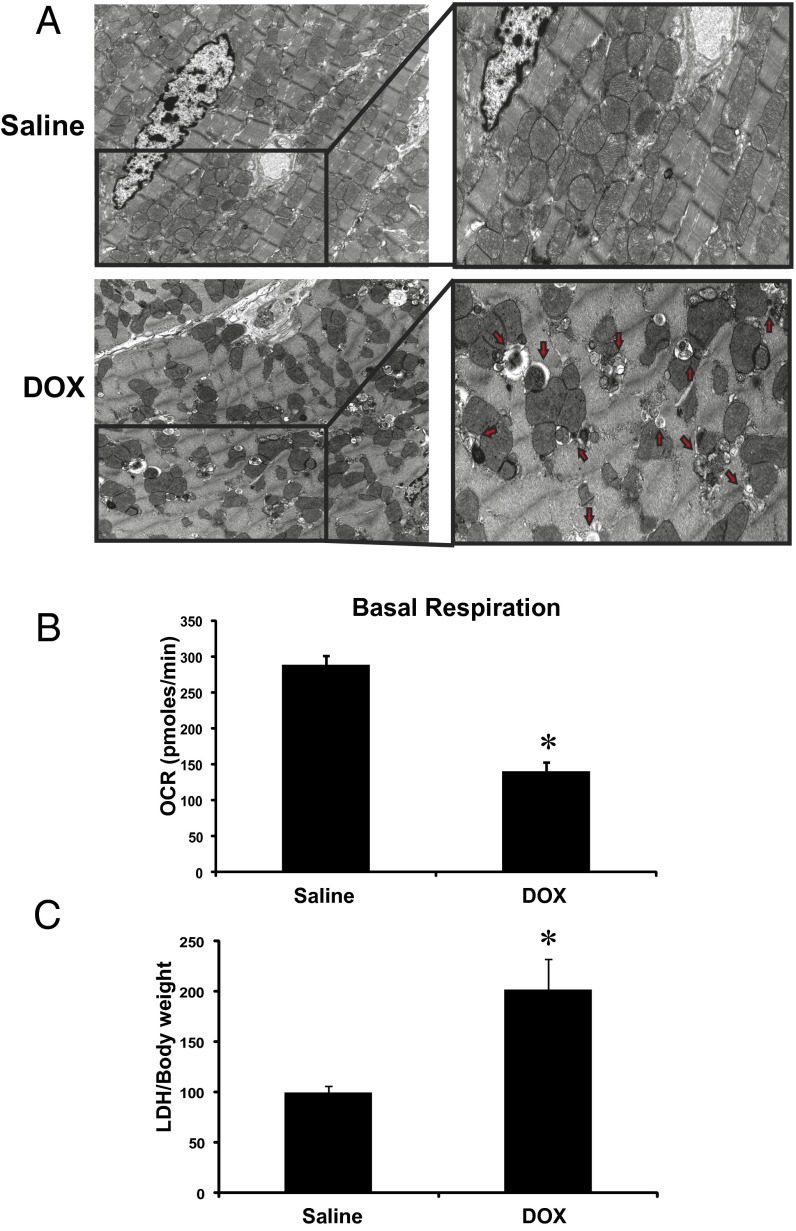
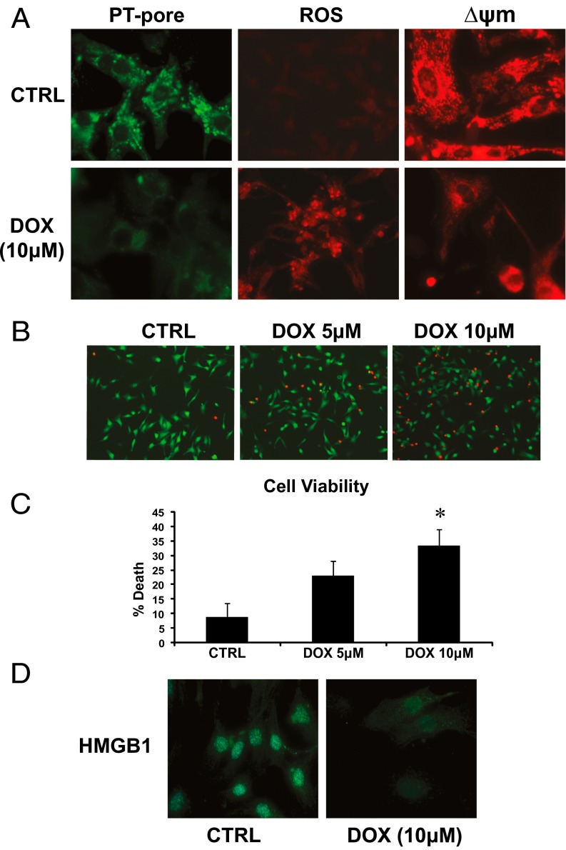
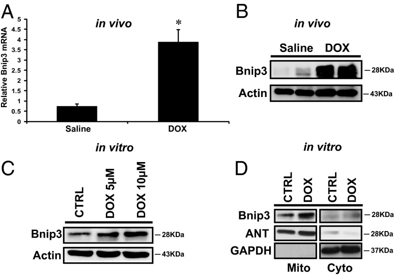
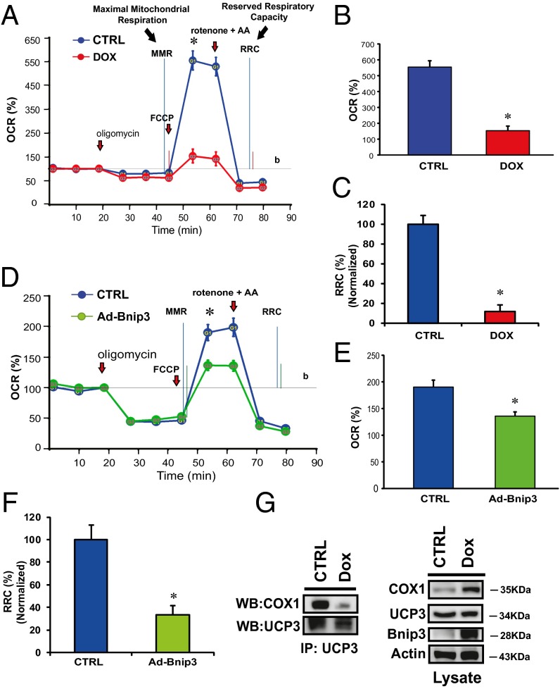
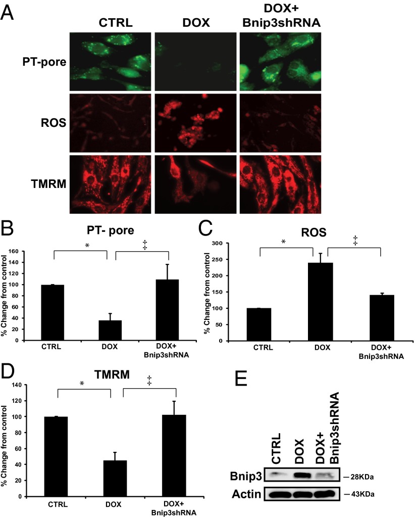
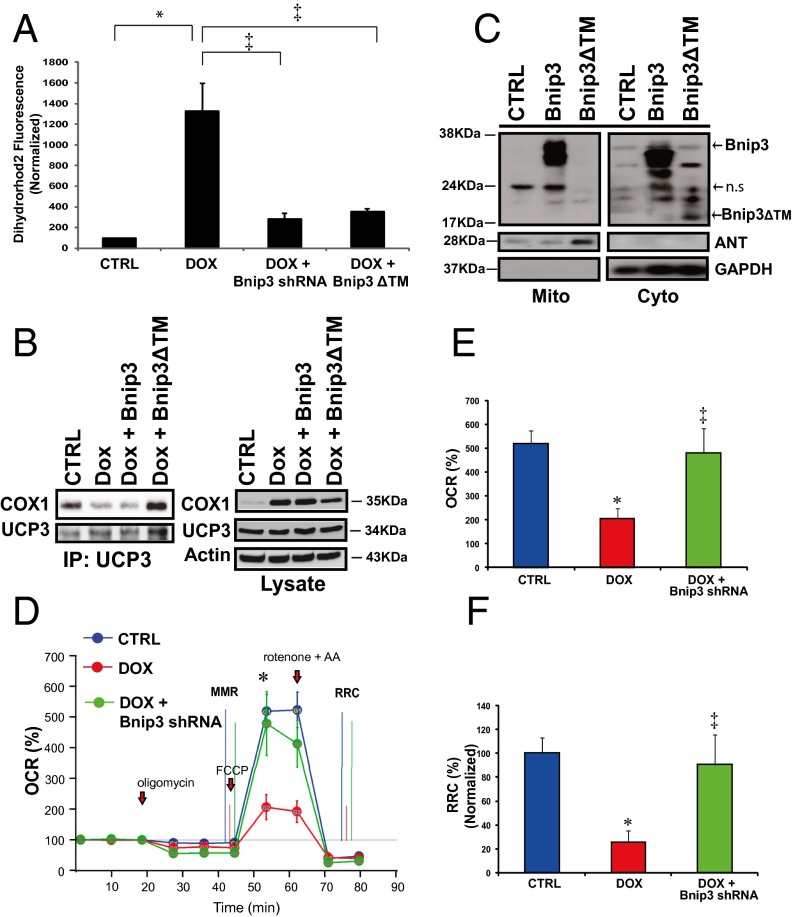
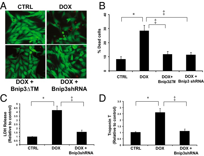
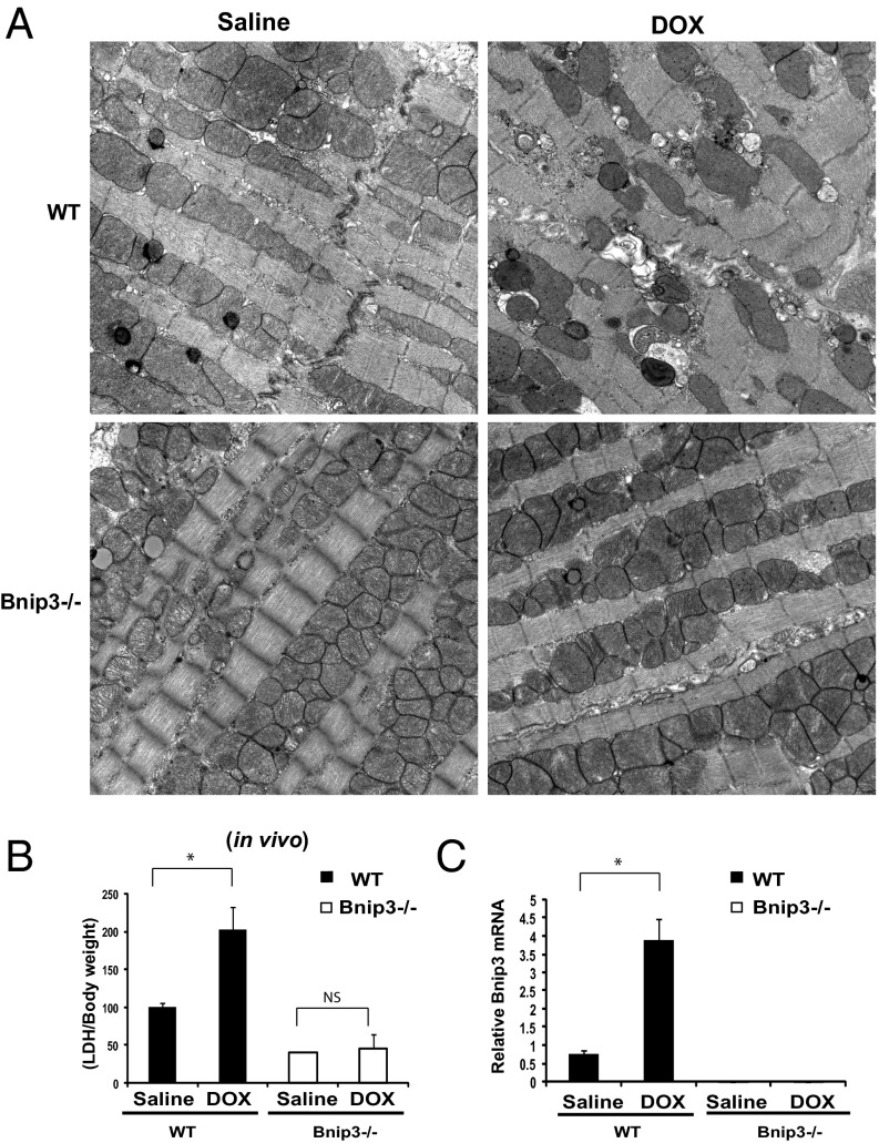
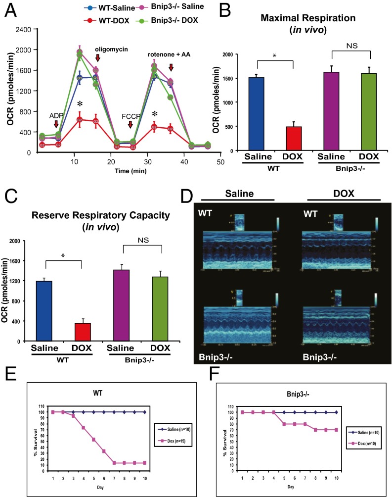
Doxorubicin (DOX) is widely used for treating human cancers, but can induce heart failure through an undefined mechanism. Herein we describe a previously unidentified signaling pathway that couples DOX-induced mitochondrial respiratory chain defects and necrotic cell death to the BH3-only protein Bcl-2-like 19 kDa-interacting protein 3 (Bnip3). Cellular defects, including vacuolization and disrupted mitochondria, were observed in DOX-treated mice hearts. This coincided with mitochondrial localization of Bnip3, increased reactive oxygen species production, loss of mitochondrial membrane potential, mitochondrial permeability transition pore opening, and necrosis. Interestingly, a 3.1-fold decrease in maximal mitochondrial respiration was observed in cardiac mitochondria of mice treated with DOX. In vehicle-treated control cells undergoing normal respiration, the respiratory chain complex IV subunit 1 (COX1) was tightly bound to uncoupling protein 3 (UCP3), but this complex was disrupted in cells treated with DOX. Mitochondrial dysfunction induced by DOX was accompanied by contractile failure and necrotic cell death. Conversely, shRNA directed against Bnip3 or a mutant of Bnip3 defective for mitochondrial targeting abrogated DOX-induced loss of COX1-UCP3 complexes and respiratory chain defects. Finally, Bnip3(-/-) mice treated with DOX displayed relatively normal mitochondrial morphology, respiration, and mortality rates comparable to those of saline-treated WT mice, supporting the idea that Bnip3 underlies the cardiotoxic effects of DOX. These findings reveal a new signaling pathway in which DOX-induced mitochondrial respiratory chain defects and necrotic cell death are mutually dependent on and obligatorily linked to Bnip3 gene activation. Interventions that antagonize Bnip3 may prove beneficial in preventing mitochondrial injury and heart failure in cancer patients undergoing chemotherapy.

Keywords: Bnip3; cell death; heart failure; mitochondria; ventricular myocytes.


Conflict of interest statement

The authors declare no conflict of interest.

Figures

Fig. 1.
Fig. 1.
DOX provokes ultrastructural defects and mitochondrial injury in vivo. (A) Representative electron micrograph images of murine cardiac muscle derived from mice treated with saline or DOX (single i.p injection, 20 mg/kg) at day 5 postinjection. (Upper Left) Saline-treated control mice. (Upper Right) Magnified section showing normal cardiac ultrastructure. (Lower Left) Representative mouse hearts after DOX treatment. (Lower Right) Magnified section showing ultrastructural defects including disrupted sarcomeres, mitochondrial swelling, and vacuolization. Red arrows denote membrane structures indicative of autophagosomes. (Magnification: 5,800×.) (B) Basal respiration of cardiac mitochondria derived from vehicle- and DOX-treated mouse hearts. OCR was measured with a Seahorse metabolic analyzer (Materials and Methods). (C) Serum LDH release from mice treated with saline or DOX. Data are presented as mean ± SEM. P < 0.05. *Statistically different from saline treatment.
Fig. 2.
Fig. 2.
DOX triggers mitochondrial perturbations and necrotic death of cardiac myoctyes. (A) Epifluorescence microscopy of control (CTRL) and DOX-treated cells for mPTP opening (Left), ROS (Center), and mitochondrial ΔΨm (Right); see Materials and Methods for details. (B) Cell viability of ventricular myocytes stained with vital dyes calcein-AM and ethidium-homodimer to detect the live (green) and dead (red) cells, respectively, in the absence and presence of DOX treatment (5 and 10 μM) for 18 h. (C) Histogram of the quantitative data in B. Data are expressed as mean ± SEM from at least four independent cell culture experiments counting ≥200 cells for each condition tested. P < 0.05. *Statistically different from control. (D) Epifluorescence microscopy of cardiac myocytes stained for nuclear HMGB1 protein (green nuclear staining) in vehicle-treated control cells and cells treated with DOX (10 μM) for 18 h.
Fig. 3.
Fig. 3.
DOX induces Bnip3 expression in ventricular myocytes. (A) Bnip3 mRNA expression in hearts from saline-treated control (CTRL) and DOX-treated mice; see Materials and Methods for details. P < 0.05. *Statistically different from saline control. (B) Western blot analysis of cardiac lysate for Bnip3 protein for conditions detailed in A. (C) Western blot analysis of cell lysate derived from isolated ventricular myocytes treated with DOX (5 and 10 μM) for 18 h or vehicle-treated control. The filter was probed with antibodies directed against Bnip3 and β-actin as a loading control for the Western blot analysis. (D) Western blot analysis of mitochondrial (Mito) and cytoplasmic s-100 (Cyto) fractions from cardiac myocytes in the absence and presence of DOX treatment. The filter was probed with antibody directed against Bnip3. Antibodies directed against mitochondrial protein adenine nucleotide transporter (ANT) and cytosolic protein GAPDH were used to verify the purity of cell fractionation.
Fig. 4.
Fig. 4.
DOX disrupts the interaction between mitochondrial COX1 and UCP3 and impairs respiration. (A and B) OCR was measured with a Seahorse metabolic analyzer. Oligomycin (1 μM), FCCP (1 μM), and rotenone (1 μM) combined with antimycin (1 μM) were added sequentially to saline-treated control (CTRL) (blue) or DOX-treated cardiomyocytes (red). b, baseline. (B and C) Histograms showing %OCR (B) and %RRC (C) data for A. Values are mean ± SEM from three to five replicates. P < 0.05. *Statistically different from control. (D) Respiration measurements in cardiomyocytes infected with adenoviral control vector (CTRL; blue) or adenovirus encoding Bnip3 cDNA (Ad Bnip3; green). (E and F) Histograms showing %OCR (E) and %RRC (F). P < 0.05. *Statistically different from control. (G) Effects of DOX on COX1 and UCP3 complex. (Left) Protein lysate derived from control and DOX-treated ventricular myocytes was immunoprecipitated with an antibody directed against UCP3 (1 μg/mL) and blotted with antibody directed against COX1. (Right) Western blot analysis of cell lysate used for immunoprecipitation and analyzed for expression of COX1, UCP3, Bnip3, and α-actin proteins.
Fig. 5.
Fig. 5.
DOX provokes mitochondrial perturbations contingent on Bnip3. Shown are mitochondrial perturbations induced by DOX in the absence or presence of Bnip3 knockdown. (A, Top) Epifluorescence microscopy of cardiac myocytes assessed for mPTP by mitochondrial calcein-AM-CoCl2 (green). Loss of green fluorescence is indicative of mPTP opening. (Middle) Epifluorescence microscopy of ROS as assessed by dihydroethidine (red). (Bottom) Epifluorescence microscopy of cardiac myocytes assessed for mitochondrial ∆Ψm by TMRM (red); see Methods for details. (B–D) Histograms for quantitative data for conditions shown in A. Data were obtained from three independent myocyte isolations using two replicates for each condition tested. P < 0.05. *Statistically different from control. Statistically different from DOX-treated. (E) Western blot analysis of cardiac cell lysate derived from vehicle-treated control or DOX-treated cells in the absence and presence of shRNA directed against endogenous Bnip3 (14). The filter was probed with a murine antibody directed against Bnip3 to verify Bnip3 knockdown.
Fig. 6.
Fig. 6.
Inhibition of Bnip3 rescues DOX-induced mitochondrial dysfunction. (A) Mitochondrial calcium content was assessed by epifluorescence microscopy using dihydrorhodamine 2 fluorescence for control (CTRL) or cells treated with DOX in the absence and presence of shRNA directed against Bnip3 or a carboxyl-terminal transmembrane mutant of Bnip3 defective for mitochondrial targeting (Bnip3∆TM). The histogram shows relative integrated optical fluorescence. P < 0.05. *Statistically different from control. Statistically different from DOX-treated. (B, Left) Immunoprecipitation of UCP3 was performed from the lysate derived from vector control cardiac myocytes and myocytes treated with DOX in the absence and presence of eukaryotic expression vectors for Bnip3 WT (Bnip3WT) or Bnip3ΔTM mutant. Cell lysate was immunoprecipitated with an antibody directed against UCP3 (Sigma-Aldrich). The filter was probed with antibody directed against COX1. (Right) Western blot analysis of cell lysate used for immunoprecipitation. The filter was probed for COX1, UCP3, and α-actin. (C) Western blot analysis of mitochondrial and cytoplasmic s-100 fractions of cells expressing Bnip3WT and Bnip3∆TM mutant. Adenine nucleotide translocase (ANT) and GAPDH were used to verify the completeness of mitochondrial and cytoplasmic fractions, respectively. ns, nonspecific. (D) Mitochondrial respiration in vehicle control (CTRL, blue), DOX (red), and DOX + Bnip3shRNA (green). b, baseline. (E and F) Histograms showing %OCR (E) and %RRC (F) for the data in D. Values are mean ± SEM from two or three experiments using five replicates. P < 0.05. *Statistically different from control. Statistically different from DOX-treated.
Fig. 7.
Fig. 7.
Bnip3 gene silencing suppresses DOX-induced necrotic cell death. (A) Epifluorescence microscopy for cell viability, living cells (green), dead cells (red) for ventricular myocytes in the absence or presence of DOX (10 μM) with and without Bnip3 knockdown with shRNA directed against Bnip3 or Bnip3ΔTM mutant defective for mitochondrial targeting and vehicle-treated control cells (CTRL). (B) Histogram showing quantitative data for the conditions in A. Data were obtained from at least three independent myocyte isolations of ≥200 for each condition tested. P < 0.05. *Statistically different from control. Statistically different from DOX-treated. (C and D) LDH and c-TnT release from supernatants was assessed in control and DOX-treated ventricular myocytes in the absence or presence of Bnip3shRNA. P < 0.05. *Statistically different from control. Statistically different from DOX-treated.
Fig. 8.
Fig. 8.
DOX provokes mitochondrial injury contingent on Bnip3. (A) Representative electron micrographs of cardiac tissue derived from WT and Bnip3−/− mice treated with saline or DOX. (Upper, Left) Saline-treated WT mice. (Upper, Right) DOX-treated WT mice. (Lower, Left) Saline- treated Bnip3−/− mice. (Lower, Right) DOX-treated Bnip3−/− mice. (Magnification: 10,500×.) (B) Serum LDH release, expressed as LDH/body weight, from WT and Bnip3−/− mice treated with saline or DOX. (C) qPCR analysis of Bnip3 mRNA in WT mice and Bnip3−/− mice treated with saline or DOX. Data are expressed as mean ± SEM, fold change from saline-treated control. P < 0.05. *Statistically different from saline. ns, statistically nonsignificant.
Fig. 9.
Fig. 9.
Bnip3−/− mice are resistant to DOX-induced mitochondrial respiratory chain defects and cardiac dysfunction. (A) Respiratory rates of heart mitochondria isolated from surviving WT mice and Bnip3 −/− mice treated with vehicle or DOX after 10 d of treatment. OCR was measured with a Seahorse metabolic analyzer; details are provided in Fig. 4. Saline-treated WT mice are shown in blue; DOX-treated WT mice, in red; saline-treated Bnip3−/− mice, in pink; DOX-treated Bnip3−/− mice, in green. (B and C) Histograms showing relative MMR and RRC from data shown in A. Data are expressed as mean ± SEM. OCR (pmol/min) from 20 μg of isolated heart mitochondria from each condition tested, n = 5 replicates; P < 0.05. *Statistically different from vehicle-treated control. NS, statistically nonsignificant. (D, Upper) Representative M-mode echocardiography images of saline- and DOX-treated WT and Bnip3−/− mice at 5 d posttreatment. (Lower) M-mode images of Bnip3−/− mice from saline- and DOX-treated age-matched littermates. (E and F) Survival curves for vehicle- and DOX-treated WT (E) and Bnip3−/− (F) mice.

References

    1. Singal PK, Iliskovic N. Doxorubicin-induced cardiomyopathy. N Engl J Med. 1998;339(13):900–905. - PubMed
    1. Singal PK, Li T, Kumar D, Danelisen I, Iliskovic N. Adriamycin-induced heart failure: Mechanism and modulation. Mol Cell Biochem. 2000;207(1-2):77–86. - PubMed
    1. Ichikawa Y, et al. Cardiotoxicity of doxorubicin is mediated through mitochondrial iron accumulation. J Clin Invest. 2014;124(2):617–630. - PMC - PubMed
    1. Simůnek T, et al. Anthracycline-induced cardiotoxicity: Overview of studies examining the roles of oxidative stress and free cellular iron. Pharmacol Rep. 2009;61(1):154–171. - PubMed
    1. Berthiaume JM, Wallace KB. Adriamycin-induced oxidative mitochondrial cardiotoxicity. Cell Biol Toxicol. 2007;23(1):15–25. - PubMed

Publication types

MeSH terms

LinkOut - more resources