. 2014 Nov 4:20:1480-90.
eCollection 2014.

Lack of dystrophin protein Dp71 results in progressive cataract formation due to loss of fiber cell organization

Affiliations

Lack of dystrophin protein Dp71 results in progressive cataract formation due to loss of fiber cell organization

Patrice E Fort et al. Mol Vis. .

Abstract

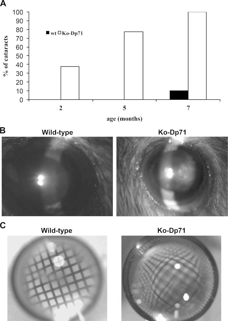
Purpose: Dp71 is the main product of the Duchenne muscular dystrophy (DMD) gene in the central nervous system. While studying the impact of its absence on retinal functions, we discovered that mice lacking Dp71 also developed a progressive opacification of the crystalline lens. The purpose of this study was to perform a detailed characterization of the cataract formation in Dp71 knockout (KO-Dp71) mice.

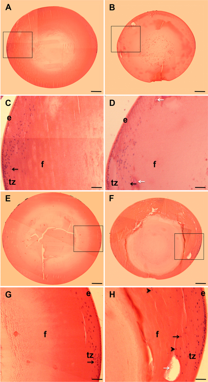
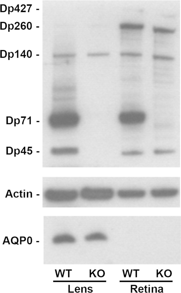
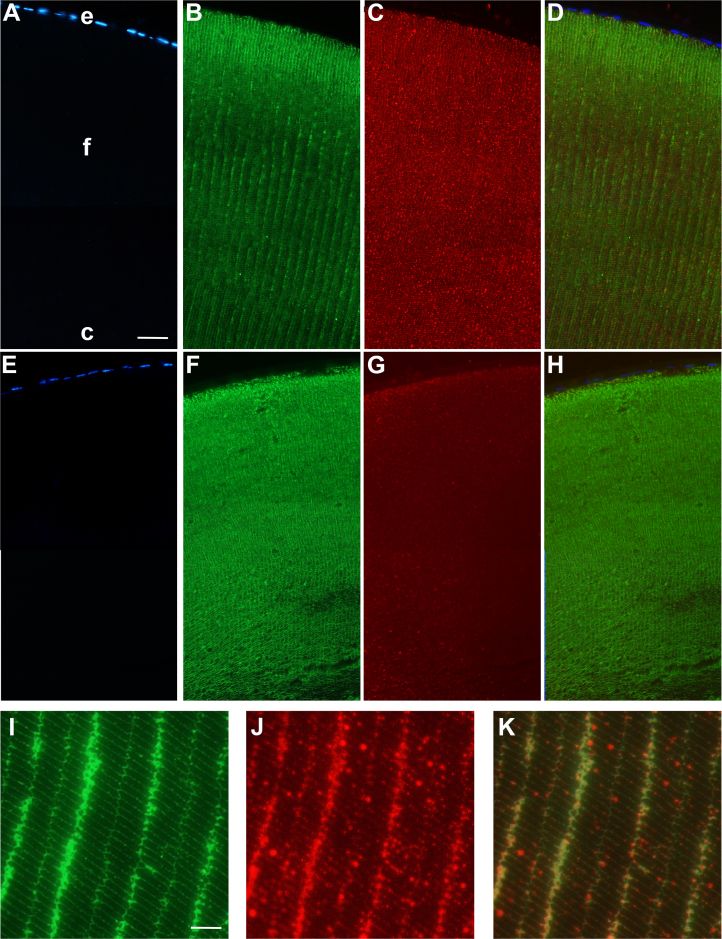
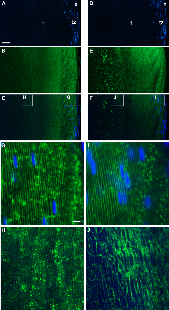
Methods: Cataract formations in KO-Dp71 mice and wild-type (wt) littermates were assessed in vivo by slit-lamp examination and ex vivo by histological analysis as a function of aging. The expression and cellular localization of the DMD gene products were monitored by western blot and immunohistochemical analysis. Fiber cell integrity was assessed by analyzing the actin cytoskeleton as well as the expression of aquaporin-0 (AQP0).

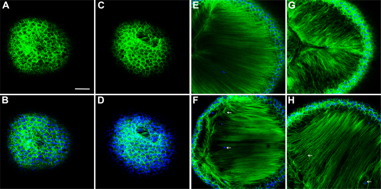
Results: As expected, a slit-lamp examination revealed that only one of the 20 tested wt animals presented with a mild opacification of the lens and only at the most advanced age. However, a lack of Dp71 was associated with a 40% incidence of cataracts as early as 2 months of age, which progressively increased to full penetrance by 7 months. A subsequent histological analysis revealed an alteration in the structures of the lenses of KO-Dp71 mice that correlated with the severity of the lens opacity. An analysis of the expression of the different dystrophin gene products revealed that Dp71 was the major DMD gene product expressed in the lens, especially in fiber cells. The role of Dp71 in fiber cells was also suggested by the progressive disorganization of the lens fibers, which was observed in the absence of Dp71 and demonstrated by irregular staining of the actin network and the aqueous channel AQP0.

Conclusions: While its role in the retina has been well characterized, this study demonstrates for the first time the role played by Dp71 in a different ocular tissue: the crystalline lens. It primarily demonstrates the role that Dp71 plays in the maintenance of the integrity of the secondary lens fibers.


Figures

Figure 1
Figure 1
A dramatic increase of the incidence of cataracts in the absence of Dp71. A: Time-course analysis of cataract formation in Dp71 knockout (KO-Dp71) mice. Observations were performed at 2, 5, and 7 months in wild-type (wt; black; n = 20) and KO-Dp71 mice (white; n = 31). While virtually no cataracts were observed in wt mice at any of the ages tested, the incidence of cataracts increased dramatically with age in KO-Dp71 mice until affecting all the KO animals tested at 7 months. B: Representative photographs of the eyes of sedated wt and KO-Dp71 mice at 7 months of age using a slit lamp. Whereas the lenses of the wt mice are entirely transparent, a strong opacification is observed in the lenses of the 7-month-old KO-Dp71 mice. C: Representative photographs of isolated crystalline lenses of wt and KO-Dp71 mice at 7 months of age on top of an EM microscopy grid showing the undistorted image obtained through a wt lens; the image is strongly distorted in the KO-Dp71 mice, indicating a refractive defect.
Figure 2
Figure 2
The absence of Dp71 is associated with histological alterations of the crystalline lens. Cross-sections of crystalline lenses from wt and KO-Dp71 mice eyes were stained with hematoxylin and eosin at 2 (A-D) and 7 (E-H) months of age. No major alterations in the histology of the crystalline lenses were observed with aging in wt mice (A and E); alternatively, a progressive disorganization was obvious in lenses from KO-Dp71 mice (B and F). Higher magnification images suggest alterations in the ultrastructure of the lenses in KO-Dp71 mice (D and H, white arrows) along with a disorganized transition zone at both 2 and 7 months of age (C versus D and G versus H, respectively; black arrows; e: epithelial cells; f: fiber cells; tz: transition zone). Scale bars A, B, E, and F: 300 μm; Scale bars C, D, G, and H: 80 μm.
Figure 3
Figure 3
Dp71 is the major dystrophin isoform expressed in the crystalline lens. Levels of the expression of DMD gene products and AQP0 in lenses and retinas from wt and KO-Dp71 mice were analyzed by immunoblotting. Actin levels were used as a loading control. As in the retina, Dp71 is the main DMD gene product expressed in the lens. We also detected Dp427, Dp260, and Dp140 in the retinas, as well as Dp260 and Dp140 in the lenses from both strains.
Figure 4
Figure 4
Dp71 localizes at the membrane of the lens fiber cells and its absence is associated with a loss of fiber cell organization. Immunolocalization of dystrophins (red) and AQP0 (green) in the anterior pole of the lens from wt (A-D) and KO-Dp71 (E-H) mice. Nuclei were counterstained with DAPI. Dystrophins are localized at the membrane of the secondary fiber together with the AQP0. As expected, total dystrophin staining in KO-Dp71 mice lenses was strongly decreased, confirming that Dp71 is the main product of the DMD gene in the crystalline lens. A comparison of the staining obtained for AQP0 in wt (B) and KO-Dp71 (F) mice confirms a strong disorganization of the ultrastructure of the lens fibers in the absence of Dp71. A weak staining remained in the epithelium of KO-Dp71 mice, suggesting that the other products of the DMD gene are mainly expressed by lens epithelial cells. Higher magnification images in wt crystalline lenses (I-K) clearly reveal a colocolalization of both proteins (K: merged) in the membranes of secondary fibers close to the anterior pole of the lens (e: epithelial cells; f: fiber cells; c: core). Scale bar A: 50 μm; Scale bar I:15 μm.
Figure 5
Figure 5
The absence of Dp71 leads to a dramatic disorganization of the secondary fibers close to the germinative zone of the lens. Immunolocalization of AQP0 (green) in the germinative zones of the lenses from wt (A-C) and KO-Dp71 (D-F) mice. Nuclei were counterstained with DAPI (blue). As expected, AQP0 is localized at the membrane of the secondary lens fibers in wt mice. In the absence of Dp71, the regular consistent staining of AQP0 is dramatically altered. Higher magnification images of the periphery of the germinative zone of wt (G) and KO-Dp71 (I) mice show limited disruptions to AQP0 staining. As well, images from the deeper regions of the KO-Dp71 (J) mice show an almost complete loss of organization of the secondary lens fibers when compared to similar regions in wt animals (H) (e: epithelial cells; f: fiber cells; tz: transition zone). Scale bar A: 50 μm; Scale bar I: 8 μm.
Figure 6
Figure 6
The absence of Dp71 leads to a profound disorganization of the actin filaments. Images of the F-actin network (green) and the cell nuclei (blue) at the level of the epithelium of 9-month-old wt (A-B) and KO-Dp71 mice (C-D). Similar to the results obtained by immunostaining, the network of actin filaments in lens epithelial cells was not different in wt (A-B) versus KO-Dp71 (C-D) mice. However, images of the actin network deep within the lens demonstrate subtle but noticeable changes in the lens fiber cell region at 3 months (E: wt-F:KO-Dp71), as well as changes that were exacerbated with aging, as demonstrated at 9 months (G: wt-H:KO-Dp71). Scale bar: 300 μm.

References

    1. Wride MA. Lens fibre cell differentiation and organelle loss: many paths lead to clarity. Philos Trans R Soc Lond B Biol Sci. 2011;366:1219–33. - PMC - PubMed
    1. Koenig M, Hoffman EP, Bertelson CJ, Monaco AP, Feener C, Kunkel LM. Complete cloning of the Duchenne muscular dystrophy (DMD) cDNA and preliminary genomic organization of the DMD gene in normal and affected individuals. Cell. 1987;50:509–17. - PubMed
    1. Gee SH, Madhavan R, Levinson SR, Caldwell JH, Sealock R, Froehner SC. Interaction of muscle and brain sodium channels with multiple members of the syntrophin family of dystrophin-associated proteins. J Neurosci. 1998;18:128–37. - PMC - PubMed
    1. Holt KH, Campbell KP. Assembly of the sarcoglycan complex. Insights for muscular dystrophy. J Biol Chem. 1998;273:34667–70. - PubMed
    1. Neely JD, Amiry-Moghaddam M, Ottersen OP, Froehner SC, Agre P, Adams ME. Syntrophin-dependent expression and localization of Aquaporin-4 water channel protein. Proc Natl Acad Sci USA. 2001;98:14108–13. - PMC - PubMed

Publication types

MeSH terms